.webp&w=384&q=75)

Loading, please wait...
Loading, please wait...
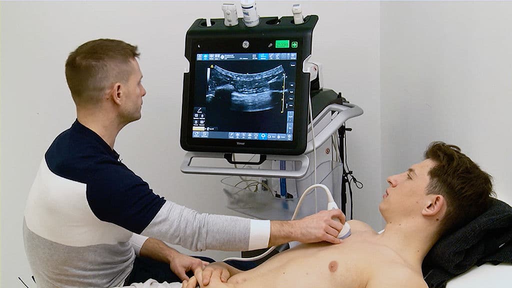
The Bedside Lung Ultrasound in Emergency (BLUE) protocol demonstrated that structured lung ultrasonography can help differentiate major causes of acute respiratory failure. In a cohort of patients with acute dyspnea, combinations of A-lines, B-lines, lung sliding, consolidation and venous findings were used to generate diagnostic profiles. The study showed high sensitivity and specificity for several common causes, including pulmonary edema, asthma or COPD and pneumothorax-related patterns. For internists, the value of POCUS is that it can be performed at the bedside without ionizing radiation and repeated when the patient's condition changes. Lung ultrasound should not replace a full clinical assessment or formal imaging when indicated, but it can accelerate diagnostic reasoning and support early treatment decisions. Training needs to emphasize probe positioning, artifact recognition and integration with cardiac and vascular findings. In resource-constrained settings, the portability of ultrasound is particularly attractive. The evidence supports incorporating basic lung ultrasound into the diagnostic toolkit of acute-care physicians rather than limiting ultrasound to radiology or critical-care specialists.

Read summarized clinical updates, watch expert medical content, and earn CME certifications right from your smartphone.
Open your smartphone camera to scan and install.





The Bedside Lung Ultrasound in Emergency (BLUE) protocol demonstrated that structured lung ultrasonography can help differentiate major causes of acute respiratory failure. In a cohort of patients with acute dyspnea, combinations of A-lines, B-lines, lung sliding, consolidation and venous findings were used to generate diagnostic profiles. The study showed high sensitivity and specificity for several common causes, including pulmonary edema, asthma or COPD and pneumothorax-related patterns. For internists, the value of POCUS is that it can be performed at the bedside without ionizing radiation and repeated when the patient's condition changes. Lung ultrasound should not replace a full clinical assessment or formal imaging when indicated, but it can accelerate diagnostic reasoning and support early treatment decisions. Training needs to emphasize probe positioning, artifact recognition and integration with cardiac and vascular findings. In resource-constrained settings, the portability of ultrasound is particularly attractive. The evidence supports incorporating basic lung ultrasound into the diagnostic toolkit of acute-care physicians rather than limiting ultrasound to radiology or critical-care specialists.
4 weeks back

A bioengineered oral nanozyme microsupplement converts pathological ROS into a protective polydopamine hydrogel shield. By curbing neutrophil extracellular traps and balancing T cells, this platform outperforms standard 5-ASA in preclinical colitis models, opening new paths for sustained remission.
3 days back
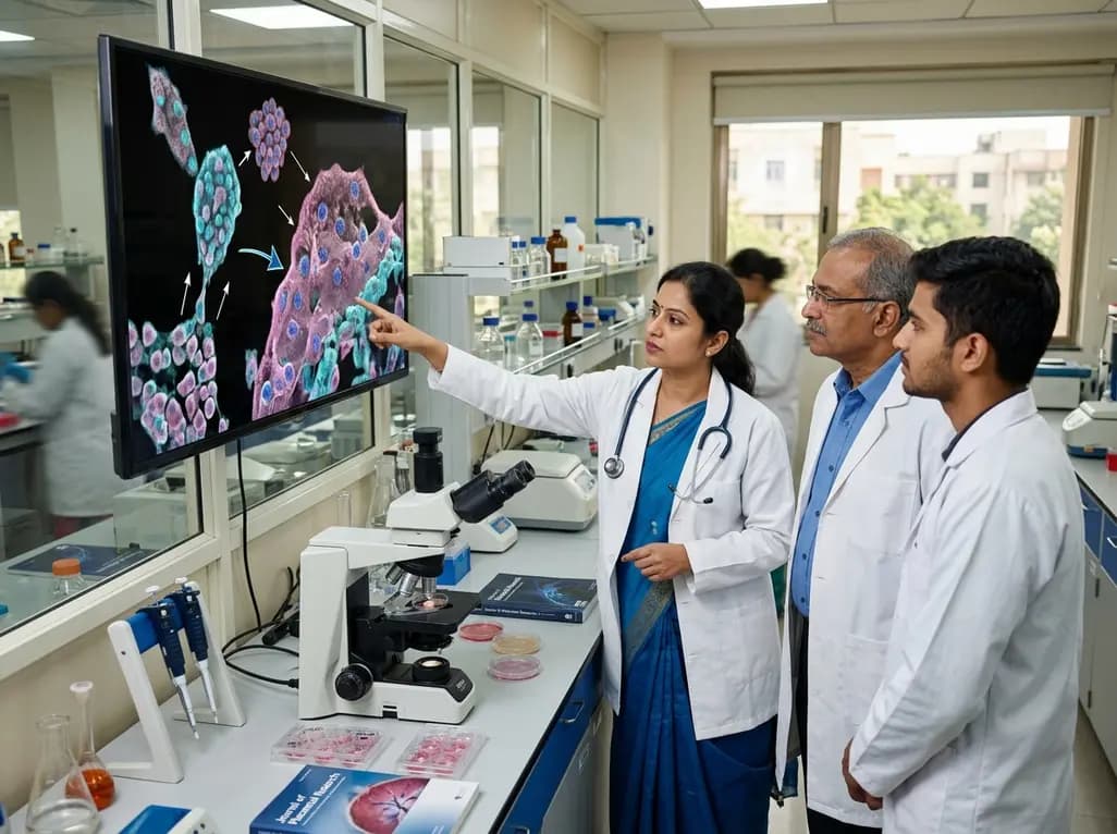
Recent research reveals that downregulating YAP1/TAZ-TEAD via Hippo signaling triggers spontaneous human trophoblast syncytialization in 3D cultures, illuminating placental biology and preeclampsia.
4 weeks back

Abdominal aortic aneurysm rupture remains a catastrophic vascular emergency. Groundbreaking research reveals that USP53 accelerates aneurysm progression by reprogramming smooth muscle cell metabolism toward aerobic glycolysis, highlighting a promising target beyond conventional diameter-based surveillance.
4 days back

A breakthrough study leverages natural language processing to accurately identify incident medication-related osteonecrosis of the jaw and track antiresorptive drug holidays from clinical narratives in electronic health records, advancing osteoporosis pharmacovigilance and dental safety.
4 days back

Recent research defines ferro-aging as a conserved iron-lipid axis driving organ decline across primates. Age-related iron dyshomeostasis stimulates ACSL4-mediated lipid peroxidation, promoting cellular senescence. Notably, vitamin C directly inhibits ACSL4, suppressing phospholipid oxidation and multi-organ decay.
5 days back
