.webp&w=384&q=75)

Loading, please wait...
Loading, please wait...
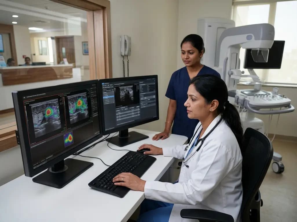
Evaluating indeterminate breast masses remains a persistent diagnostic challenge in modern radiological practice. Specifically, lesions categorized under the Breast Imaging Reporting and Data System (BI-RADS) as Category 3 (probably benign) or Category 4 (suspicious) frequently present overlapping morphological features. Consequently, clinicians often face difficult decisions regarding whether to recommend serial short-interval imaging surveillance or invasive tissue sampling. To address this clinical uncertainty, integrating automated breast ultrasound AI systems into diagnostic algorithms has emerged as a promising solution. By providing reproducible, automated quantitative analysis of lesion morphology across multiplanar reconstructions, machine-learning algorithms assist radiologists in making precise risk assessments. Recent clinical evidence demonstrates that applying artificial intelligence to automated breast ultrasound scans significantly enhances diagnostic performance for these challenging intermediate lesions.
Breast ultrasound is an essential modality for identifying focal tissue abnormalities, particularly in patients with dense parenchymal patterns. However, conventional hand-held ultrasound depends heavily on operator skill, transducer positioning, and subjective image interpretation. This variability often leads to inconsistent classification of ambiguous masses, particularly BI-RADS 3 and BI-RADS 4 lesions. While BI-RADS 3 findings carry a lower than two percent probability of malignancy, BI-RADS 4 lesions possess a broad risk spectrum ranging from three percent to over ninety-five percent suspicion. Consequently, radiologists frequently err on the side of caution to avoid delayed cancer diagnoses.
This conservative approach triggers a massive volume of core needle biopsies. Furthermore, tissue sampling procedures introduce physical discomfort, emotional anxiety, and financial burdens for patients. Additionally, the vast majority of core needle biopsies performed on BI-RADS 3 and BI-RADS 4A lesions ultimately yield benign histological diagnoses. Therefore, modern breast imaging requires objective diagnostic tools that can reliably differentiate malignant neoplasms from benign structural changes. Computer-aided diagnostic systems leverage advanced machine-learning algorithms to analyze intricate image characteristics that may elude human vision. Ultimately, refining the pre-biopsy stratification of intermediate lesions can optimize patient management and streamline clinical workflows across comprehensive care centers.
Automated breast ultrasound technology utilizes a standardized broad-bandwidth transducer that systematically captures full-volume images of the entire breast tissue. Subsequently, the system generates three-dimensional dataset reconstructions across axial, coronal, and sagittal planes. When paired with machine-learning algorithms, automated breast ultrasound AI evaluates key structural features including lesion margins, acoustic shadowing, posterior echogenicity, and spatial orientation. Radiologists manually delineate a region of interest around the target lesion, enabling the computer-aided diagnostic tool to instantaneously perform an automated feature analysis.
Furthermore, machine-learning models trained on extensive histological data assess subtle acoustic patterns with high mathematical precision. Consequently, the software outputs a standardized score reflecting the likelihood of malignancy. This objective score helps standardise image interpretation across institutions, reducing inter-observer variability between junior trainees and experienced breast specialists. Moreover, multiplanar volume acquisition ensures that coronal plane retraction architectural features are thoroughly evaluated, providing a complete structural profile. By serving as an intelligent second reader, artificial intelligence enhances confidence during real-time image interpretation. As a result, radiologists can make more accurate triage recommendations, ensuring high-risk lesions are promptly biopsied while truly benign lesions are managed conservatively.
A recently published clinical study investigated the diagnostic performance of a machine-learning computer-aided diagnostic tool applied to automated breast ultrasound images in fifty-four patients with sixty-eight BI-RADS 3 and 4 lesions. Histopathological verification via core needle biopsy established forty positive malignant lesions and twenty-eight negative benign findings. Remarkably, the artificial intelligence system accurately identified thirty-eight suspicious lesions, exhibiting only six false-positive results and two false-negative classifications. These results yielded a sensitivity of ninety-five percent, a specificity of seventy-nine percent, and an overall diagnostic accuracy of eighty-eight percent.
Additionally, the positive predictive value reached eighty-six percent, while the negative predictive value achieved an impressive ninety-two percent. Notably, a high negative predictive value is clinically vital because it gives practitioners high confidence when ruling out malignancy in low-suspicion lesions. Furthermore, achieving seventy-nine percent specificity within a cohort restricted exclusively to intermediate BI-RADS categories demonstrates robust discrimination capabilities. In typical clinical practice, unassisted ultrasound interpretation for BI-RADS 4 lesions often exhibits lower specificity due to overlapping morphological features. Therefore, these quantitative findings validate the artificial intelligence algorithm as an exceptionally sensitive and reliable adjunct tool for characterizing complex breast masses.
The clinical integration of high-precision diagnostic tools significantly alters patient care trajectories by curbing unnecessary invasive interventions. Core needle biopsy, although safe, remains an invasive procedure associated with risks such as hematoma formation, localized infection, post-procedure pain, and tissue scarring. Moreover, waiting for histological confirmation causes substantial psychological stress for patients and their families. Consequently, reducing false-positive interpretations in BI-RADS 3 and BI-RADS 4 lesions directly reduces the burden of unnecessary invasive diagnostics on healthcare systems.
By correctly identifying true-negative lesions with a ninety-two percent negative predictive value, artificial intelligence software empowers clinicians to comfortably assign low-risk lesions to imaging surveillance rather than immediate biopsy. Furthermore, the impressive ninety-five percent sensitivity ensures that true malignancies are rarely overlooked, preserving diagnostic safety. Consequently, patients with confirmed suspicious features proceed rapidly to definitive tissue sampling and prompt oncological intervention. Additionally, minimizing unnecessary core needle biopsies reduces hospital expenditures, optimizes pathology department throughput, and improves patient satisfaction scores. Ultimately, intelligent decision-support tools harmonise clinical accuracy with patient-centered care principles, fostering a more efficient and compassionate diagnostic environment.
Implementing artificial intelligence into daily radiology operations requires careful consideration of clinical workflow integration and operational efficiency. Automated breast ultrasound systems generate large three-dimensional imaging datasets that can require extended reading times for busy radiologists. However, integrating automated computer-aided diagnostic tools accelerates feature analysis by immediately highlighting critical morphological attributes once a region of interest is designated. Consequently, clinicians spend less time measuring complex margins and evaluating subtle acoustic shadows across multiple orthogonal planes.
Moreover, artificial intelligence acts as an invaluable educational and diagnostic aid in community hospitals or resource-limited settings where dedicated breast imaging specialists may be unavailable. Less experienced practitioners can utilize algorithmic outputs as a standardized second opinion, thereby narrowing the diagnostic performance gap between general radiologists and subspecialized mammographers. Additionally, standardized quantitative analysis facilitates clearer communication between radiologists, surgical oncologists, and referring primary care physicians. Seamless integration into picture archiving and communication systems allows seamless reporting and automated data entry. Therefore, adopting computer-aided software into routine automated ultrasound protocols enhances both diagnostic consistency and institutional operational efficiency.
As deep learning and artificial intelligence architectures continue to evolve, computer-aided diagnostic systems for automated breast ultrasound will become increasingly sophisticated. Future software iterations may incorporate automated lesion detection directly from full-volume scans, eliminating the initial manual region-of-interest drawing step. Furthermore, combining multi-modal imaging data—such as digital mammography, contrast-enhanced mammography, magnetic resonance imaging, and patient clinical risk profiles—into unified multi-parametric artificial intelligence models will further elevate diagnostic precision.
Simultaneously, prospective multi-center clinical trials with larger, diverse patient cohorts are essential to validate these machine-learning tools across varied ethnic populations and equipment manufacturers. Establishing robust regulatory oversight and standardized validation frameworks will ensure safe, ethical, and equitable deployment across clinical practice settings. Additionally, ongoing physician training will remain essential so that healthcare providers interpret artificial intelligence scores as supportive decision-making instruments rather than autonomous diagnostic replacements. Ultimately, pairing advanced automated ultrasound imaging with artificial intelligence heralds a new era in precision breast medicine, where early cancer detection and minimized intervention coexist harmoniously.
Automated breast ultrasound utilizes a wide-field transducer to automatically acquire comprehensive three-dimensional volumetric images of the entire breast tissue. Unlike hand-held ultrasound, which depends heavily on operator technique and manual probe manipulation, automated system scans produce highly reproducible, standardized datasets in axial, coronal, and sagittal planes. Consequently, this standardized acquisition allows seamless integration with artificial intelligence decision-support software for uniform lesion characterization across different clinical settings.
BI-RADS 3 lesions are classified as probably benign with a low risk of cancer, whereas BI-RADS 4 lesions carry suspicious features requiring biopsy consideration. However, overlapping structural characteristics like subtle margin irregularities or micro-lobulations often create interpretation uncertainty among radiologists. Consequently, clinicians frequently perform core needle biopsies on benign lesions to avoid missing cancers, leading to higher rates of unnecessary invasive procedures and patient anxiety.
No, artificial intelligence software is designed to function as an advanced computer-aided diagnostic assistant rather than an autonomous reader. Radiologists retain full clinical responsibility for drawing regions of interest, interpreting algorithmic findings within the broader clinical context, and making final management decisions. Ultimately, artificial intelligence serves as an intelligent second opinion that improves diagnostic accuracy, reduces inter-observer variability, and minimizes unnecessary invasive tissue biopsies.
Disclaimer: This content is for informational and educational purposes only... Refer to the latest local and national guidelines for clinical practice.
References

Read summarized clinical updates, watch expert medical content, and earn CME certifications right from your smartphone.
Open your smartphone camera to scan and install.





A clinical study shows that automated breast ultrasound paired with artificial intelligence accurately classifies BIRADS 3-4 lesions, reaching 95% sensitivity and 79% specificity. This diagnostic advance promises to reduce unnecessary core needle biopsies and refine clinical workflows in breast imaging.
last month
A randomized trial shows oral DL-3-n-butylphthalide (200 mg thrice daily) significantly slows functional decline in spinocerebellar ataxia type 3 over 12 months, improving SARA and SCAFI scores with favorable safety and zero serious adverse events.
Last week
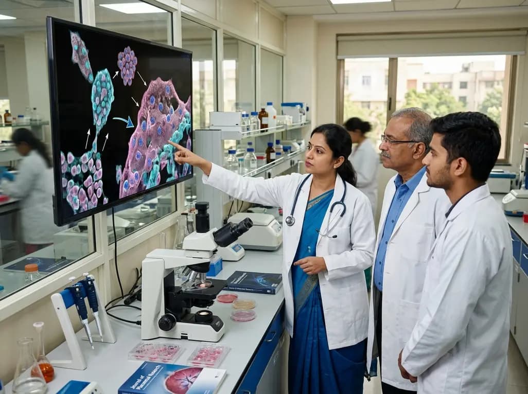
Recent research reveals that downregulating YAP1/TAZ-TEAD via Hippo signaling triggers spontaneous human trophoblast syncytialization in 3D cultures, illuminating placental biology and preeclampsia.
2 weeks back
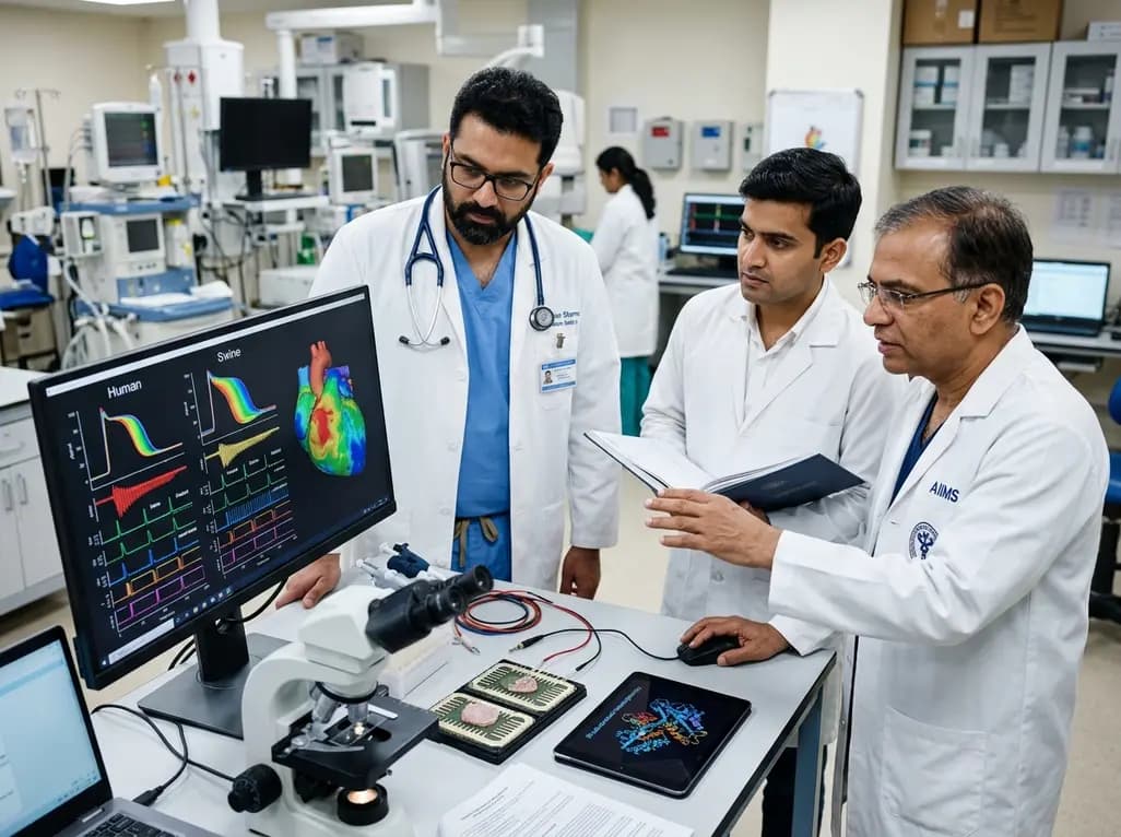
A landmark comparative study evaluates atrial electrophysiology across humans, swine, and rodents. By linking cellular recordings with transcriptomics and in silico modeling, researchers resolve cross-species gaps, explaining translational failures to advance targeted therapies for atrial arrhythmias.
2 weeks back
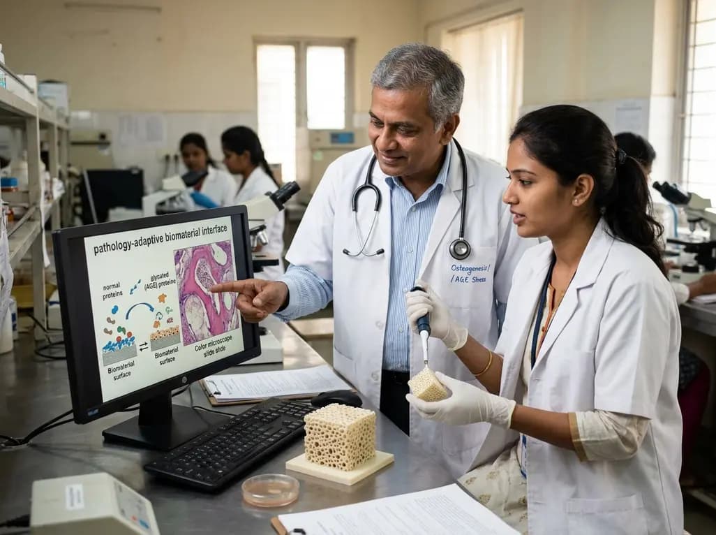
A novel pathology-adaptive surface engineering strategy uses functionalized plasma polymer coatings to selectively modulate AGE adsorption, reducing oxidative stress and restoring bone formation in diabetic and aging microenvironments.
last month
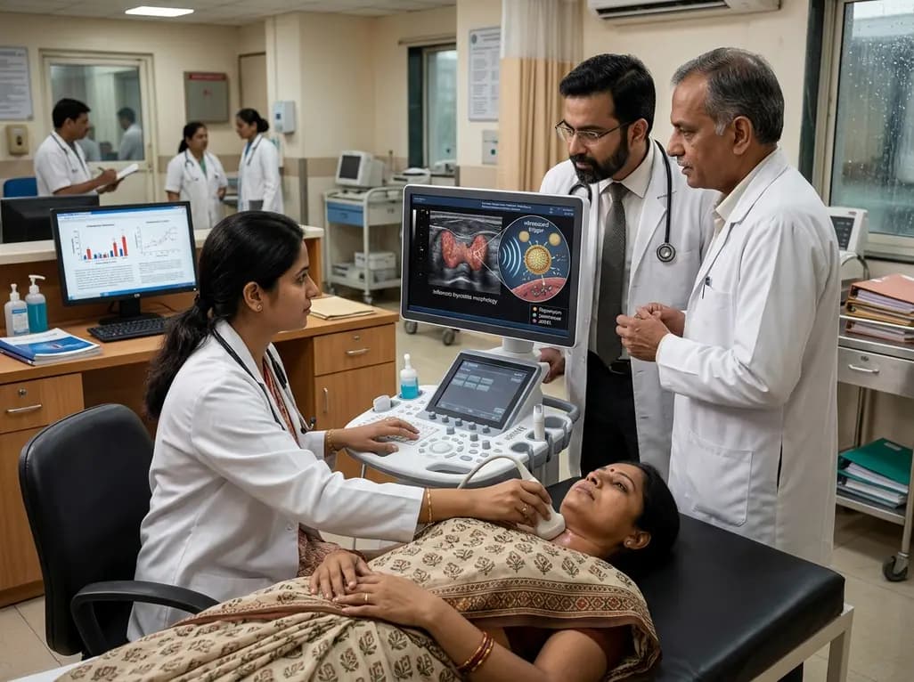
Researchers have developed an innovative ultrasound-responsive nano-contrast agent that co-delivers CIITA-siRNA and rapamycin directly to inflamed thyroid tissue, significantly reducing autoimmune injury and thyroid autoantibodies in preclinical models of Hashimoto thyroiditis.
2 weeks back
