.webp&w=384&q=75)

Loading, please wait...
Loading, please wait...
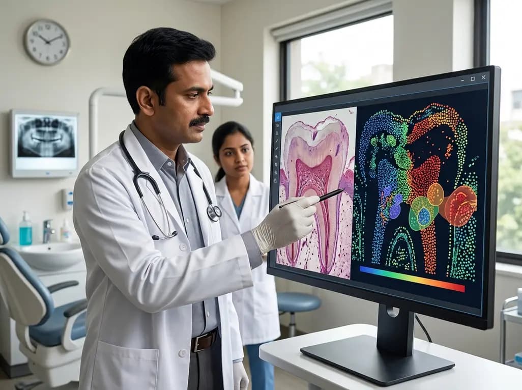
For decades, clinicians have relied on subjective pain reports and thermal sensibility tests to determine the fate of an inflamed dental pulp. Traditionally, the classification of pulpitis into reversible (RP) and irreversible (IP) stages has served as the primary guide for treatment decisions. However, this clinical dichotomy often fails to reflect the true histological and molecular landscape of the tissue. Recent advancements in spatial transcriptomics in pulpitis research are beginning to highlight a significant discordance between what the patient feels and the actual healing potential of the pulp. This technological leap allows scientists to map gene expression directly onto tissue sections, providing a spatially resolved view of cellular responses. Consequently, we are moving toward a more nuanced understanding of pulpal biology that could revolutionize endodontic practice. Understanding these molecular underpinnings is essential for dentists who wish to prioritize vital pulp therapy over more invasive root canal procedures. As the field evolves, the integration of high-resolution transcriptomic data may eventually provide the precision tools needed to distinguish between a pulp that is truly beyond repair and one that merely requires a conservative intervention. This shift in perspective is critical for improving long-term tooth retention and patient outcomes in clinical dentistry.
In daily dental practice, the transition from reversible to irreversible pulpitis is frequently treated as a definitive threshold. Clinicians typically diagnose IP when a patient presents with lingering pain to cold or spontaneous nocturnal throbbing. While these symptoms are distressing, they do not always correlate with a total loss of pulpal regenerative capacity. Furthermore, studies have shown that histological evidence of necrosis or severe inflammation can be localized to a small portion of the coronal pulp while the radicular tissue remains healthy. Therefore, labeling an entire tooth as "irreversible" based on localized symptoms can lead to overtreatment. The current diagnostic protocols are inherently limited because they lack the ability to assess real-time molecular signaling or immune cell interactions within the pulp chamber. Consequently, many teeth that could have been saved through pulpotomy or other conservative measures are instead subjected to complete devitalization via root canal therapy. This biological disconnect highlights the urgent need for diagnostic refinements that go beyond pulp sensibility. By exploring the transcriptomic state of the tissue, researchers hope to identify signatures that accurately predict whether the pulp can resolve inflammation after the removal of the primary irritant, such as dental caries.
The emergence of spatial transcriptomics in pulpitis research provides a powerful method for analyzing the complex cellular environment of the tooth. Unlike traditional bulk RNA sequencing, which grinds up tissue and loses all spatial context, spatial transcriptomics preserves the geographical location of gene expression. In this pilot study, researchers utilized the Visium-CytAssist-V2 platform to analyze human dental pulp tissues. This technology allows for the precise mapping of transcripts across different zones of the pulp, including the area directly adjacent to a carious lesion. By using cell deconvolution techniques, scientists can identify exactly which cell types—such as fibroblasts, odontoblasts, or immune cells—are active in specific regions. Moreover, this approach enables the identification of pathway activation patterns, such as those related to neuroinflammation or toll-like receptor 4 (TLR4) signaling. This level of detail is unprecedented in endodontic research and provides a high-resolution "map" of the inflammatory process. Notably, the study revealed that some samples clinically diagnosed as irreversible actually possessed molecular profiles nearly identical to reversible cases. This finding suggests that spatial transcriptomics can uncover hidden healing potential that is currently invisible to the clinician’s eye, potentially shifting the standard of care toward more biological, pulp-preserving strategies.
One of the most compelling findings from recent transcriptomic research is the role of immune-fibroblast interactions in determining pulpal fate. Historically, the focus of inflammation was primarily on immune cell infiltration, such as macrophages and neutrophils. However, spatial data suggests that fibroblasts are not merely passive structural cells but are active participants in the inflammatory milieu. The ratio of immune cells to fibroblasts appears to be a critical indicator of whether the pulpitis is likely to be reversible. In samples that exhibited a "reversible-like" molecular profile, the interaction between these two cell populations was characterized by specific activation patterns in TLR4 and neuroinflammation pathways. Furthermore, the study identified that even in cases of symptomatic IP, the molecular landscape could still reflect a state of manageable inflammation rather than total tissue breakdown. Therefore, the crosstalk between fibroblasts and the immune system may dictate the success of vital pulp therapy. By understanding these spatial relationships, clinicians can better appreciate how the pulp attempts to wall off infection and initiate repair. This insight underscores the importance of the pulp's microenvironment in the clinical decision-making process, suggesting that preserving the immune-fibroblast balance may be the key to avoiding unnecessary tooth devitalization.
To translate transcriptomic findings into clinical utility, identifying specific molecular markers is essential. The pilot study highlighted several candidate genes that were differentially expressed in reversible-like profiles compared to severely inflamed IP. For instance, increased expression of Pleiotrophin (PTN) and CXCL14 emerged as potential indicators of a favorable healing environment. Conversely, a lower expression of genes such as ENG (CD105), SELE, and CXCL13 was associated with tissues that retained a reversible-like status despite clinical symptoms. These markers are involved in various processes, including cell migration, tissue remodeling, and immune signaling. Consequently, these genes could serve as the foundation for future diagnostic tools, such as chairside biomarkers or specialized pulp-testing kits. In addition, the downregulation of specific inflammatory mediators like COL4A1 and CXCL1 in RP-like samples provides further evidence that the molecular state of the pulp is a continuum rather than a set of discrete stages. While these findings are preliminary and require validation in larger patient cohorts, they offer a promising roadmap for the future of endodontics. If clinicians can eventually test for these specific markers during a procedure, they can make far more informed decisions about whether to perform a full root canal or a conservative pulpotomy.
The push toward precision medicine is gaining momentum in India, and endodontics is no exception. With a high burden of dental caries and a growing demand for tooth-preserving treatments, the insights gained from spatial transcriptomics in pulpitis are highly relevant. Many patients in India seek affordable and effective ways to save their natural teeth, and vital pulp therapy represents a cost-effective alternative to complex root canal treatments. However, the success of these therapies depends entirely on accurate case selection. Therefore, integrating molecular insights into clinical practice could significantly reduce the rate of endodontic failures and unnecessary extractions. Furthermore, as biotechnology infrastructure continues to grow in India, the possibility of incorporating biomarker-based diagnostics into specialized clinics becomes more feasible. Moving forward, Indian dental schools and research institutions could play a vital role in validating these transcriptomic markers within diverse populations. This research not only enhances our biological understanding but also empowers dentists to provide personalized care based on the actual health of the pulp rather than subjective symptoms alone. Ultimately, the transition to a more molecularly-informed diagnostic framework will help preserve the "heart" of the tooth for more patients across the country.
Traditional pulp testing, such as thermal or electric sensibility tests, only assesses the sensory response of the pulpal nerves or the presence of blood flow. It provides a limited 'yes/no' answer regarding vitality but tells us little about the actual health of the tissue. In contrast, spatial transcriptomics maps the expression of thousands of genes across the tissue, revealing the specific immune and cellular activities occurring in different regions of the pulp.
The ratio of immune cells to fibroblasts is a critical marker because it reflects the balance between active inflammation and tissue repair capacity. Fibroblasts are essential for structural maintenance and healing, while an excessive influx of immune cells often signals severe, non-resolving inflammation. A balanced ratio, as seen in reversible-like profiles, suggests that the pulp still possesses the necessary cellular machinery to recover if the irritant is removed.
While we are not yet at the stage of routine chairside genetic testing, these findings should encourage clinicians to consider vital pulp therapy (VPT) more frequently, even when symptoms seem severe. The research shows that clinical signs of 'irreversible' pulpitis do not always mean the tissue is dead. By using high-quality bioceramic materials and careful pulpotomy techniques, dentists can potentially save many teeth that were previously destined for root canal therapy.
Disclaimer: This content is for informational and educational purposes only. It is not intended to be a substitute for professional medical or dental advice, diagnosis, or treatment. Always seek the advice of your physician, dentist, or other qualified healthcare provider with any questions you may have regarding a medical condition. Refer to the latest local and national guidelines for clinical practice.
References
Zaky S et al. Pilot spatial transcriptomics of dental pulpitis suggests immune-fibroblast profiling linked to reversibility. J Transl Med. 2026 Jul 03. doi: 10.1186/s12967-026-08520-4. PMID: 42399991.
Ricucci D et al. Correlation between clinical and histologic diagnosis of pulp diseases: a critical review. J Endod. 2014;40(12):1932-1939.
Zhu et al. Success rate of full pulpotomy for teeth with irreversible pulpitis: A systematic review and meta-analysis. J Clin Med. 2024;13(4):1120.

Read summarized clinical updates, watch expert medical content, and earn CME certifications right from your smartphone.
Open your smartphone camera to scan and install.





A pilot study using spatial transcriptomics reveals that clinical diagnoses of pulpitis often mismatch underlying molecular profiles. Researchers identified immune-fibroblast interactions as potential markers for pulpal reversibility, offering hope for more conservative, tooth-preserving treatments.
3 months ago

Depemokimab offers twice-yearly dosing for severe eosinophilic asthma and chronic rhinosinusitis with nasal polyps, significantly cutting exacerbations and hospital admissions.
3 days back

A novel copper metal-organic framework hydrogel remodels the uterine microenvironment to stimulate robust endometrial regeneration, reduce intrauterine fibrosis, and restore reproductive fertility, offering a groundbreaking strategy for managing refractory endometrial damage and Asherman syndrome.
3 days back

New research reveals that non-ablative radiotherapy suppresses ventricular tachycardia by increasing conduction velocity via NaV1.5 upregulation and action potential prolongation. However, sustained sodium current elevation and depressed calcium handling highlight the need for careful clinical translation.
5 days back

A single-cell transcriptomic atlas reveals how peripheral nerve injuries cause dorsal root ganglion remodeling. By evaluating proximal nerve crush and resection, researchers identify distinct glial and immune programs that resolve neuropathic pain and promote regeneration.
3 days back

A breakthrough study demonstrates that fullerol-primed extracellular vesicles generated in a 3D SECCS bioreactor attenuate ferroptosis and restore myogenic regeneration in diabetes via the FOXO3 pathway, offering a cell-free therapeutic strategy for diabetic musculoskeletal degeneration.
3 days back
