.webp&w=384&q=75)

Loading, please wait...
Loading, please wait...
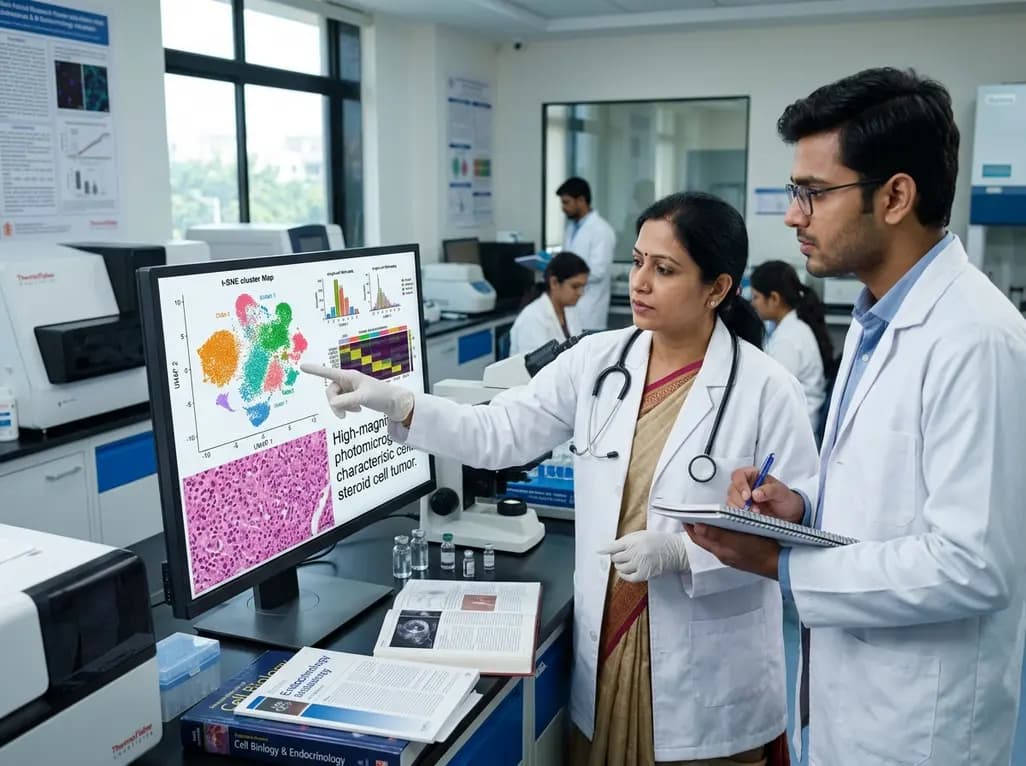
Ovarian steroid cell tumors-not otherwise specified (SCT-NOS) represent an exceptionally rare subset of sex cord-stromal tumors. These neoplasms account for less than 0.1% of all ovarian tumors, making them a significant diagnostic challenge for clinicians. Patients typically present with symptoms of hyperandrogenism, such as hirsutism, voice deepening, and other virilization signs. In a recent case study, a 41-year-old woman presented with severe virilization, leading to the diagnosis of this rare tumor. While clinicians often encounter polycystic ovary syndrome (PCOS) as the primary cause of androgen excess, the rapid onset of virilization often points toward a more aggressive pathology like SCT-NOS. Consequently, understanding the molecular drivers behind these tumors is essential for effective management. Traditional histopathological analysis identifies these tumors by their characteristic polygonal cells and positive staining for markers like inhibin. Ovarian SCT-NOS transcriptome analysis now provides a deeper look into the cellular landscape of these malignancies. By examining the tumor at a single-cell level, researchers identify specific cell subtypes driving hormonal overproduction and tumor growth.
Recent breakthroughs in genomic technologies have revolutionized our approach to rare oncology cases. Single-cell RNA sequencing (scRNA-seq) allows scientists to dissect the heterogeneity within a single tumor specimen. This precision is particularly valuable for ovarian SCT-NOS, where various cell types interact within a complex microenvironment. Researchers recently utilized this technology to create a comprehensive cellular atlas of an SCT-NOS case. By sequencing individual cells, they successfully identified seven distinct cell types within the tumor. This granular view surpasses the capabilities of bulk RNA sequencing, which often masks rare but important cell populations. Furthermore, the study highlighted aberrant gene expression patterns previously uncharacterized in this tumor category. Notably, this transcriptomic approach revealed how different cell clusters contribute to the overall clinical presentation. Understanding these interactions is vital for developing targeted therapies. For instance, identifying specific pathways helps clinicians understand why some patients respond better to surgery. Moreover, this high-resolution mapping sets the stage for future investigations into these rare malignancies, allowing for more precise diagnostic markers.
The hallmark of SCT-NOS is the production of steroid hormones leading to significant clinical symptoms. The recent single-cell study identified four subtypes of keratin 19 (KRT19)+ steroidogenic cells within the tumor microenvironment. These KRT19+ cells are central to the tumor's ability to synthesize hormones. Specifically, these subtypes showed significant enrichment in genes related to ovarian steroidogenesis and steroid biosynthesis pathways. This molecular finding correlates perfectly with the hyperandrogenism observed in the clinical setting. By focusing on these steroidogenic clusters, researchers can pinpoint the exact enzymatic pathways that are upregulated. Consequently, this information could lead to the use of specific enzyme inhibitors as adjunct treatments in recurrent cases. Furthermore, the identification of KRT19 provides a new perspective on the origin of these tumor cells. While these tumors were thought to arise from the ovarian stroma, the transcriptomic signature suggests a more specialized origin. Understanding these pathways aids in diagnosis and provides a roadmap for monitoring therapeutic response through systemic hormonal levels, specifically androgens like testosterone.
The tumor microenvironment (TME) of SCT-NOS contains a complex array of immune cells. The study identified five distinct immune cell types within the tumor, with tumor-associated macrophages (TAMs) and dendritic cells (DCs) being the most prevalent. This finding is significant because the immune landscape often dictates the tumor's progression and its response to treatment. TAMs, in particular, are known to play a dual role in cancer, sometimes promoting growth and other times acting against it. In the context of SCT-NOS, the predominance of these cells suggests a highly active immune interface. Moreover, the presence of dendritic cells indicates the tumor is interacting with the body's adaptive immune system. This transcriptomic insight into the microenvironment opens the door for potential immunotherapy applications in rare ovarian tumors. Consequently, this study provides a foundational blueprint for future research into the immunobiology of sex cord-stromal tumors. Understanding how immune cells interact with steroidogenic cells might reveal how the hormonal environment influences immune evasion and local tumor spread.
For clinicians in India, the diagnosis of SCT-NOS requires a high index of suspicion in women with rapid-onset virilization. While the gold standard remains histopathological analysis, the insights from Ovarian SCT-NOS transcriptome analysis are beginning to influence clinical thinking. Specifically, the identification of key genes and cell subtypes assists in the differential diagnosis from other sex cord-stromal tumors. Since roughly one-third of SCT-NOS cases exhibit malignant behavior, identifying molecular markers of aggression is paramount. Furthermore, the study's findings on the immune microenvironment could lead to more personalized management strategies. Currently, surgical resection remains the primary treatment, but the high recurrence rate in some patients necessitates better follow-up protocols. Monitoring systemic androgen levels continues to be a vital part of post-operative care. However, adding molecular profiling could help stratify patients into different risk categories. Consequently, this research encourages a multi-disciplinary approach involving pathologists, endocrinologists, and oncologists to optimize patient outcomes and ensure that every aspect of the tumor's biology is considered during therapy planning.
The study of SCT-NOS through single-cell transcriptomics marks a significant step toward precision medicine in gynecological oncology. By providing a detailed cellular atlas, researchers have moved beyond descriptive pathology into functional molecular analysis. This transition is essential for rare diseases where large-scale clinical trials are difficult to conduct. Instead, we must rely on high-fidelity data from individual cases to guide decisions. Furthermore, the discovery of KRT19+ steroidogenic cell subtypes provides a new focus for laboratory investigations. Understanding the developmental origins of these cells could reveal why these tumors form. Additionally, the transcriptomic data highlights the heterogeneity of the tumor microenvironment, suggesting that a one-size-fits-all approach is likely insufficient. As we integrate these findings into practice, the goal is to develop more effective and less toxic therapies for patients. Moreover, the methodologies used in this study can be applied to other rare ovarian cancers, uncovering similar hidden complexities. Thus, this single-cell analysis enhances our understanding and serves as a model for future genomic research in oncology, specifically within the Indian healthcare context.
Ovarian steroid cell tumors-not otherwise specified often present with dramatic symptoms related to hyperandrogenism. Patients frequently experience rapid-onset hirsutism, acne, and menstrual irregularities. In more severe cases, virilization occurs, characterized by clitoromegaly, voice deepening, and male-pattern baldness. These clinical signs result from the tumor's excessive production of androgens, which can be identified through blood tests showing significantly elevated testosterone levels. Early clinical recognition is essential for timely surgical intervention.
Single-cell RNA sequencing allows researchers to examine the gene expression of thousands of individual cells within a single tumor. In the case of SCT-NOS, this technology revealed seven distinct cell types and four steroidogenic subtypes that were previously indistinguishable. By mapping these cells, scientists identified the exact pathways responsible for steroid biosynthesis. This level of detail helps clinicians understand the tumor's heterogeneity and identifies potential molecular targets for future personalized therapies beyond traditional surgery.
Most ovarian steroid cell tumors-not otherwise specified are benign; however, approximately one-third of cases exhibit malignant behavior. Indicators of potential malignancy include large tumor size, advanced patient age, and specific histological features like high mitotic activity or hemorrhage. Because of this risk, surgical resection is the primary treatment, followed by close long-term monitoring of androgen levels. Molecular profiling is becoming increasingly important for identifying markers that may predict tumor recurrence or progression in affected patients.
Disclaimer: This content is for informational and educational purposes only. It is not intended as medical advice or a substitute for professional clinical judgment. Diagnostic and treatment decisions should always be made by qualified healthcare professionals based on individual patient needs. Refer to the latest local and national guidelines for clinical practice.
References
Huang F et al. Single-cell transcriptome analysis in ovarian steroid cell tumors-not otherwise specified. J Ovarian Res. 2026 Jul 17. doi: undefined. PMID: 42469914.
Laga T et al. Single-cell profiling in ovarian germ cell and sex cord-stromal tumours. Br J Cancer. 2025 Jun 15. doi: 10.1038/s41416-025-03012-6. PMID: 40269311.
Hayes MC, Scully RE. Ovarian steroid cell tumors (not otherwise specified). A clinicopathological analysis of 63 cases. Am J Surg Pathol. 1987 Nov;11(11):835-45. doi: 10.1097/00000478-198711000-00002. PMID: 2823622.
"
Read summarized clinical updates, watch expert medical content, and earn CME certifications right from your smartphone.
Open your smartphone camera to scan and install.





A groundbreaking study utilizes single-cell RNA sequencing to map the tumor microenvironment of ovarian steroid cell tumors-not otherwise specified (SCT-NOS), identifying key steroidogenic subtypes and immune cell distributions that drive hyperandrogenism and tumor progression.
3 months ago

Depemokimab offers twice-yearly dosing for severe eosinophilic asthma and chronic rhinosinusitis with nasal polyps, significantly cutting exacerbations and hospital admissions.
2 days back

A novel copper metal-organic framework hydrogel remodels the uterine microenvironment to stimulate robust endometrial regeneration, reduce intrauterine fibrosis, and restore reproductive fertility, offering a groundbreaking strategy for managing refractory endometrial damage and Asherman syndrome.
3 days back

New research reveals that non-ablative radiotherapy suppresses ventricular tachycardia by increasing conduction velocity via NaV1.5 upregulation and action potential prolongation. However, sustained sodium current elevation and depressed calcium handling highlight the need for careful clinical translation.
5 days back

A single-cell transcriptomic atlas reveals how peripheral nerve injuries cause dorsal root ganglion remodeling. By evaluating proximal nerve crush and resection, researchers identify distinct glial and immune programs that resolve neuropathic pain and promote regeneration.
3 days back

A breakthrough study demonstrates that fullerol-primed extracellular vesicles generated in a 3D SECCS bioreactor attenuate ferroptosis and restore myogenic regeneration in diabetes via the FOXO3 pathway, offering a cell-free therapeutic strategy for diabetic musculoskeletal degeneration.
3 days back
