.webp&w=384&q=75)

Loading, please wait...
Loading, please wait...
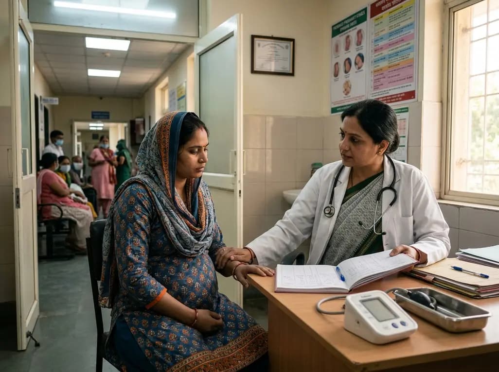
A groundbreaking national research project published in The Lancet Regional Health Southeast Asia has provided robust epidemiological evidence establishing a direct link between maternal anaemia stillbirth risk in India. Researchers analyzed data from the Indian Council of Medical Research (ICMR) Stillbirth Pooled Indian Cohort Consortium (SPIC). The comprehensive dataset incorporated 214,709 single-fetus pregnancies with known birth outcomes and gestational hemoglobin measurements across ten Indian states. Notably, the study revealed that pregnant women experiencing moderate-to-severe anaemia at any stage during gestation face a 27 per cent higher risk of stillbirth after 28 weeks of pregnancy. Overall, more than 52 per cent of women in the cohort had moderate to severe anaemia, defined as hemoglobin levels below 10 grams per deciliter. Among the overall cohort, clinicians documented 3,595 stillbirths, representing a rate of 1.7 per cent. Consequently, this study underscores maternal anaemia as one of the most critical modifiable risk factors affecting obstetric outcomes in South Asia. Collaborators from Oxford University, the London School of Hygiene and Tropical Medicine, and the Indian Institute of Public Health Bengaluru led the investigation. They highlighted that systematic early screening and aggressive management could significantly alter these figures nationwide.
The research team performed detailed stratification based on hemoglobin concentration to assess how varying degrees of severity impact pregnancy outcomes. Specifically, severe maternal anaemia—characterized by hemoglobin levels below 7 grams per deciliter—tripled the risk of stillbirth compared to women with normal or mild anaemia. Meanwhile, moderate anaemia elevated the risk approximately 1.2-fold across all gestational ages. Furthermore, the investigators observed a significant shift in the gestational timing of late fetal loss. Among pregnant women diagnosed with severe anaemia, stillbirths occurred earlier, clustering around the 29th week of gestation. In contrast, stillbirths among non-anaemic women or those with mild disease occurred later, around the 31st week. This two-week acceleration in fetal loss highlights the acute physiological stress that severe maternal hypoxia exerts on the developing fetus during the third trimester. Therefore, obstetricians must recognize that delay in treating low hemoglobin levels increases the probability of early intrauterine fetal demise. Moreover, the study demonstrates that even moderate reductions in maternal hemoglobin pose a continuous threat throughout pregnancy. Consequently, clinicians should not view moderate anaemia as a benign baseline finding. Instead, medical teams must implement proactive diagnostic protocols to catch declining hemoglobin trends before severe decompensation develops.
Understanding the biological pathways through which maternal anaemia induces fetal death is essential for designing targeted therapeutic protocols. Primarily, chronic maternal hypoxia reduces oxygen supply to the placenta and fetus, compromising basic metabolic functions. When maternal hemoglobin drops significantly, reduced oxygen transport can lead directly to embryonic and fetal anaemia. Subsequently, this severe hypoxic environment triggers elevated oxidative stress within developing fetal tissues and placental vascular structures. Furthermore, sustained fetal hypoxia induces profound circulatory adaptations, including alterations in organ perfusion and cellular metabolism. Over time, these compensatory mechanisms can fail, leading to fetal cardiovascular failure or congenital vascular defects. Additionally, maternal systemic response to chronic iron deficiency often includes elevated systemic inflammation and compromised immune function. Consequently, maternal anaemia increases susceptibility to silent intrauterine infections and placental insufficiency, both of which strongly correlate with stillbirth. Furthermore, chronic maternal hypoxia impairs placental growth, leading to structural abnormalities in placental villi and reduced nutrient exchange. As a result, fetal growth restriction develops, leaving the fetus vulnerable to sudden intrauterine asphyxia during late pregnancy. Therefore, prompt clinical restoration of optimal hemoglobin concentrations protects delicate placental hemodynamics and preserves vital fetal organ perfusion throughout the third trimester.
Given that maternal anaemia affects over half of pregnant women in India, public health frameworks must aggressively prioritize comprehensive prevention and clinical management strategies. Historically, national programs like Anaemia Mukt Bharat have raised awareness and expanded access to routine oral iron-folic acid supplementation. However, the high prevalence of moderate-to-severe anaemia reported in the ICMR-SPIC study indicates persistent implementation gaps. Consequently, health authorities must strengthen point-of-care hemoglobin testing at rural primary health centers and urban clinics alike. Furthermore, public health policies should shift toward early first-trimester screening to establish baseline hemoglobin levels before fetal metabolic demands increase. When oral iron therapy proves inadequate or leads to poor patient compliance due to gastrointestinal side effects, clinical protocols should facilitate rapid escalation to intravenous iron formulations, such as ferric carboxymaltose. Additionally, nutritional counseling must emphasize dietary diversification and iron absorption enhancers to address chronic nutritional deficiencies. Moreover, integrating community health workers into follow-up monitoring ensures higher adherence to prescribed treatments. By transforming routine antenatal care into an active, risk-stratified management system, healthcare infrastructure across Indian states can substantially reduce preventable stillbirths and improve overall maternal survival rates.
Effective clinical management requires clear differentiation between modifiable nutritional or infectious causes of anaemia and underlying genetic disorders. Nutritional iron deficiency, folate deficiency, and chronic parasitic infections represent highly modifiable factors that respond rapidly to standard public health interventions. Consequently, administering prophylactic anti-helminthic therapy and targeted nutritional supplementation can successfully reverse mild to moderate anaemia in most pregnant patients. However, genetic hemoglobinopathies, including thalassemia minor, sickle cell trait, and metabolic blood disorders, present distinct clinical challenges that cannot be resolved through iron supplementation alone. In fact, empirical iron therapy in patients with unidentified thalassemia traits can cause iron overload without correcting hemoglobin levels. Therefore, researchers advocate for routine hemoglobin electrophoresis screening during early antenatal visits, particularly in regions with high genetic hemoglobinopathy prevalence. When non-modifiable genetic causes are identified, multidisciplinary care involving hematologists and specialized obstetricians becomes essential. Subsequently, clinicians must tailor surveillance protocols to monitor fetal growth and well-being continuously. By distinguishing between nutritional deficits and genetic blood disorders early in pregnancy, medical teams can deliver precise, individualized care that mitigates fetal risk while avoiding unnecessary or ineffective treatments.
Q1: What defines moderate-to-severe anaemia during pregnancy under clinical guidelines in India?
In clinical practice across India, maternal anaemia is classified based on blood hemoglobin levels. Moderate anaemia corresponds to a hemoglobin concentration between 7.0 and 9.9 grams per deciliter, while severe anaemia is defined as hemoglobin levels below 7.0 grams per deciliter. Any hemoglobin measurement under 10.0 grams per deciliter during pregnancy warrants immediate clinical evaluation, tailored nutritional or intravenous intervention, and frequent ongoing monitoring to prevent severe fetal complications.
Q2: How does severe maternal anaemia alter the gestational timing of stillbirth?
Research demonstrates that severe maternal anaemia shifts the timing of fetal loss to earlier gestational ages. Stillbirths in severely anaemic mothers tend to cluster around the 29th week of gestation, compared to the 31st week in non-anaemic mothers. This two-week shift highlights severe fetal hypoxia during peak developmental periods. Consequently, healthcare providers must perform rigorous fetal monitoring and aggressive hemoglobin correction before the late second and early third trimesters.
Q3: Why is intravenous iron therapy recommended for moderate-to-severe gestational anaemia?
Intravenous iron, such as ferric carboxymaltose, provides rapid restoration of maternal iron stores and hemoglobin levels. Oral iron supplements often cause gastrointestinal side effects, leading to poor patient adherence and slow therapeutic response. In moderate-to-severe gestational anaemia, rapid hemoglobin correction is necessary to restore oxygen supply to the fetus, improve placental perfusion, and reduce the risk of adverse outcomes like stillbirth and preterm birth.
Disclaimer: This content is for informational and educational purposes only. It does not constitute medical advice or replace professional judgment. Refer to the latest local and national guidelines for clinical practice.
References

Read summarized clinical updates, watch expert medical content, and earn CME certifications right from your smartphone.
Open your smartphone camera to scan and install.





A major study published in The Lancet Regional Health Southeast Asia reveals that pregnant women in India with moderate-to-severe anaemia face a 27% higher risk of stillbirth after 28 weeks of gestation. Evaluating over 210,000 pregnancies, researchers emphasize the urgent need for targeted antenatal interventions.
last month

The phase 3 ACACIA-HCM trial reveals that aficamten improves cardiac structure, diastolic relaxation, functional capacity, and symptom burden in symptomatic nonobstructive hypertrophic cardiomyopathy, marking a major milestone in targeted myosin inhibition.
Last week

A clinical study shows that automated breast ultrasound paired with artificial intelligence accurately classifies BIRADS 3-4 lesions, reaching 95% sensitivity and 79% specificity. This diagnostic advance promises to reduce unnecessary core needle biopsies and refine clinical workflows in breast imaging.
4 weeks back
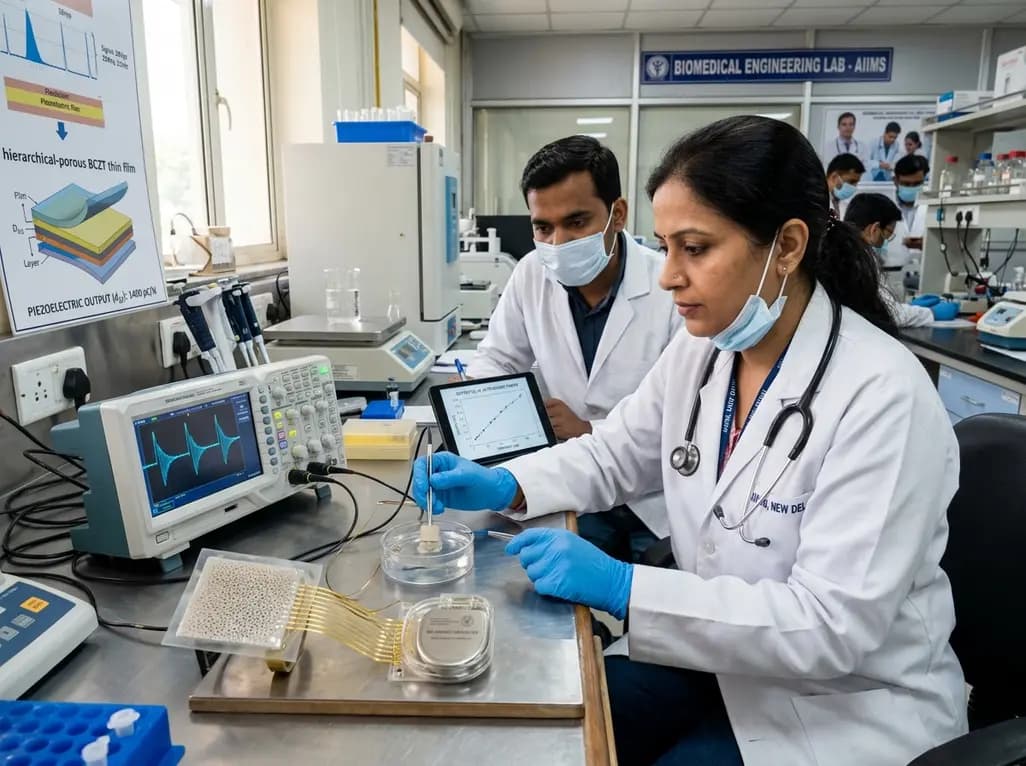
Researchers have engineered freestanding hierarchical-porous BCZT thin films that resist cracking and enhance ultrasonic energy harvesting in soft tissue. Achieving high piezoelectric output and acoustic matching, this lead-free material offers transformative potential for implantable bioelectronics.
Last week
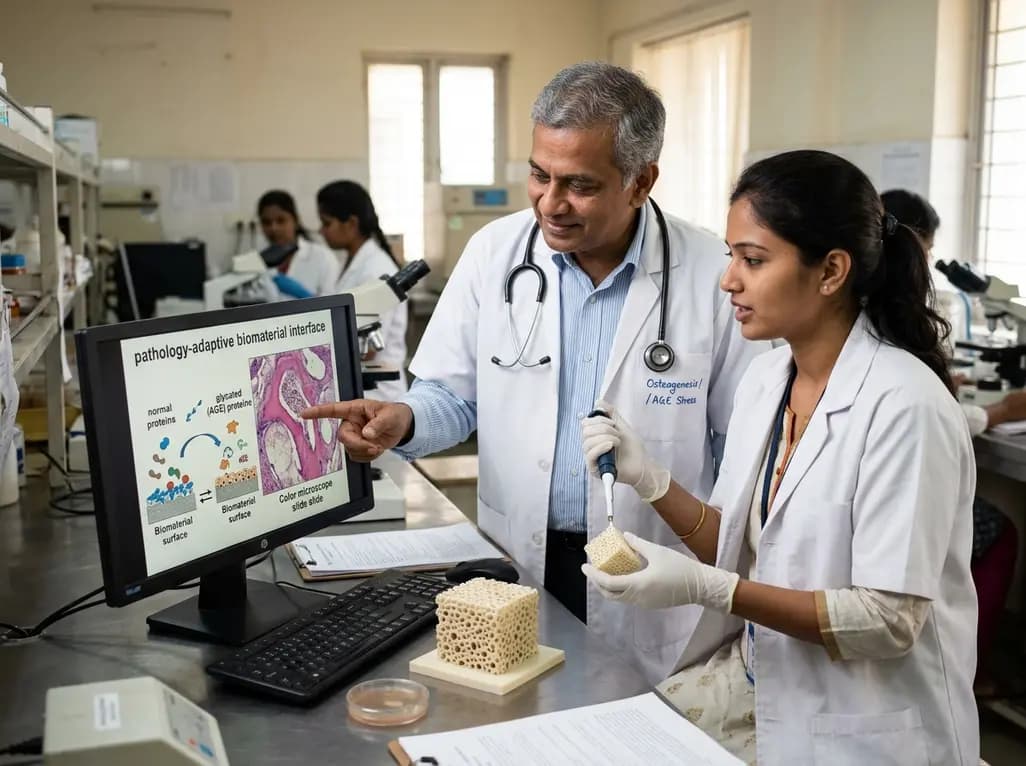
A novel pathology-adaptive surface engineering strategy uses functionalized plasma polymer coatings to selectively modulate AGE adsorption, reducing oxidative stress and restoring bone formation in diabetic and aging microenvironments.
4 weeks back
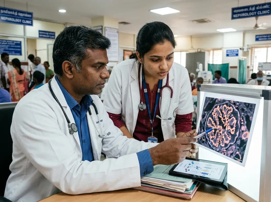
A breakthrough study identifies the Klotho/PKCα/CUX1/SPARC/TGFβ-RII axis as a critical driver of podocyte mitochondrial injury and ferroptosis in diabetic kidney disease, unveiling promising molecular targets to halt renal disease progression.
Last week
