.webp&w=384&q=75)

Loading, please wait...
Loading, please wait...
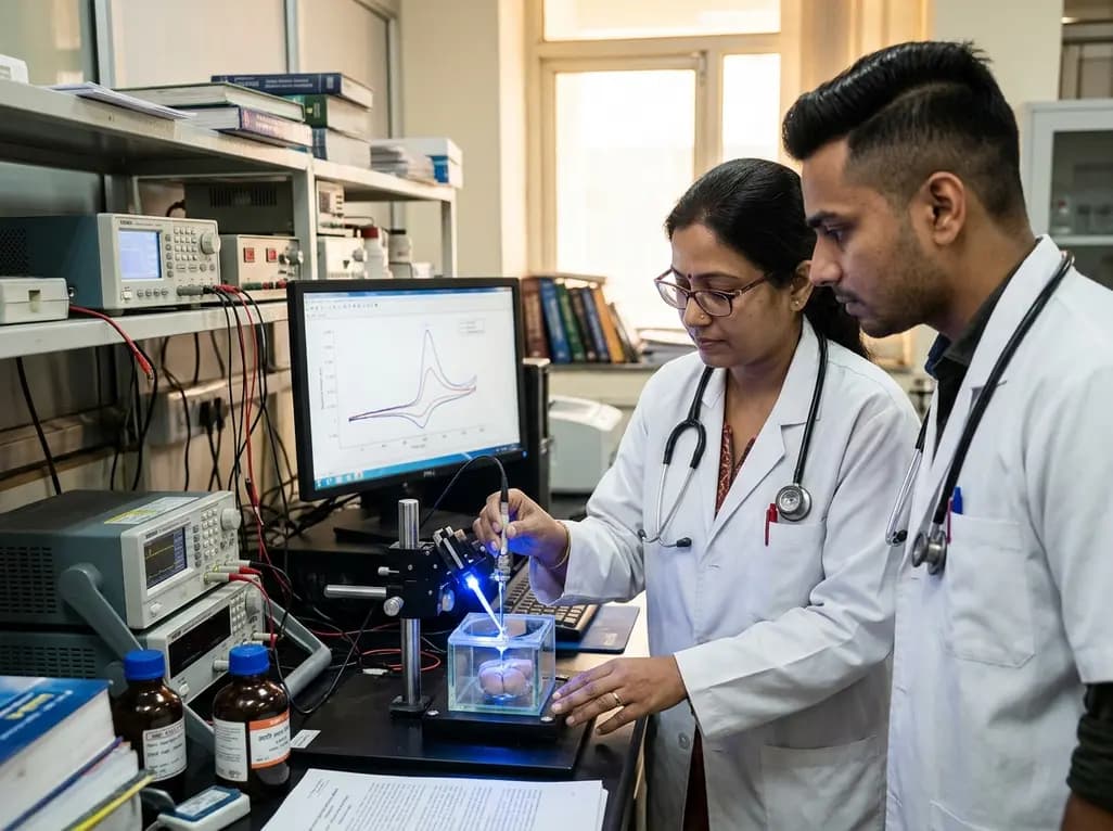
Modern neuroscientific research frequently combines advanced techniques to understand the complex signaling within the brain. One of the most prevalent combinations involves the use of optogenetics alongside electrochemical measurements. Fast-scan cyclic voltammetry (FSCV) serves as a cornerstone for dopamine monitoring in neuroscience, allowing researchers to track neurotransmitter dynamics with high temporal resolution. However, the introduction of light from optogenetic or fluorescent techniques often creates unintended artifacts on the recording electrodes. Specifically, carbon-fiber microelectrodes (CFMEs) exhibit sensitivity to light exposure that can skew experimental results. Understanding these interactions is vital for ensuring the accuracy of neurochemical data. Recent investigations have sought to systematically characterize how different light wavelengths influence the background current and sensitivity of these electrodes. This research provides a roadmap for mitigating interference and leveraging light-induced enhancements to improve detection limits. By examining the electrochemical shifts during blue and green light exposure, scientists are now able to refine their methodologies. Consequently, these insights allow for more robust interpretations of dopamine release in various brain regions, including the nucleus accumbens core.
The primary challenge when exposing carbon-fiber microelectrodes to light during FSCV is the alteration of the background charging current. Researchers analyzed this phenomenon by focusing on three critical potential regions of the dopamine waveform. These included the area near the switching potential (1.2-1.3 V), the dopamine oxidation potential (0.6-0.7 V), and a lower potential baseline (0.2 V). Notably, blue light exposure produced the most significant changes in background current. This shift was most pronounced at the switching potential, where the voltage changes direction. Such fluctuations occur both during in vitro calibrations and within live tissue environments. Moreover, the magnitude of these changes suggests that the electrode surface undergoes rapid physical or chemical transitions when illuminated. Scientists observed that the background current does not remain stable if light is introduced abruptly. Therefore, identifying the exact voltage points where light exerts its greatest influence is essential for data correction. By characterizing these shifts, researchers can better distinguish between true neurotransmitter signals and light-induced capacitive changes. This detailed mapping ensures that the high sensitivity of FSCV remains reliable even in complex optical setups.
Wavelength selection plays a pivotal role in the degree of interference observed during neurochemical recordings. When researchers compared blue light to green light, they found substantial differences in electrode response. Specifically, green light exposure resulted in a background current change that was approximately 50% less than that caused by blue light. Despite this reduction, the largest shift still occurred near the switching potential of the waveform. This finding suggests that longer, more red-shifted wavelengths are inherently less disruptive to the electrochemical environment of the CFME. Consequently, shifting toward green or red light sources in optogenetic experiments could significantly enhance the signal-to-noise ratio. Furthermore, the sensitivity of the electrode to the neurotransmitter itself varies with light exposure. While blue light causes more significant background shifts, it also contributes to a notable enhancement in dopamine detection sensitivity. This paradox requires researchers to balance the need for low noise with the potential for increased signal amplitude. Understanding these wavelength-dependent effects allows laboratories to choose optical parameters that minimize artifacts while maintaining the necessary stimulation for their biological targets.
The mechanism behind light-induced enhancement of dopamine signals is likely rooted in photothermal effects. Researchers hypothesize that the absorption of light energy by the carbon fiber leads to localized heating at the electrode-solution interface. This temperature increase generates several important changes in the electrochemical environment. Primarily, it appears to shrink the width of the electric double layer surrounding the electrode. Furthermore, this localized heating alters the thermodynamics of analyte adsorption. Since dopamine detection in FSCV relies heavily on the adsorption of molecules onto the carbon surface, any factor that improves this process will enhance the recorded current. Additionally, the photothermal effect may increase the rate of electron transfer, leading to sharper and larger oxidation peaks. This hypothesis explains why blue light, which carries higher energy than green light, produces a more significant boost in sensitivity. However, this enhancement must be accounted for during calibration to avoid overestimating the concentration of dopamine released in tissue. By recognizing the role of thermal energy, researchers can better predict how different environmental factors will impact their electrochemical sensors.
To validate these findings in a biological context, researchers performed experiments in mouse brain slices, specifically targeting the nucleus accumbens core (NAcC). This region is central to the brain\'s reward circuitry and is a primary focus of dopamine monitoring in neuroscience. During these tests, CFMEs were exposed to blue light for one minute prior to dopamine detection. The results were striking, showing a 33% increase in the current recorded for electrically stimulated dopamine release. This enhancement was consistent with the results observed during pre-experiment calibrations. Consequently, it confirms that light exposure significantly impacts the quantitative interpretation of dopamine levels in live tissue. Without proper adjustments, the 33% boost could lead to inaccurate conclusions regarding the efficacy of neural stimulation or the effects of pharmacological agents. Moreover, the tissue environment itself may influence how the photothermal effect manifests, making it crucial to perform calibrations under the same light conditions used in the experiment. These findings highlight the need for rigorous control protocols when utilizing multi-modal neuroimaging and stimulation techniques in CNS research.
Based on the systematic characterization of light effects, researchers have developed clear guidelines for performing FSCV alongside optical techniques. First, it is recommended that the light source be activated at least 30 seconds before starting the electrochemical measurements. This lead time allows the background current to reach a new steady state, minimizing drift during the actual recording period. Second, all electrode calibrations must be conducted under identical lighting conditions to those used in the tissue experiments. This ensures that the sensitivity factors used for data conversion accurately reflect the photothermal enhancements. Additionally, the use of red-shifted wavelengths should be prioritized whenever the biological model allows, as this reduces the baseline interference. Furthermore, researchers should be mindful of the switching potential in their waveforms, as this is where the most significant background fluctuations occur. Implementing these strategies will lead to more precise and reproducible neurochemical data. Ultimately, these refinements empower scientists to explore the nuances of dopamine signaling with greater confidence, bridging the gap between optical stimulation and electrochemical readout.
Disclaimer: This content is for informational and educational purposes only... Refer to the latest local and national guidelines for clinical practice.
References
Donarski ED et al. Blue light enhances background current and dopamine sensitivity of carbon-fiber microelectrodes during fast-scan cyclic voltammetry. Anal Methods. 2026 Jul 03. doi: 10.1039/d6ay00403b. PMID: 42397697.
Heien ML, Phillips PE, Stuber GD, Seipel AT, Wightman RM. Overoxidation of carbon-fiber microelectrodes enhances dopamine adsorption and increases sensitivity. Analyst. 2003;128(12):1413-1419.
Bucher ES, Wightman RM. Electrochemical analysis of neurotransmitters. Annu Rev Anal Chem. 2015;8:171-190.
Blue light shifts the background charging current of carbon-fiber microelectrodes, particularly near the switching potential. This shift can mask or mimic real neurotransmitter signals if not properly managed. Furthermore, blue light increases the sensitivity of the electrode to dopamine by approximately 33%. Consequently, researchers must calibrate their electrodes under the same lighting conditions used in the experiment to ensure that concentration calculations remain accurate and consistent across different trials.
The photothermal effect occurs when the carbon fiber absorbs light energy, causing localized heating. This process is thought to shrink the electric double layer at the electrode surface and alter the thermodynamics of dopamine adsorption. Because dopamine detection in FSCV depends on the molecule sticking to the electrode, these changes lead to enhanced current signals. However, this effect varies with wavelength, with higher-energy blue light producing more significant enhancements than lower-energy green light.
To minimize artifacts, researchers should turn on the light source at least 30 seconds before beginning their experiments. This allows the background current to stabilize. Additionally, it is essential to perform all calibrations with the light on if the tissue experiment involves light exposure. Using more red-shifted wavelengths, such as green or red light, can also help reduce the magnitude of background current shifts, leading to cleaner data and more reliable neurochemical measurements.

Read summarized clinical updates, watch expert medical content, and earn CME certifications right from your smartphone.
Open your smartphone camera to scan and install.





New research reveals that blue light exposure significantly enhances the dopamine sensitivity of carbon-fiber microelectrodes during fast-scan cyclic voltammetry (FSCV). This finding is crucial for accurate neurochemical monitoring in experiments involving optogenetics or fluorescence.
3 months ago

Depemokimab offers twice-yearly dosing for severe eosinophilic asthma and chronic rhinosinusitis with nasal polyps, significantly cutting exacerbations and hospital admissions.
4 days back

A novel copper metal-organic framework hydrogel remodels the uterine microenvironment to stimulate robust endometrial regeneration, reduce intrauterine fibrosis, and restore reproductive fertility, offering a groundbreaking strategy for managing refractory endometrial damage and Asherman syndrome.
4 days back

New research reveals that non-ablative radiotherapy suppresses ventricular tachycardia by increasing conduction velocity via NaV1.5 upregulation and action potential prolongation. However, sustained sodium current elevation and depressed calcium handling highlight the need for careful clinical translation.
6 days back

A single-cell transcriptomic atlas reveals how peripheral nerve injuries cause dorsal root ganglion remodeling. By evaluating proximal nerve crush and resection, researchers identify distinct glial and immune programs that resolve neuropathic pain and promote regeneration.
4 days back

A breakthrough study demonstrates that fullerol-primed extracellular vesicles generated in a 3D SECCS bioreactor attenuate ferroptosis and restore myogenic regeneration in diabetes via the FOXO3 pathway, offering a cell-free therapeutic strategy for diabetic musculoskeletal degeneration.
4 days back
