.webp&w=384&q=75)

Loading, please wait...
Loading, please wait...
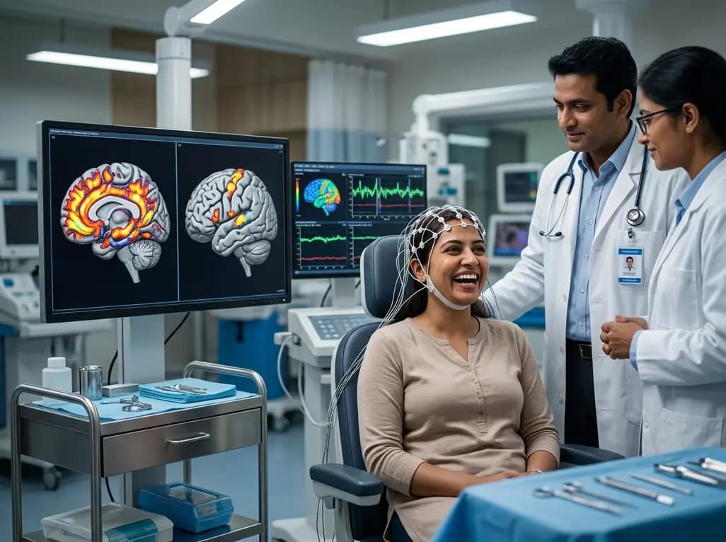
Human laughter serves as a fundamental pillar of social communication, yet its neurological mechanisms are surprisingly complex. In clinical settings, researchers distinguish between two primary forms: spontaneous, involuntary laughter and precisely timed voluntary laughter. Spontaneous laughter represents those helpless outbursts of mirth that we cannot control when exposed to humor or tickling. Consequently, this uncontrollable response can sometimes manifest pathologically in patients with seizure disorders, severe mood disorders, Alzheimer's disease, and schizophrenia. In contrast, the second form is the volitional kind, which represents the vast majority of our daily social interactions. This conversational vocalization starts and stops with remarkable speed, allowing individuals to coordinate their breathing and speech dynamically. Furthermore, this deliberate coordination points to a high degree of cortical control that is entirely lacking in spontaneous, involuntary outbursts. Neuroscientists struggle to study these laughter types because evoking genuine mirth in laboratory environments is exceptionally difficult. Therefore, researchers analyzed pre-surgical brain mapping reports to untangle these distinct neural networks. Ultimately, this analytical approach provided clear insights into the functional architecture of our brains. Additionally, the team identified the exact cortical regions that coordinate conversational smiling.
To map the intricate pathways of human laughter, investigators turned to historical reports of pre-surgical brain stimulation in epilepsy patients. During these delicate procedures, neurosurgeons must identify precise cortical and subcortical regions to target for surgical resection. Because the patients remain awake, clinicians can electrically stimulate specific brain areas using highly localized probes to map functional boundaries safely. Interestingly, these targeted electrical impulses often unintentionally evoke laughing or smiling, providing a rare opportunity for real-time observation. Furthermore, because patients are awake, they can describe their immediate subjective feelings and emotional states during the stimulation. By aggregating these clinical reports with veterinary and functional imaging studies, researchers mapped two entirely distinct anatomical networks. This unique methodology bypassed the classic laboratory bottleneck of trying to elicit genuine, unforced mirth in sterile research settings. Consequently, the research team successfully localized the functional nodes that generate either physiological laughter or complete emotional euphoria. Ultimately, these clinical insights from awake brain mapping have provided an unprecedented look into the dual-network architecture of human non-verbal vocalizations. Clinicians now possess a clearer map of how electrical stimulation activates human vocal systems.
The researchers discovered that spontaneous, involuntary laughter is governed by an emotionally charged neural network located deep within the brain. Specifically, this ancient network consists of regions highly involved in motor control and complex emotional regulation. These key structures include the pregenual anterior cingulate cortex, the nucleus accumbens, and the temporal pole. When clinicians electrically stimulate these deep regions, patients experience intense positive emotions and genuine, uncontrollable mirth. Therefore, this pathway is directly tied to our internal reward systems and authentic emotional states. Moreover, evolutionary biologists believe that this spontaneous network is an ancient mammalian survival pathway. For instance, this system likely arose during rough-and-tumble play in early animal ancestors. Specifically, these laughter-like vocalisations served to prevent aggression and promote social bonding. Consequently, this deep emotional circuit remains vital for human connection, helping us share genuine joy and reinforce communal relationships. In contrast to more modern cortical structures, this emotional pathway operates outside of our conscious, deliberate planning. Neuroscientists notes that this subconscious pathway coordinates both our physical laughter and our psychological well-being. Thus, the deep brain centers ensure that genuine laughter remains a powerful tool for social survival and cohesion.
Unlike the deep emotional pathways of spontaneous mirth, the voluntary network relies purely on motor execution areas. Specifically, this voluntary laughter network comprises the rolandic operculum, the globus pallidus, and the presupplementary motor area. When neurosurgeons stimulate these specific regions, patients laugh and smile physically without feeling any positive emotions. Thus, this motor-driven pathway generates the mechanical actions of laughter without triggering any underlying feelings of happiness or euphoria. Furthermore, researchers discovered that this voluntary network heavily overlaps with the brain regions responsible for speech production. This anatomical overlap strongly supports the scientific theory that volitional laughter is an evolutionary extension of human language. Consequently, this system acts as a highly controlled, purpose-driven tool to navigate complex conversational dynamics and strengthen social agreements. While spontaneous laughter erupts from primal play signals, volitional vocalizations represent a sophisticated cortical mechanism. This conversational system allows humans to sync their breathing and vocalizations during rapid, real-time social alignment. Therefore, we can consciously control this social vocalization to convey politeness, agreement, and shared attention. Ultimately, this motor circuit serves as the primary physiological framework for modern human conversation.
These groundbreaking neuroanatomical findings carry profound clinical implications for both neurology and psychiatry. For example, understanding these distinct pathways helps explain why patients with certain neurological conditions display pathological laughter. Specifically, in disorders like gelastic epilepsy or pseudobulbar affect, patients experience uncontrollable laughing fits. These symptoms can also occur in patients suffering from Alzheimer's disease and schizophrenia. These clinical manifestations likely stem from dysfunction or abnormal excitation within the deep spontaneous network, bypassing cortical control entirely. Conversely, studying the voluntary motor network provides valuable insights into how humans modulate social communication and perceive authenticity. When a patient struggles to produce social laughter, clinicians can pinpoint potential disruptions in speech-associated motor pathways. Furthermore, this dual-network framework assists neurosurgeons during pre-surgical planning. Specifically, it helps them preserve vital emotional and conversational circuits during tumor resections. Ultimately, mapping these systems enhances our diagnostic accuracy and therapeutic strategies for complex neuropsychiatric conditions. By recognizing laughter as a dual-natured neurological phenomenon, medical professionals can better address patient challenges. Thus, these discoveries pave the way for more targeted interventions in cognitive rehabilitation. Additionally, future psychiatric research can explore how pharmacological agents affect these two distinct brain networks.
Q1: What are the main differences between spontaneous and voluntary laughter?
Spontaneous laughter is an uncontrollable, involuntary reaction to humor that triggers deep emotional brain regions, resulting in genuine feelings of mirth and euphoria. In contrast, voluntary laughter is a highly controlled, conversational vocalization used in social interactions. It relies strictly on motor control areas of the brain and occurs without any underlying positive emotions, acting primarily as a social tool.
Q2: How did researchers identify the distinct brain regions responsible for these two types of laughter?
The research team analyzed clinical reports of pre-surgical brain stimulation in awake epilepsy patients. During these delicate procedures, neurosurgeons electrically stimulated specific brain areas to map functional boundaries. Because the patients were fully awake, they could describe their immediate emotional feelings. These observations, combined with animal studies, allowed researchers to successfully map the distinct motor and emotional networks.
Q3: Why does voluntary laughter overlap with the brain regions that control speech?
Voluntary laughter serves as an essential conversational lubricant that requires extremely precise timing during social dialogues. Because of this communicative role, the voluntary network overlaps with speech-producing areas of the brain, including the rolandic operculum. This anatomical link supports the evolutionary theory that volitional laughter developed as a highly sophisticated, purpose-driven mechanism to facilitate human communication and social bonding.
Disclaimer: This content is for informational and educational purposes only. It does not constitute medical advice or replace professional judgment. Refer to the latest local and national guidelines for clinical practice.
References

Read summarized clinical updates, watch expert medical content, and earn CME certifications right from your smartphone.
Open your smartphone camera to scan and install.





An international study analyzes awake brain mapping data from epilepsy patients to reveal two completely distinct neural networks responsible for spontaneous emotional mirth and controlled, voluntary conversational laughter.
3 months ago

Discover the crucial role of glymphatic dysfunction in stroke, where perivascular clearance failure and neuroinflammation exacerbate cerebral edema and post-stroke brain injury.
Today

A prenatal case highlights severe glutaric acidemia type II presenting as bilateral hyperechoic cystic kidneys at 18 weeks. Whole-exome sequencing revealed a novel homozygous ETFDH frameshift variant, demonstrating the power of rapid genetic testing in fetal renal anomalies.
Today

Review how tailored exercise programs benefit frail older patients with heart failure. Explore pathophysiological mechanisms, individualized training prescriptions, and strategies to overcome adherence barriers in clinical practice.
Yesterday

This comparative study evaluates posterior labral tears with and without glenohumeral instability, highlighting distinct demographic patterns, MRI underestimation in stable shoulders, and successful arthroscopic repair outcomes.
Yesterday

A clinical study demonstrates that early intervention with tocilizumab, either as monotherapy or bridged with corticosteroids, achieves significant proptosis reduction and resolves active inflammation in treatment-naive thyroid eye disease.
Yesterday
