.webp&w=384&q=75)

Loading, please wait...
Loading, please wait...

Acute cerebrovascular events frequently produce severe cognitive impairments that complicate clinical recovery. Among these debilitating deficits, visuospatial neglect in stroke represents a profound failure to attend, respond, or orient toward contralesional stimuli. Historically, hospital registries substantially underestimated its prevalence because standard bedside screenings were inadequate. However, recent evidence from a landmark prospective Swiss cohort demonstrates that the actual burden is far greater than clinicians previously assumed.
Diagnostic evaluations often overlook subtle cognitive disturbances during hyperacute hospital management. Historically, observational registries excluded stroke patients who presented with severe aphasia, profound hemiparesis, or acute delirium. Consequently, earlier literature suggested that spatial attention deficits occurred only in a small fraction of stroke admissions. A recent prospective study evaluating an unselected cohort of 221 consecutive acute stroke patients within 72 hours of symptom onset has fundamentally revised these clinical assumptions.
The investigators discovered an overall neglect incidence of approximately 38% across all stroke presentations. Furthermore, the frequency of neglect varied substantially depending on lesion location. Patients with right hemispheric infarctions displayed neglect in roughly 61% of cases. Meanwhile, approximately 22% of patients with left hemispheric injuries also exhibited demonstrable spatial neglect. In addition, the research established that neglect occurs across less typical vascular territories, including infratentorial and multifocal lesions in 14% to 37% of cases. Therefore, recognizing this substantial disease burden allows multidisciplinary teams to anticipate complications early and optimize individual rehabilitation pathways.
Arterial territory plays a crucial role in predicting the emergence of spatial attention deficits. In hemispheric stroke, territorial distribution directly dictates clinical presentation. Specifically, infarctions involving the middle cerebral artery territory produced the highest burden, with spatial neglect developing in 64% of right-sided and 21% of left-sided strokes. Similarly, posterior cerebral artery infarctions yielded spatial neglect in 53% of right-sided and 25% of left-sided events.
Furthermore, multivariate analyses identified distinct vascular and demographic profiles strongly associated with neglect development. Affected individuals systematically presented with significantly higher baseline National Institutes of Health Stroke Scale scores. Moreover, these patients exhibited a higher prevalence of preexisting atrial fibrillation and required mechanical thrombectomy more frequently. Consequently, large-vessel occlusions and cardioembolic mechanisms clearly heighten neglect risk. In contrast, patients with undetermined stroke etiologies showed a lower likelihood of spatial inattention. In addition, patient age served as a steady, independent risk predictor. Each single year of advancing age increased the odds of visuospatial neglect by approximately 4%. Therefore, elderly patients suffering large-vessel cardioembolic strokes warrant heightened clinical suspicion during initial triage.
For decades, clinicians relied almost exclusively on conventional paper-pencil cancellation, line bisection, and copying tests. While these bedside assessments provide familiar metrics, they suffer from fundamental diagnostic weaknesses. First, paper-pencil tools require intact motor performance and manual dexterity. When a patient presents with dense hemiplegia, examiners cannot administer these tests successfully. Second, standard tests often demand substantial verbal comprehension. Consequently, clinicians routinely exclude aphasic patients from cognitive assessments, creating severe diagnostic ascertainment bias.
Furthermore, standard paper-pencil assessments lack adequate sensitivity for subtle or compensated attention deficits. Patients often learn compensatory scanning strategies when completing stationary sheets resting directly on a small hospital table. However, these brief paper tasks fail to replicate dynamic, three-dimensional real-world visual exploration. As a result, bedside clinicians frequently discharge patients with unrecognized attention failure, mistaking persistent neglect for uncooperative behavior or simple motor fatigue. In addition, conventional scoring thresholds lack uniform standardization across acute centers. Therefore, bedside teams consistently underreport milder forms of neglect, leaving vulnerable patients exposed to unexpected fall hazards and diminished rehabilitation potential.
To overcome traditional diagnostic limitations, researchers implemented video oculography during free visual exploration. This sophisticated, non-invasive eye-tracking technique captures spontaneous ocular fixations and saccadic gaze distribution across broad visual fields. Crucially, the protocol requires minimal verbal instructions and demands zero active upper-limb motor engagement. Thus, examiners can successfully administer the evaluation even to bedridden individuals with severe paresis or expressive language barriers.
The observational cohort confirmed that video oculography provided excellent clinical feasibility in acute stroke units. Moreover, eye-tracking detected spatial neglect far more reliably than standard paper-pencil tests. By recording continuous visual search paths, video oculography captures subtle asymmetric exploration patterns that paper sheets easily miss. For example, affected individuals show skewed mean horizontal gaze positions and restricted contralesional exploration areas. Furthermore, automated gaze tracking delivers objective, quantitative metrics devoid of examiner subjectivity. Consequently, incorporating objective oculomotor exploration into acute stroke pathways provides a robust, reproducible diagnostic standard. In the future, portable eye-tracking systems could bring this advanced diagnostic precision directly to bedside stroke monitoring.
Unrecognized visuospatial neglect produces severe, multidimensional barriers during acute and subacute recovery. Patients with spatial neglect experience prolonged hospitalizations, slower functional improvement, and significantly greater rates of accidental falls. Because patients fail to perceive stimuli in contralesional space, they frequently bump into doorways, ignore meal trays, and neglect their affected limbs. Furthermore, recent kinematic investigations demonstrate that neglect actively suppresses spontaneous upper-limb motor recovery, even when motor pathways remain physically intact.
Fortunately, earlier identification opens critical therapeutic windows for neurorehabilitation. Once clinicians identify spatial deficits, interdisciplinary teams can immediately institute structured compensatory and restorative interventions. For instance, staff can employ visual scanning training, prism adaptation therapies, optokinetic stimulation, and limb activation protocols. In addition, nurses and occupational therapists can arrange patient rooms to stimulate exploratory gaze toward the neglected field safely. Early family counseling also proves indispensable, as it educates caregivers regarding safety hazards and spatial inattention. Therefore, implementing sensitive diagnostic screening during the hyperacute window directly optimizes functional trajectories, reduces preventable hospital complications, and supports long-term patient independence.
Visuospatial neglect represents a higher-order cognitive disorder of spatial attention rather than a sensory ocular deficit. Patients with hemianopia cannot see contralesional stimuli due to optic pathway damage, yet they compensate spontaneously. In contrast, patients with neglect fail to direct attention contralesionally, frequently remaining completely unaware of their spatial impairment.
Yes, spatial neglect occurs in approximately 22% of acute left hemispheric strokes. Although clinicians traditionally associate neglect with right parietal lesions, left hemispheric damage also induces neglect. Healthcare providers often miss this impairment because concurrent aphasia limits bedside verbal testing, making objective assessments like video oculography particularly valuable for accurate diagnosis.
Video oculography provides an objective, motor-free, and language-independent assessment of spontaneous visual exploration. Conventional paper-pencil tasks require upper-limb motor dexterity and verbal comprehension, automatically excluding severely disabled acute stroke patients. Furthermore, eye-tracking detects subtle, asymmetric gaze trajectories that static bedside testing consistently overlooks, providing superior diagnostic sensitivity in acute clinical environments.
Disclaimer: This content is for informational and educational purposes only, and should not be taken as professional medical advice. Healthcare professionals should rely on their clinical judgment and verify details independently. Refer to the latest local and national guidelines for clinical practice.
References

Read summarized clinical updates, watch expert medical content, and earn CME certifications right from your smartphone.
Open your smartphone camera to scan and install.





New prospective cohort data reveals that visuospatial neglect affects nearly 38% of acute stroke patients. Utilizing video oculography during free visual exploration dramatically improves detection across right, left, and atypical stroke presentations compared to conventional paper-pencil tests.
Today
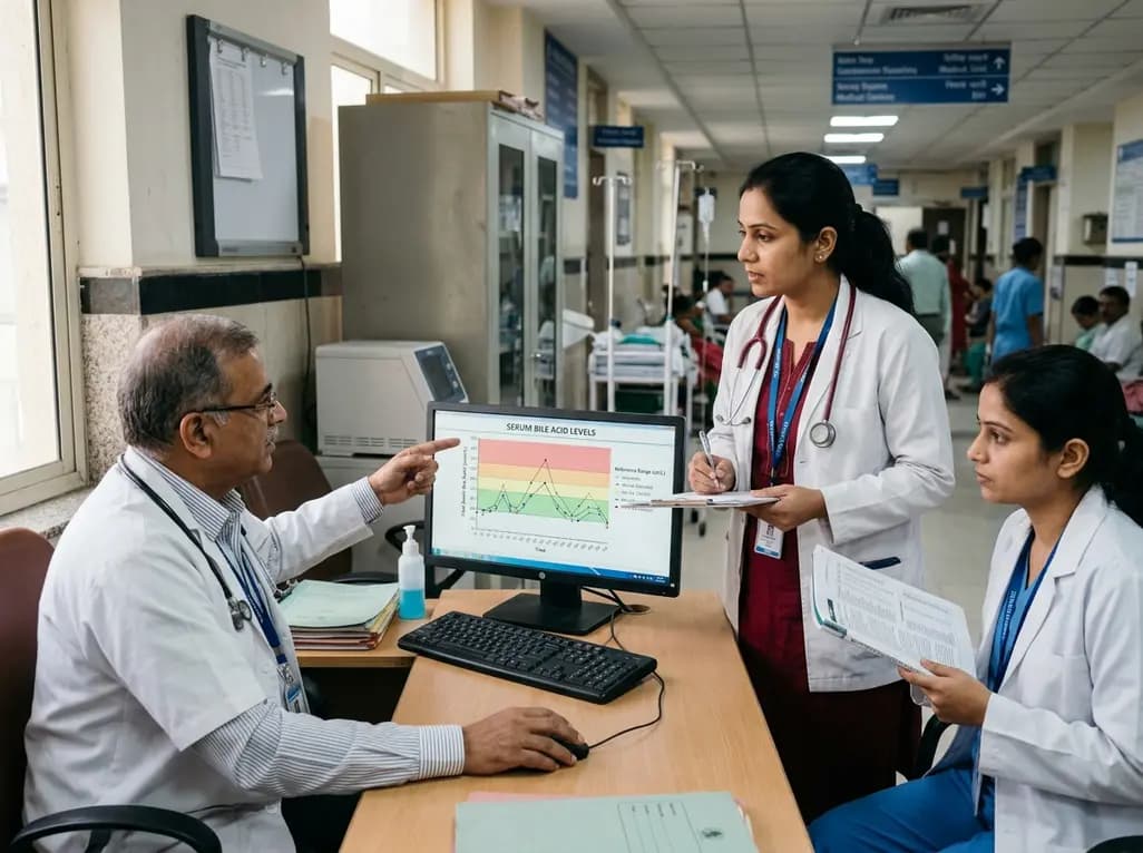
A comprehensive analysis of serum bile acid variability establishes robust reference ranges and reveals how sex, age, BMI, and smoking alter circulating profiles.
Today

A retrospective cost-minimization study of 16,695 Medicare patients reveals that pelvic floor muscle therapy and pessaries incur less than half the 1-year costs of primary sling surgery, supporting conservative management as initial therapy.
Today
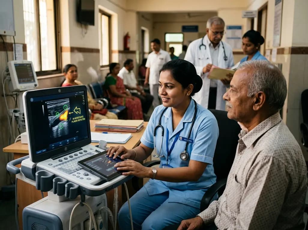
A user-centered study demonstrates that AI-assisted carotid ultrasound enables nonexpert primary care staff to detect subclinical atherosclerosis. With real-time guidance and workflow optimization, this technology supports early cardiovascular risk communication and task-shifting in routine clinical practice.
Today

A hyaluronic acid-modified metal-polyphenol nanocomposite successfully eliminates reactive oxygen species in chondrocytes, halts cartilage breakdown, and promotes tissue anabolism, presenting a novel disease-modifying strategy for early osteoarthritis.
Today

A pilot randomized trial demonstrates that digital wellness applications and medically tailored meals significantly attenuate rapid weight regain following GLP-1 receptor agonist discontinuation, providing valuable transitional support for long-term obesity management.
Today
