

Loading, please wait...
Loading, please wait...

Cerebrovascular neurosurgeons continually pursue higher precision during the microsurgical clipping of complex unruptured intracranial aneurysms. Recently, patient-specific virtual reality clip planning has emerged as a promising tool to enhance preoperative spatial orientation and clip selection. Although immersive platforms allow clinicians to manipulate digital anatomy, empirical validation against real-world surgical outcomes has remained limited. A groundbreaking retrospective validation study led by Saemann and colleagues rigorously evaluated whether simulated clip choices match actual operative constructs and postoperative vascular imaging. Their findings provide vital benchmarks for cerebrovascular specialists aiming to integrate immersive simulation into routine clinical workflows.
Modern neurosurgery increasingly utilizes three-dimensional computational modeling to navigate intricate cranial anatomy. Historically, surgeons relied solely on two-dimensional digital subtraction angiography and computed tomography angiography slices. These conventional modalities require substantial mental rendering to anticipate complex neck morphology and perforator anatomy. Consequently, immersive visualization environments now bridge this spatial gap. These systems allow operators to inspect aneurysmal architecture from arbitrary viewing angles before opening the dura. Furthermore, virtual reality clip planning enables surgical teams to select, position, and adjust virtual aneurysm clips within reconstructed vascular trees. Investigators initially observed significant reductions in operative duration and improved craniotomy tailoring with stereoscopic planning systems. However, most prior studies focused primarily on subjective user satisfaction rather than objective geometric fidelity. True clinical translation demands verifiable concordance between simulated clip configurations and the final constructs placed in vivo. Therefore, validating virtual environments against postoperative imaging represents an essential step toward establishing simulation as a dependable standard of care in vascular neurosurgery. Without rigorous metric benchmarks, surgeons cannot reliably predict whether a preselected implant will achieve definitive obliteration without compromising parent vessel patency.
The validation study evaluated ten consecutive patients who underwent elective microsurgical clipping for unruptured anterior circulation aneurysms. To ensure objective appraisal, three experienced vascular neurosurgeons performed independent, anonymized planning sessions at least six months postoperatively. Crucially, the investigators blinded the raters to clinical charts, original operative documentation, physical clip logs, and postoperative radiological studies. The virtual simulation platform incorporated life-sized Yasargil clip models that directly mirrored the surgical center's physical armamentarium. During each session, raters selected the primary clip geometry, exact blade length, and overall strategy, choosing between single-clip application or multi-clip reconstruction. Additionally, surgeons noted any anticipated dog-ear remnants and recorded their total active planning duration. The researchers established the primary reference standard using intraoperative records alongside day-one postoperative computed tomography angiography. They quantified procedural agreement utilizing a structured four-point Virtual Reality Clip Concordance Index. Ultimately, this rigorous protocol generated thirty independent simulation datasets, providing robust statistical power to contrast preprocedural virtual choices with verified surgical execution. Moreover, this rigorous blinding method eliminated observational recall bias, ensuring genuine reflection of each surgeon's spatial appraisal.
The simulation platform demonstrated exceptional workflow efficiency, requiring an average planning time of only 2.23 minutes per aneurysm. However, overall construct agreement revealed important practical limitations. The cohort achieved a mean Virtual Reality Clip Concordance Index score of 2.00 out of 4 points. Specifically, only 33.3 percent of simulated sessions reached high concordance with a score of three or four. When evaluating broad technical strategy, raters demonstrated solid alignment, achieving 70.0 percent agreement on single-clip versus multi-clip reconstruction. Similarly, primary clip type concordance reached 56.7 percent, and remnant expectation aligned in 60.0 percent of trials. In sharp contrast, exact primary blade length agreement occurred in merely 13.3 percent of simulated cases. Broadening the margin of error to a 0.5-millimeter tolerance only modestly improved blade length concordance to 16.7 percent. Furthermore, an exploratory 1.0-millimeter tolerance produced only 33.3 percent agreement. Thus, while virtual environments reliably guide overarching clipping strategies, they struggle to replicate the minute microvascular measurements required for final implant sizing. Consequently, operating surgeons must maintain physical intraoperative flexibility rather than rigidly adhering to virtual measurements.
Postoperative computed tomography angiography confirmed technical success across the cohort, achieving complete occlusion in nine patients and leaving a minimal remnant in one. Nevertheless, detailed spatial analysis uncovered notable discrepancies between the simulated clip trajectories and actual postoperative clip poses. The researchers documented a median positional translation deviation of 2.41 millimeters between virtual and real-world positions. Even more striking, rotational alignment showed widespread divergence, exhibiting a median rotational deviation of 44.42 degrees. This angular variation stems from several biomechanical realities that static imaging algorithms currently fail to reproduce. For instance, real microsurgery involves dynamic brain relaxation, arachnoid dissection, and mechanical tissue manipulation. In addition, applying an actual titanium clip exerts significant closing force, which compresses the aneurysmal sac and alters neighboring vessel geometry. Virtual models frequently display rigid vessel walls, neglecting the natural elasticity and calcification of vascular tissue. As a result, virtual trajectories often diverge from the final construct chosen under the operating microscope. Therefore, understanding these biomechanical discrepancies helps clinicians contextualize virtual projections against dynamic intraoperative shifts.
These objective findings establish a clear roadmap for advancing surgical simulation technology prior to routine prospective adoption. First, developers must integrate non-linear finite element modeling to simulate biomechanical soft-tissue deformation accurately. Simulating how an aneurysm neck flattens during blade closure will substantially enhance blade length precision. Second, platforms require improved collision detection and force-feedback algorithms to reflect resistance from surrounding perforating vessels and bony structures. Furthermore, software systems should incorporate advanced algorithms that recognize nonstraight clip geometries and facilitate complex tandem clipping configurations. In Indian neurosurgical centers, where high caseloads demand cost-effective resource utilization, validated simulation tools could dramatically augment operative training and patient safety. However, engineers must refine these spatial tools before clinicians rely on them for precise implant selection. Until platforms reflect true tissue biomechanics, virtual reality will serve primarily as an orientation guide rather than a definitive prescription. Ultimately, combining immersive 3D visualization with tactile surgical judgment offers the safest paradigm for cerebrovascular interventions. Thus, multidisciplinary collaboration between neurosurgeons and biomedical software engineers remains pivotal for transforming digital rehearsals into flawless real-world interventions.
Virtual reality platforms enhance aneurysm surgery by converting static two-dimensional angiographic scans into dynamic three-dimensional stereoscopic models. This immersive environment allows neurosurgeons to examine complex vascular anatomy, inspect branching vessels, and anticipate surgical corridors prior to craniotomy. Consequently, clinicians gain superior spatial awareness and can preselect appropriate clip trajectories. While simulation accelerates operative familiarity and reduces procedural time, surgeons must continue confirming final implant dimensions intraoperatively under direct microscopic visualization.
The low concordance in blade length stems primarily from the absence of dynamic biomechanical simulation in current virtual systems. In live surgery, closing clip blades compresses the aneurysm neck, displacing tissue and altering vessel morphology. Furthermore, real vessels possess distinct wall elasticity, atheroma, or calcifications that change closure behavior. Because static imaging models treat vascular structures as rigid objects, virtual measurements frequently underestimate or overestimate the actual blade span required for complete transmural closure.
Engineering platforms must incorporate non-linear finite element modeling to reproduce tissue elasticity, wall compliance, and vascular pulsation. Additionally, developers must refine collision detection algorithms so simulated clip blades interact realistically with surrounding cranial nerves, bone, and perforating arteries. Incorporating complex multi-clip reconstructions and tactile haptic feedback will further bridge the translational gap. Once platforms accurately simulate tissue displacement and closing forces, virtual reality can evolve from a visualization adjunct into a reliable prescriptive planner.
Disclaimer: This content is for informational and educational purposes only, and does not substitute professional medical advice, diagnosis, or treatment. Healthcare professionals should utilize their clinical judgment alongside these findings. Refer to the latest local and national guidelines for clinical practice.
References

Read summarized clinical updates, watch expert medical content, and earn CME certifications right from your smartphone.
Open your smartphone camera to scan and install.





A validation study assessed patient-specific virtual reality clip planning for unruptured anterior circulation aneurysms. Comparing simulated selections against operative records and postoperative CTA revealed strong strategy agreement but highlighted rotational deviations and blade length sizing challenges.
Today
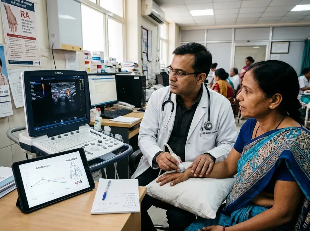
Subjective components in DAS28 often inflate disease activity scores due to central sensitization and emotional distress. Integrating the DAS28-P index, musculoskeletal ultrasound, and emerging digital biomarkers allows clinicians to differentiate true synovitis from non-inflammatory pain and prevent futile escalation.
Today

AIIMS researchers suggest yoga may alleviate gut dysbiosis in Polyendocrine Metabolic Ovarian Syndrome (PMOS) by modulating the gut-brain axis, lowering chronic inflammation, and enhancing parasympathetic tone to support metabolic and hormonal equilibrium.
Today
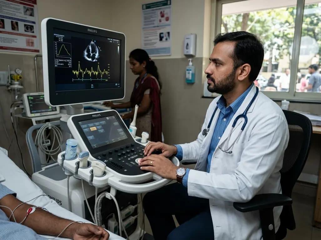
A structured practical guide to left atrial echocardiographic assessment covering biplane volumetry, phasic physiology, speckle-tracking strain, and hemodynamic filling pressure estimation for routine cardiovascular practice.
Today

Age-related spinal kyphosis is driven by vertebral fragility, disc degeneration, and paraspinal muscle dysfunction. Coexisting with locomotive syndrome and frailty, its management requires combined anti-osteoporotic therapy, spinal extensor rehabilitation, and comprehensive nutritional optimization.
Today

A longitudinal feasibility pilot study highlights that combining polygenic risk score disclosure with in-person lifestyle counseling significantly enhances genomic literacy in individuals at risk for type 2 diabetes, overcoming educational disparities and reinforcing patient motivation.
Today
