

Loading, please wait...
Loading, please wait...

Airway management forms the absolute cornerstone of anaesthesiology training and patient safety in operative environments. Securing the airway swiftly on the initial attempt prevents critical complications such as profound arterial desaturation, cardiovascular instability, and airway trauma. For decades, traditional direct laryngoscopy using the standard Macintosh blade served as the universal teaching modality. However, modern optical devices have fundamentally revolutionized perioperative practice. The routine adoption of videolaryngoscopy for tracheal intubation by anaesthesia resident doctors offers substantial technical advantages, yet large pragmatic multicentre evidence has remained limited until now.
Securing a patent airway represents one of the most stressful technical procedures for early-career doctors. When trainees perform direct laryngoscopy, they must align the oral, pharyngeal, and laryngeal axes mechanically to attain a direct line of sight. Consequently, anatomical variations, prominent incisors, cervical immobility, or limited mouth opening frequently impair glottic exposure. Multiple failed attempts escalate the danger of soft-tissue damage, vocal cord oedema, and emergency surgical airway rescue. In contrast, videolaryngoscopy overcomes these strict anatomical line-of-sight constraints by incorporating a high-resolution miniature camera near the blade tip. Furthermore, the video screen displays an unobstructed panoramic view of the larynx, transforming airway visualization into an objective experience. Supervisors can observe the identical visual field simultaneously, offering real-time verbal guidance and correcting blade insertion depth before desaturation occurs. Although several smaller single-centre trials highlighted higher success rates with video blades, sceptics argued that routine use might impair trainee mastery of direct laryngoscopy. Therefore, clinicians required robust, multicentre randomised evidence to confirm whether first-line videolaryngoscopy systematically benefits novice practitioners in everyday hospital surgical theatres.
To provide clear clinical evidence, investigators conducted the VILARE trial across multiple tertiary surgical hospitals. This pragmatic, multicentre randomised controlled trial enrolled 1,001 adult surgical patients requiring endotracheal intubation under general anaesthesia. The trial design closely mirrored daily operative practice to maximize ecological validity. Investigators randomly assigned participants to receive tracheal intubation with either a videolaryngoscope or a conventional direct laryngoscope for the primary attempt. Specifically, 501 patients underwent videolaryngoscopy, whereas 500 patients underwent direct laryngoscopy. Anaesthesia resident doctors of varying training years performed all initial intubation attempts under direct consultant supervision. The primary outcome evaluated whether residents achieved successful tracheal intubation on the very first attempt. Meanwhile, secondary outcomes evaluated key perioperative parameters, including glottic visualisation quality, procedural duration, and total intubation attempts. Additionally, researchers tracked the requirement for airway adjuncts, overall procedural difficulty scores, and immediate airway-related complications. By deliberately maintaining pragmatic operating room conditions without excessive exclusion criteria, the trial generated pragmatic data that clinicians can directly extrapolate to standard academic teaching hospitals worldwide.
The VILARE trial results demonstrated decisive technical advantages for videolaryngoscopy when performed by resident physicians. Specifically, residents achieved successful first-attempt tracheal intubation in 397 of 501 patients (79.2%) allocated to the videolaryngoscopy arm. In contrast, residents achieved first-attempt success in only 351 of 500 patients (70.2%) in the direct laryngoscopy arm. This difference represented an absolute risk difference of 9.0 percentage points, demonstrating statistically significant and clinically meaningful superiority. Furthermore, videolaryngoscopy dramatically enhanced anatomical glottic exposure. Trainees consistently attained Cormack-Lehane grade 1 views far more frequently with video technology than with conventional Macintosh blades. Poor glottic visualisation occurred rarely in the videolaryngoscopy group, eliminating the blind, uncertain tube advancement that often plagues early training experiences. Consequently, residents experienced markedly lower rates of multiple failed passes. When novice operators achieve immediate glottic clarity, their procedural confidence increases, and patients experience smooth anaesthetic inductions. These robust multicentre results firmly establish that videolaryngoscopy provides junior anaesthesia doctors with a superior initial technical advantage during general surgical cases.
Beyond improving primary intubation success, the VILARE trial provided crucial data regarding airway safety and operator workload. Residents reported significantly lower procedural difficulty scores when employing videolaryngoscopes compared to traditional direct blades. Because the video screen projects a magnified view of the vocal cords, trainees could easily guide the endotracheal tube without applying excessive force against the maxillary incisors. In addition, supervising consultants intervened far less frequently to rescue failed intubation attempts in the videolaryngoscopy cohort. Notably, the requirement for rescue airway adjuncts, such as flexible bronchoscopes or supraglottic rescue devices, decreased alongside video adoption. Traumatic complications also demonstrated favourable patterns. Inadvertent oesophageal intubations, dental damage, and pharyngeal mucosal bleeding occurred less frequently when operators visualised the airway structures clearly. Prolonged apnoea and desaturation episodes dropped as first-pass success climbed. Therefore, videolaryngoscopy not only facilitates smoother procedural execution for training physicians, but it also fortifies essential clinical safety margins for anaesthetised surgical patients.
The VILARE findings carry profound implications for modern anaesthesiology training curricula and clinical airway governance. For decades, training programs mandated direct laryngoscopy as the mandatory primary skill before allowing trainees access to advanced optical devices. However, growing multicentre evidence suggests that educators should reconsider this traditional dogma. Defaulting to videolaryngoscopy from day one protects patients while trainees develop foundational eye-hand coordination. Furthermore, screen-based guidance allows faculty instructors to correct subtle errors instantaneously, transforming airway management into a shared collaborative procedure rather than a solitary guessing exercise. In Indian healthcare institutions and resource-diverse settings, investing in durable videolaryngoscopes can significantly enhance patient safety across high-volume tertiary teaching hospitals. Nevertheless, academic departments must preserve balanced simulation training so residents retain manual direct laryngoscopy competency during unexpected equipment failures or battery outages. Ultimately, establishing videolaryngoscopy as the standard initial approach creates a safer surgical environment, optimizes postgraduate education, and minimizes life-threatening airway emergencies.
Researchers designed the VILARE trial specifically around resident doctors because training clinicians face the steep learning curve of airway management. Early procedural success directly impacts patient safety during induction. Although seasoned consultants frequently rescue failed airways, trainees perform countless initial intubations during hospital shifts. Consequently, identifying whether videolaryngoscopy accelerates procedural success helps academic centres refine residency training curricula, standardise operating room protocols, and substantially reduce avoidable patient trauma during early clinical training.
Videolaryngoscopy provides superior glottic visualization, but it does not completely replace traditional direct laryngoscopy skills. Anaesthetists must maintain competency in direct laryngoscopy because equipment failures, dead batteries, lens contamination, and limited institutional resources can occur unexpectedly. Furthermore, developing sound tactile feedback and understanding physical airway axes remains essential for foundational medical training. Therefore, educators recommend teaching videolaryngoscopy as the primary clinical tool while ensuring residents also retain proficient manual direct laryngoscopy capabilities for backup scenarios.
Videolaryngoscopy fundamentally improves clinical teaching by sharing the operator's real-time field of view on an external monitor. During direct laryngoscopy, supervisors only observe external hand movements and cannot assess vocal cord views simultaneously. Conversely, screen sharing allows supervising consultants to offer timely guidance regarding blade repositioning, suctioning, or tube delivery before hypoxemia develops. Thus, the video screen provides an invaluable pedagogical shared visual mental model, substantially enhancing both patient safety and resident clinical instruction.
Disclaimer: This content is for informational and educational purposes only... Refer to the latest local and national guidelines for clinical practice.
References

Read summarized clinical updates, watch expert medical content, and earn CME certifications right from your smartphone.
Open your smartphone camera to scan and install.





The multicentre VILARE trial evaluated videolaryngoscopy versus direct laryngoscopy for tracheal intubation performed by anaesthesia resident doctors. Videolaryngoscopy significantly increased first-attempt success rates, improved glottic visualisation, and reduced procedural difficulty during routine surgical airway management.
Today
A large cohort study evaluating 1,245 patients demonstrates that unsedated gastroscopy is safe, feasible, and achieves high completion rates, offering an efficient alternative to sedated procedures.
Today
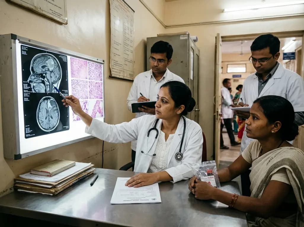
An intracranial EWSR1::CREM fusion sarcoma in a 31-year-old female manifested as a paraneoplastic inflammatory syndrome with anemia and secondary infertility. The dural mass mimicked meningioma, but gross-total resection resolved her systemic inflammation and restored natural fertility within one month.
Today
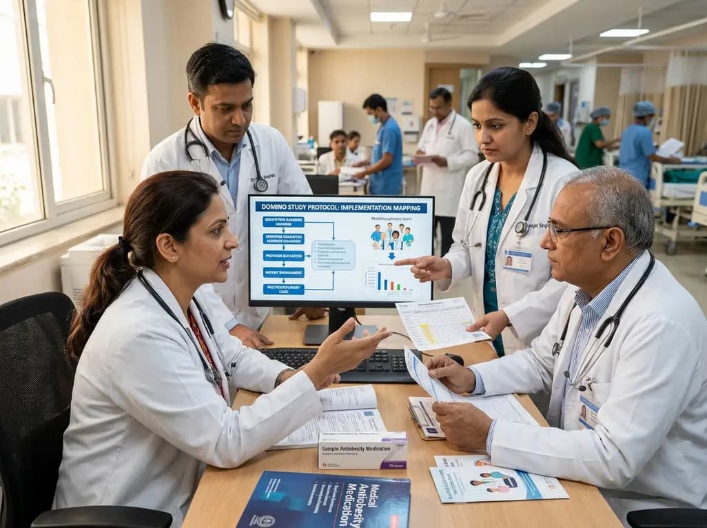
The DOMINO study protocol presents a multidisciplinary implementation mapping approach to overcome provider, patient, and payer barriers to antiobesity medications, offering vital insights for Indian clinicians managing the growing diabesity burden.
Today
An age-matched cross-sectional study reveals that lower muscle quality and impaired lower-extremity function strongly correlate with an increased fear of falling in older women, highlighting the necessity of targeted resistance training and balance interventions in geriatric care.
Today
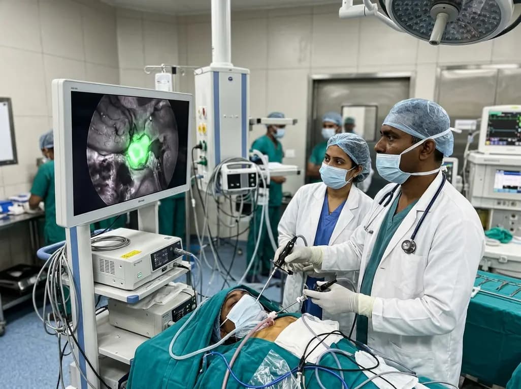
Near-infrared autofluorescence provides real-time label-free optical guidance during endoscopic transsphenoidal pituitary surgery. By highlighting stark signal contrasts between normal pituitary gland and adenomas, this technique aids gross-total tumor resection while safeguarding critical endocrine function.
Today
