.webp&w=384&q=75)

Loading, please wait...
Loading, please wait...

Chronic wound management remains a formidable clinical challenge across global healthcare settings, particularly when multidrug-resistant pathogens establish persistent infections. Bacterial aggregates encased in a dense extracellular matrix thwart standard topical therapies and conventional systemic antimicrobials. Recently, therapeutic ultrasound cavitation has emerged as a compelling modality to overcome these biophysical barriers. By destabilizing protective extracellular matrices, acoustic energy significantly enhances local drug penetration. Consequently, clinicians and researchers are actively evaluating biophysical adjunctive techniques. Understanding how acoustic emissions correlate with microbiological clearance provides vital insights for future clinical translations in wound care.
Pathogenic biofilms present a formidable hurdle in chronic skin ulcers, diabetic foot infections, and post-surgical wounds. Bacteria such as methicillin-resistant Staphylococcus aureus produce a self-secreted polymeric matrix consisting of polysaccharides, extracellular DNA, and structural proteins. Therefore, this dense slime layer acts as a mechanical shield that severely restricts antibiotic diffusion. Furthermore, bacteria nestled within deep biofilm layers enter a metabolically quiescent state known as persister phenotype. As a result, standard bactericidal agents that target active cell division exhibit drastically reduced efficacy against these dormant organisms. Clinicians frequently encounter recurrent infections despite adhering strictly to established antimicrobial protocols. In addition, aggressive surgical debridement often fails to eradicate residual microscopic clusters lodged within deep tissue crevices. Consequently, persistent bacterial colonization sustains chronic local inflammation, degrading essential growth factors and impairing normal tissue granulation. In India, where diabetic ulcer complications remain rampant, refractory biofilm infections cause substantial morbidity and elevate limb amputation risks. Hence, developing targeted mechanical strategies that disrupt biofilm architecture without inflicting collateral tissue injury represents an urgent surgical and dermatological priority.
Acoustic cavitation describes the formation, oscillation, and collapse of vapor or gas-filled microcavities within a liquid medium under cyclic acoustic pressure. Specifically, ultrasound cavitation exerts potent mechanical forces that cleave the structural backbone of mature bacterial matrices. Non-inertial or stable cavitation occurs at moderate acoustic pressures, generating microstreaming currents and localized shear stresses adjacent to vibrating bubbles. These hydrodynamic currents loosen intercellular bonds and enhance fluid convective transport across the biofilm boundary layer. In contrast, inertial cavitation involves rapid bubble expansion followed by violent bubble collapse. This implosion produces high-velocity microjets, localized shockwaves, and transient mechanical micro-fissures within tissue interfaces. Furthermore, these acoustic shockwaves disintegrate dense polysaccharide polymers, exposing embedded persister bacteria to surrounding bactericidal compounds. As a result, co-administered antimicrobial agents penetrate deep recesses that passive diffusion could never reach. Moreover, mechanical permeabilization alters bacterial cell membrane permeability, facilitating intracellular influx of aminoglycosides and fatty acids. Therefore, ultrasound cavitation transforms impenetrable bacterial fortresses into vulnerable, accessible targets.
Conventional gas microbubbles exhibit limited tissue penetration because their relatively large micron size prevents entry into cramped intercellular gaps. In response, biomedical engineers developed submicron phase change contrast agents utilizing low-boiling point perfluorocarbon cores. For instance, liquid octafluoropropane droplets maintain stable submicron diameters during routine storage and initial topical administration. Because these droplets are tiny, they readily infiltrate dense microscopic fissures throughout infected wound beds. Subsequently, targeted ultrasound waves vaporize the liquid perfluorocarbon core through a phenomenon termed acoustic droplet vaporization. This thermal and mechanical phase transition rapidly expands the droplet into an active, highly responsive microbubble. Additionally, low-boiling point formulations vaporize at significantly lower acoustic energy thresholds than traditional high-boiling perfluorocarbons. Consequently, clinicians can achieve robust bubble expansion without generating excessive thermal injury in fragile granulation tissue. Once vaporized, these microbubbles oscillate vigorously under acoustic irradiation, generating sustained cavitation signatures. Therefore, perfluorocarbon phase change agents serve as exquisite acoustic amplifiers, concentrating destructive mechanical micro-forces directly at the bacterial-host interface.
Recent preclinical investigations have successfully quantified real-time acoustic emissions during topical antibiotic treatment of methicillin-resistant Staphylococcus aureus infections. In a standardized murine wound model, researchers combined topical gentamicin and palmitoleic acid with octafluoropropane phase-change agents. Investigators delivered ultrasound exposures at 1.1 megahertz utilizing peak acoustic pressures of either 700 kilopascals or 1700 kilopascals. Notably, passive cavitation detectors recorded harmonic and broadband emissions every fifteen seconds to quantify the cumulative acoustic dose. The experimental data revealed a striking inverse correlation between cumulative cavitation dose and final bacterial burden. Specifically, higher cavitation doses directly drove greater log-reductions in viable bacterial colony-forming units. Furthermore, high acoustic pressure sustained continuous microbubble activity throughout the five-minute treatment cycle. In contrast, acoustic cavitation signals at lower pressure decayed rapidly over time, diminishing overall therapeutic impact. However, researchers observed incomplete wound surface coverage, which constrained total bacterial clearance across the entire lesion. Thus, ensuring uniform acoustic delivery across all infected wound margins remains essential for achieving complete microbial eradication.
These preclinical breakthroughs offer profound translational relevance for dermatologists, plastic surgeons, and wound care practitioners managing chronic recalcitrant ulcers. In contemporary practice, clinicians administer broad-spectrum topical and systemic antimicrobials empirically, frequently encountering therapeutic stagnation. By incorporating passive acoustic monitoring, practitioners could objectively verify therapeutic cavitation activity during clinic-based interventions. Moreover, real-time acoustic dosimetric feedback allows physicians to titrate ultrasound energy dynamically, avoiding under-treatment or unwanted mechanical tissue disruption. In Indian hospital wards and diabetic outpatient clinics, resistant hospital-acquired infections impose heavy financial and physical strains on patients. Consequently, non-invasive biophysical adjuncts that restore sensitivity to standard antibiotics like aminoglycosides provide immense clinical utility. Additionally, pairing acoustic cavitation with adjuvant fatty acids or enzymatic cleansers may suppress emergent resistant mutations. However, translating these findings requires ergonomically designed transducer arrays that conform seamlessly to irregular anatomical contours. Ultimately, standardizing acoustic treatment parameters will empower multidisciplinary teams to eliminate resilient biofilms and accelerate wound re-epithelialization.
Acoustic cavitation generates microscopic fluid jets and intense hydrodynamic shear stresses that mechanically fragment the rigid extracellular biofilm matrix. Consequently, these micro-disruptions open transient conduits through which topical antibiotics rapidly diffuse into deep bacterial reservoirs. Furthermore, the acoustic microstreaming alters microbial membrane permeability, facilitating increased antibiotic entry into previously tolerant persister cells. As a result, standard antimicrobial agents achieve profound bactericidal efficacy at significantly lower systemic doses.
Traditional contrast microbubbles are large and unstable, preventing adequate penetration into tight intercellular matrix networks within infected tissue. In contrast, low-boiling point perfluorocarbon agents exist initially as submicron liquid droplets that readily infiltrate deep wound crevices. Upon localized ultrasound exposure, these droplets rapidly vaporize into active microbubbles via acoustic droplet vaporization. Furthermore, they activate at lower acoustic pressures, producing vigorous, sustained cavitation without causing thermal injury to fragile granulating tissues.
Passive cavitation detection records real-time acoustic harmonic and broadband emissions during therapeutic ultrasound application. Therefore, clinicians can measure the exact acoustic dose delivered to the wound bed rather than estimating treatment efficacy blindly. Additionally, this real-time dosimetric feedback allows practitioners to adjust acoustic pressures instantly, ensuring continuous microbubble activation across the entire infected surface. Consequently, clinicians can achieve reliable biofilm disruption while avoiding tissue overtreatment and accelerating clinical wound closure.
Disclaimer: This content is for informational and educational purposes only... Refer to the latest local and national guidelines for clinical practice.
References

Read summarized clinical updates, watch expert medical content, and earn CME certifications right from your smartphone.
Open your smartphone camera to scan and install.





A landmark preclinical study demonstrates that ultrasound cavitation driven by low-boiling phase-change contrast agents correlates strongly with bacterial burden reduction in MRSA biofilm wounds, paving the way for monitored acoustic biofilm therapy.
Today
A groundbreaking study highlights how elevated ISG15 fuels RIG-I/MDA5 signaling and macrophage-microglia crosstalk during blood-brain barrier disruption in neuromyelitis optica spectrum disorder, revealing novel therapeutic targets for neuroinflammation.
Today
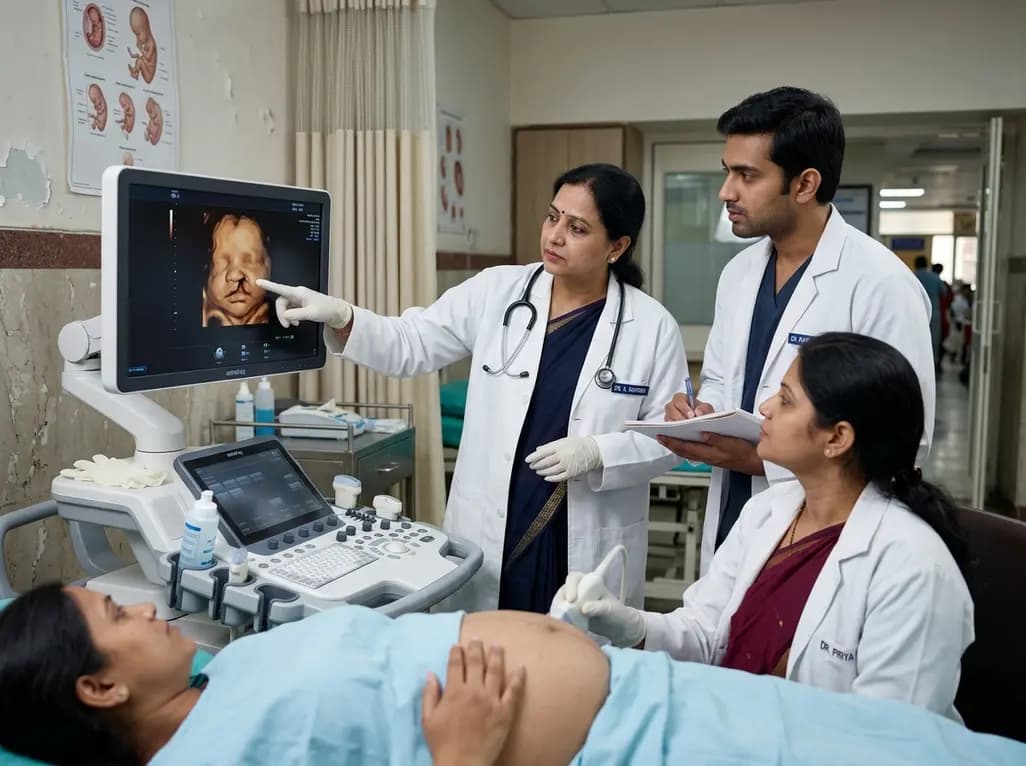
A 5-year retrospective study evaluates prenatal ultrasound detection of fetal cleft lip and palate. While overall detection reached 87%, accuracy varied from 99.3% in combined clefts to 50% in isolated cases. Combined defects showed elevated risks of concurrent structural anomalies and chromosomal abnormalities.
Today

An updated systematic review and meta-analysis of 7,875 patients reveals that routine microaxial left ventricular support during nonemergent high-risk PCI does not reduce mortality or ischemic complications, while significantly increasing major bleeding events.
Yesterday
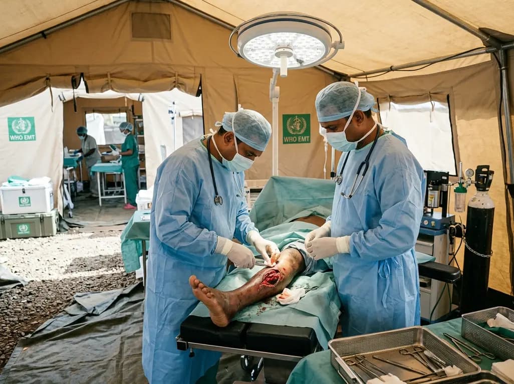
Following Cyclone Chido, a WHO EMT-2 field hospital performed 268 surgeries in Mayotte. While acute trauma is expected initially, 98% of cases were septic wounds of the extremities requiring secondary intention healing, revealing crucial operational insights for disaster surgical preparedness.
Yesterday

The SED-SEEN-SEEP consensus outlines an Integrated Care Pathway for preclinical type 1 diabetes, structuring target screening, autoantibody confirmation, metabolic staging, and proactive follow-up to prevent diabetic ketoacidosis and facilitate early disease-modifying intervention.
Yesterday
