

Loading, please wait...
Loading, please wait...

Surgical management of skull base meningiomas presents formidable technical challenges due to cramped anatomic corridors and delicate neurovascular structures. In contemporary neuro-oncology, comprehensive meningioma molecular profiling has become essential for precise risk stratification, clinical prognostication, and targeted therapeutic planning. However, obtaining adequate tumor bulk without compromising neurological function remains difficult. During complex debulking, ultrasonic surgical aspirators efficiently emulsify neoplastic tissue, yet surgical teams routinely discard the aspirated effluent. A prospective feasibility study has demonstrated that this previously discarded aspirate represents a pristine reservoir of high-molecular-weight DNA, exhibiting remarkable genomic and epigenomic concordance with standard en bloc surgical resections.
Skull base meningiomas frequently encase vital cranial nerves, compress brainstem structures, and adhere tenaciously to major intracranial blood vessels. Consequently, neurosurgeons prioritize functional preservation over aggressive gross total resection, often relying on narrow surgical corridors. In these restricted spaces, surgeons rarely perform large en bloc excisions. Instead, ultrasonic aspirators break down the fibrous tumor matrix through high-frequency vibrations while simultaneously irrigating and suctioning the particulate debris. Historically, pathology departments prioritized the few solid fragments retrieved via forceps for diagnostic histology.
As a result, minimal tissue remained available for advanced molecular diagnostics. This limitation has become increasingly problematic because recent World Health Organization classifications incorporate molecular alterations into meningioma grading. Epigenomic DNA methylation subgroups and specific somatic copy-number variations predict recurrence risk far more accurately than classical histological grading alone. Discarding aspirated tumor tissue therefore represents a significant missed opportunity. If surgical aspirates can reliably supply diagnostic nucleic acids, clinicians can overcome sample scarcity in subtotal resections, expanding access to cutting-edge precision diagnostics for complex skull base tumors.
To establish whether ultrasonic surgical aspirates provide suitable substrates for comprehensive testing, investigators conducted a prospective feasibility series comparing paired specimens. The surgical team collected matched standard resection specimens and Sonopet ultrasonic aspirate samples from seven consecutive patients undergoing craniotomies for skull base meningiomas. Rather than relying on targeted sequencing panels, the investigators implemented native, single-molecule whole-genome sequencing utilizing Oxford Nanopore Technologies PromethION R10 flow cells. This advanced approach enabled simultaneous assessment of structural genomic variants and native CpG methylation patterns without requiring chemical bisulfite conversion.
Remarkably, the ultrasonic aspirates produced sequencing yields, read length distributions, and estimated tumor purities that closely matched conventional resection specimens. High-frequency ultrasonic cavitation and mechanical suction did not cause catastrophic shearing of high-molecular-weight genomic DNA. Instead, long-read sequencing metrics remained robust across all paired comparisons. These findings confirm that ultrasonic aspirate preserves cellular and molecular integrity effectively, establishing meningioma molecular profiling from surgical washings as an authentic, high-yield diagnostic method.
The comparative analysis revealed outstanding concordance between paired aspirate and resection samples across broad genomic and epigenomic dimensions. Genome-wide copy-number variation profiles demonstrated near-perfect agreement, successfully recovering canonical meningioma alterations such as monosomy 22 and characteristic chromosomal arm losses. Quantitative concordance in total copy-number burden and genomic loss burden achieved a Lin concordance correlation coefficient of 0.99. Therefore, clinicians can confidently interpret copy-number signatures derived from ultrasonic aspirate material without fearing artifactual chromosomal deletions or distortions.
Furthermore, native epigenomic profiling revealed extraordinary fidelity between tissue procurement methods. Analysis across approximately 177,000 gene promoter regions demonstrated a Pearson correlation coefficient exceeding 0.99 between paired aspirates and solid tissue fragments. Projecting these nanopore methylation profiles onto an established reference framework of 121 EPIC array-profiled tumors yielded identical molecular group classifications in nearly all pairs. In only one patient with borderline overall tumor purity, classification proved ambiguous. Consequently, aspirate-derived DNA methylation profiling offers diagnostic reliability identical to standard surgical tissue, ensuring dependable WHO molecular grouping.
Interestingly, the investigation noted moderate concordance regarding somatic single-nucleotide variant burden, exhibiting a concordance correlation coefficient of 0.53. In two patient pairs, researchers observed divergent variant allele frequencies between the aspirate and the targeted resection biopsy. Rather than representing an assay artifact, this divergence highlights a distinct spatial advantage of ultrasonic aspiration. A standard surgical biopsy represents a single, highly localized punch taken from one accessible geographic area of the tumor.
In contrast, an ultrasonic aspirator actively pools fragmented cells from the entire operative field throughout the debulking procedure. As the surgeon works through diverse tumor coordinates, the aspirate canister accumulates a blended, representative cross-section of the entire neoplastic mass. Therefore, ultrasonic aspirates can overcome localized sampling bias by capturing subclonal mutations and branched evolutionary features that a discrete focal biopsy might overlook. This comprehensive spatial pooling provides neuro-oncologists with a more holistic view of tumor heterogeneity, which is critical for identifying rare, therapy-resistant subclonal populations.
The successful extraction of intact epigenomic and genomic data from surgical aspirates carries immediate clinical significance for multidisciplinary neuro-oncology teams. First, it addresses the persistent dilemma of the insufficient tissue sample in challenging skull base approaches. When surgeons perform cautious subtotal debulking near the cavernous sinus, optic apparatus, or petroclival region, preserving patient neurological function often results in minimal solid tissue retrieval. Capturing aspirate ensures that even subtotal resections yield ample biological substrate for full molecular risk stratification.
Second, this approach streamlines clinical translational research workflows. Standard pathology archives typically lock away precious formalin-fixed, paraffin-embedded blocks for diagnostic immunochemistry, leaving limited tissue for research biobanking. Routinely processing ultrasonic aspirate fluid creates an abundant source of high-quality tumor DNA without depleting diagnostic specimens. Consequently, centers can establish extensive biobanks of native, high-molecular-weight nucleic acids from complex intracranial tumors, accelerating genomic discoveries and clinical trial correlative studies.
Looking ahead, combining ultrasonic aspiration with real-time nanopore sequencing could revolutionize intraoperative decision-making. Single-molecule sequencing platforms read native DNA molecules in real time, streaming methylation and structural variant data within minutes of loading flow cells. If surgical teams centrifuge and isolate tumor nuclei from aspirate fluid during the early stages of craniotomy debulking, intraoperative nanopore workflows could deliver molecular classification before surgical closure.
Moreover, intraoperative epigenomic confirmation could clarify whether a challenging skull base lesion represents a benign grade 1 meningioma or an aggressive molecular subtype harboring high-risk copy-number alterations. Knowing the precise molecular risk tier intraoperatively might help neurosurgeons balance the risks of aggressive boundary resection against functional preservation. As bedside sequencing workflows continue to mature and library preparation protocols become more automated, ultrasonic aspirates will undoubtedly transition from discarded operating room waste into an essential cornerstone of precision neurosurgical diagnostics.
Ultrasonic aspirate tissue supplements rather than replaces conventional histopathology. While aspirates yield pristine high-molecular-weight DNA for genomic and epigenomic profiling, pathologists still require intact tissue architecture to evaluate histological features, mitotic indices, and brain invasion. Combining both tissue sources ensures comprehensive histological and molecular diagnostic accuracy.
Long-read single-molecule sequencing directly characterizes native, unamplified DNA. This technology simultaneously identifies complex structural chromosomal variants, copy-number alterations, and base modifications like 5-methylcytosine without requiring separate bisulfite conversion assays. Consequently, it accelerates comprehensive WHO molecular subgrouping while conserving minimal nucleic acid volumes.
Skull base meningiomas arise within narrow, delicate anatomical corridors where aggressive resection risks catastrophic cranial nerve and vascular injuries. Because surgeons prioritize patient neurological function, obtained tissue samples are often scarce. Utilizing discarded surgical aspirates provides abundant tumor material for advanced diagnostics without forcing surgeons to compromise safety.
Disclaimer: This content is for informational and educational purposes only, and does not substitute professional medical advice, diagnosis, or treatment. Refer to the latest local and national guidelines for clinical practice.
References
Unal M et al. Single-molecule whole-genome sequencing reveals genomic and epigenomic concordance between ultrasonic aspiration and surgical resection in meningioma: prospective feasibility series. J Neurooncol. 2026 Nov 20. doi: 10.1007/s11060-026-05846-3. PMID: 43062499.
Nguyen MP, Mirchia K, Braman BC, Morshed RA, Oberheim-Bush NA, Arum Z, et al. Prospective Genomic Profiling of Consecutive Meningiomas. JAMA Oncol. 2026 Jun 1;12(6):660-662. doi: 10.1001/jamaoncol.2026.0507.
Sahm F, Schrimpf D, Stichel D, Jones DTW, Hielscher T, Schefzyk S, et al. DNA methylation-based classification and grading system for meningioma: a multicentre, retrospective analysis. Lancet Oncol. 2017 May;18(5):682-694. doi: 10.1016/S1470-2045(17)30155-9.

Read summarized clinical updates, watch expert medical content, and earn CME certifications right from your smartphone.
Open your smartphone camera to scan and install.





A prospective feasibility series demonstrates that discarded ultrasonic aspirate tissue delivers high-molecular-weight DNA comparable to standard resection for long-read genomic and epigenomic profiling in skull base meningioma, unlocking comprehensive molecular diagnostics even in restricted surgical corridors.
Today

A landmark study reveals that congenital DLK1 deficiency in women with a history of central precocious puberty causes visceral adiposity, mitochondrial dysfunction, oxidative stress, and neutrophil activation, highlighting the critical need for lifelong cardiometabolic surveillance.
Today

Estetrol (E4), a native fetal estrogen, offers a unique tissue-selective profile with minimal hepatic impact and lower thrombotic risk. This review highlights its clinical use in oral contraception, menopausal symptom management, and emerging applications across oncology and neurology.
Today
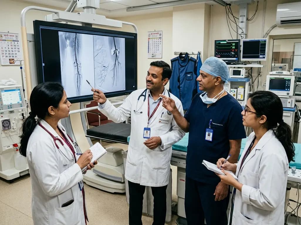
Explore revascularisation strategies in dialysis-dependent chronic limb-threatening ischaemia. This review examines surgical bypass vs endovascular therapy, BEST-CLI trial findings, procedural morbidity, conduit availability, and multidisciplinary decision-making for optimal limb salvage.
Today

A recent cross-sectional analysis reveals that over a quarter of randomized controlled trials for non-specific low back pain face discontinuation or unknown status, while more than 36% of completed trials remain unpublished, exposing notable reporting gaps in spine and musculoskeletal clinical research.
Today
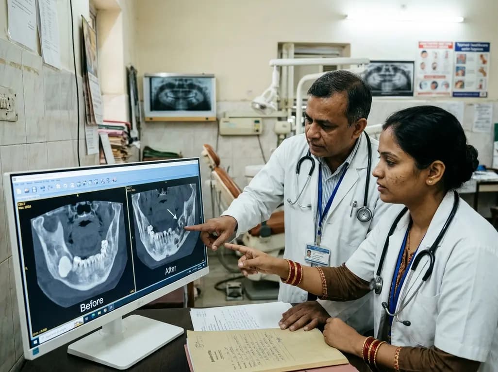
This case report examines the longitudinal CBCT evolution of Stage 2 MRONJ in a 70-year-old woman on denosumab at the site of a pre-existing mandibular dense bone island. It emphasizes the radiographic features, differential diagnosis, and multidisciplinary strategies required for high-risk bone lesions.
Today
