.webp&w=384&q=75)

Loading, please wait...
Loading, please wait...
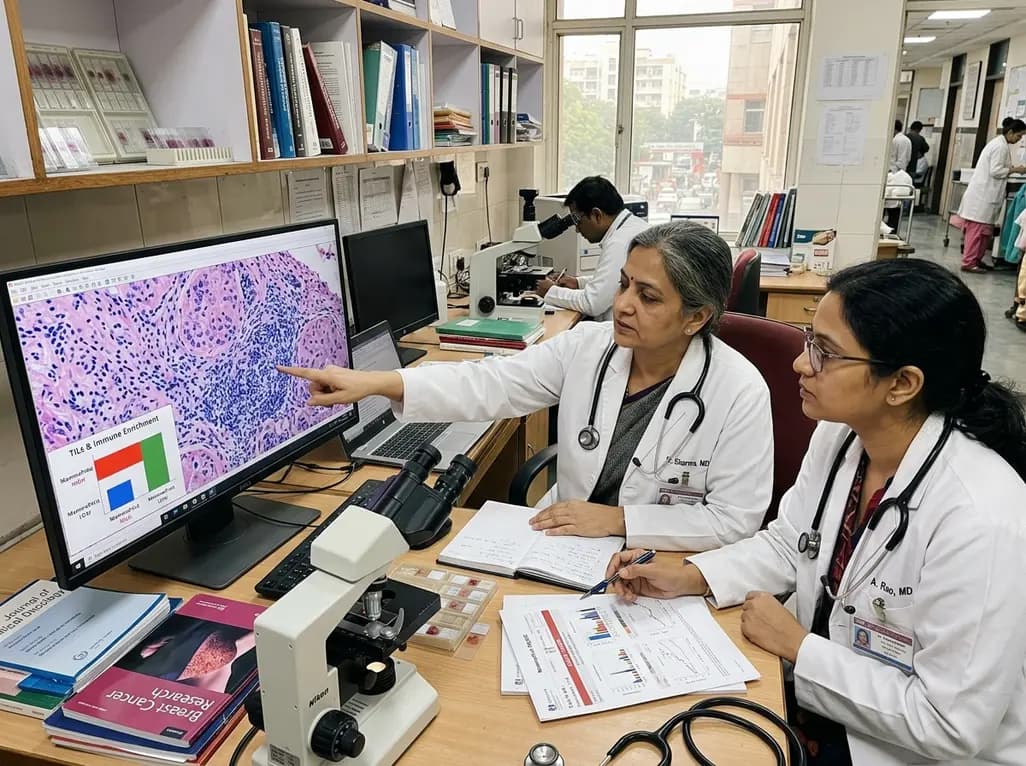
Risk stratification in early-stage breast cancer has advanced rapidly over recent years. Clinicians routinely evaluate genomic profiles alongside standard histopathological features to tailor adjuvant treatment plans. In particular, examining the presence and distribution of TILs in breast cancer provides critical biological insights into host antitumor immunity. Historically, clinicians considered estrogen receptor-positive luminal tumors to be immunologically quiescent. However, emerging research demonstrates substantial immune heterogeneity within hormone receptor-positive cohorts. A recent landmark study investigated the relationship between MammaPrint genomic classifications and immune infiltration. By assessing 492 luminal breast cancer specimens, researchers uncovered distinct immune microenvironment variations across genomic risk categories. These findings significantly enhance our understanding of tumor biology, prognosis, and therapeutic stratification in luminal disease.
The 70-gene MammaPrint signature serves as an established diagnostic tool for early estrogen receptor-positive, HER2-negative breast cancer. Initially developed as a binary classifier distinguishing low-risk from high-risk disease, the assay now provides a refined four-tier model. This updated classification delineates tumors into UltraLow, Low, High-Risk1 (H1), and High-Risk2 (H2) categories. Consequently, clinicians can identify ultralow-risk patients who can safely de-escalate endocrine therapy. Furthermore, the four-tier system highlights high-risk patients who require aggressive adjuvant chemotherapy. In routine practice, genomic assays help oncologists avoid both undertreatment and overtreatment. However, genomic risk scores do not capture the spatial architecture of the tumor microenvironment on their own. Therefore, integrating genomic data with cellular immune parameters offers a more comprehensive perspective on luminal tumor behavior. This integrative approach bridges the gap between molecular diagnostics and traditional surgical pathology.
Pathologists quantify stromal tumor-infiltrating lymphocytes using standardized criteria established by the International Immuno-Oncology Biomarker Working Group. In this context, evaluating TILs in breast cancer involves both quantitative enumeration and spatial distribution analysis. Researchers systematically classify tumors into four discrete spatial compartments: immune-deserted, stromal-restricted, immune-excluded, and inflamed phenotypes. Deserted tumors exhibit virtually no immune cell presence within the tumor boundaries. In contrast, inflamed tumors feature active immune cell infiltration across stromal and epithelial compartments. Tissue microarray analysis allows precise evaluation of CD4 helper T cells and CD8 cytotoxic lymphocytes within these subsets. Notably, high-risk luminal tumors demonstrate a significant shift toward inflamed spatial configurations. Conversely, low-risk tumors frequently present with deserted or stromal-restricted patterns. Thus, spatial categorization illuminates how tumor genomics shapes local immune infiltration.
The analysis of 492 luminal breast cancer specimens revealed strong associations between immune dynamics and adverse pathological traits. Specifically, High-Risk MammaPrint tumors constituted 41% of the cohort and exhibited distinctive aggressive characteristics. These tumors demonstrated significantly higher histologic grade, elevated Ki-67 proliferation indices, and frequent lymphovascular invasion. In addition, High-Risk tumors showed marked downregulation of progesterone receptor expression. Pathologists identified a median stromal TIL level of 15% in High-Risk cases compared to only 5% in Low-Risk tumors. Multivariable regression confirmed that elevated TIL density independently correlates with higher genomic risk. Furthermore, high-grade tumors harbored dense concentrations of CD8-positive cytotoxic T lymphocytes. Therefore, aggressive genomic profiles coincide with heightened intrinsic immunogenicity. These observations indicate that immune mobilization frequently parallels biological aggressiveness in luminal carcinomas.
Transitioning from binary to four-tier MammaPrint categorization provides granular insight into luminal tumor biology. UltraLow-risk tumors consistently exhibit an immune-quiescent microenvironment with negligible lymphocytic infiltration. Similarly, classic Low-Risk tumors show low median TIL levels and predominantly immune-deserted architectures. In sharp contrast, tumors classified as High-Risk2 display the highest infiltration of CD4 and CD8 T cells. The High-Risk1 subgroup occupies an intermediate immunophenotypic space between classic Low-Risk and High-Risk2 categories. Consequently, the four-tier genomic framework mirrors a progressive biological gradient of immune activation. Moreover, multivariable models show that the four-tier stratification captures immune variations far more accurately than binary models. This granular subtyping helps oncologists distinguish subtly aggressive tumors from truly indolent luminal neoplasms. As a result, multidisciplinary teams obtain clearer guidance for long-term clinical monitoring.
These findings carry profound implications for personalizing adjuvant therapy in hormone receptor-positive breast cancer. For patients with UltraLow risk and deserted immune profiles, clinicians can confidently pursue treatment de-escalation strategies. Such patients achieve excellent long-term survival with minimal endocrine intervention, avoiding chemotherapy toxicity. Conversely, High-Risk2 tumors with dense TILs and inflamed microenvironments may derive substantial benefit from multimodal regimens. Although luminal cancers traditionally show limited sensitivity to immune checkpoint inhibitors, inflamed High-Risk subsets might represent viable candidates for targeted immunotherapy combinations. In addition, quantifying TILs offers an inexpensive, reproducible prognostic biomarker that complements complex molecular assays. Clinicians can integrate routine histopathology with genomic data to optimize decision-making in resource-constrained environments. Ultimately, understanding tumor-immune interactions drives precise, individualized breast cancer care.
Tumor-infiltrating lymphocytes reflect the host immune response against neoplastic cells. In luminal breast cancer, higher levels of immune infiltration correlate with aggressive biological traits, such as high Ki-67 and high genomic risk scores. Identifying these lymphocytes helps pathologists and oncologists assess tumor immunogenicity, predict therapeutic response, and optimize systemic treatment planning for patients.
The four-tier MammaPrint assay divides patients into UltraLow, Low, High-Risk1, and High-Risk2 subgroups. This detailed framework identifies patients with indolent disease who can safely avoid aggressive adjuvant therapies. Furthermore, it accurately identifies highly aggressive luminal tumors that harbor elevated immune infiltration and require intensive chemotherapy or targeted regimens.
Yes, evaluating lymphocytic infiltration and spatial patterns identifies inflamed tumor microenvironments within hormone receptor-positive disease. Although most luminal cancers resist immune checkpoint inhibition, High-Risk2 tumors with high CD8 infiltration and inflamed spatial architecture may represent an immunogenic subset that benefits from clinical trials evaluating novel immunotherapy combinations.
Disclaimer: This content is for informational and educational purposes only. It is not intended to be a substitute for professional medical advice, diagnosis, or treatment. Always seek the advice of your physician or other qualified health provider with any questions you may have regarding a medical condition. Refer to the latest local and national guidelines for clinical practice.
References

Read summarized clinical updates, watch expert medical content, and earn CME certifications right from your smartphone.
Open your smartphone camera to scan and install.





A new retrospective analysis of 492 luminal breast carcinomas reveals that tumor-infiltrating lymphocytes (TILs) and inflamed immune patterns are significantly enriched in MammaPrint High-Risk tumors compared to Low-Risk subtypes, refining genomic and immunophenotypic risk stratification in early breast cancer.
last month

With post-monsoon dengue surges threatening South Asian health systems, physicians must refine triage and management. This guide covers vital warning signs, plasma leakage detection, crystalloid titration, and vector containment strategies to prevent mortality during seasonal arboviral epidemics.
Today

Findings from Project Viva indicate that sustained postpartum weight retention, rather than excessive gestational weight gain alone, directly worsens women's mid-life cardiovascular health, degrading blood pressure and glycemic parameters decades after delivery.
Today

A large UK Biobank study reveals that plasma proteomic signatures capture preclinical organ damage and improve multiorgan risk prediction across early CKM syndrome stages 0 to 2 beyond traditional PREVENT clinical models.
Today

A retrospective CBCT study reveals that superior TMJ space asymmetry correlates moderately with occipital condyle-atlas-axis asymmetry in unilateral TMJ osteoarthritis, highlighting an imaging correlate between jaw biomechanics and upper cervical alignment rather than a standalone diagnostic marker.
Today

A Bayesian multilevel meta-analysis reveals that aerobic training combined with moderate carbohydrate restriction modestly lowers HbA1c in type 2 diabetes. However, sparse data and very low certainty leave incremental benefits over exercise or diet alone unproven, highlighting the need for individualized care.
Today
