.webp&w=384&q=75)

Loading, please wait...
Loading, please wait...

Temporomandibular joint disorders frequently present complex diagnostic dilemmas to dentists, radiologists, and orthopedic surgeons. In particular, TMJ space asymmetry often reflects underlying functional disturbances, joint loading imbalances, and structural degeneration within the craniomandibular complex. Clinicians have long recognized an intimate kinematic relationship between the temporomandibular joint and the upper cervical spine. Because these anatomical regions share intricate muscular attachments and trigeminocervical pathways, alterations in one joint system inevitably influence the other. Unilateral temporomandibular joint osteoarthritis presents a valuable pathological model to examine these reciprocal biomechanical interactions. When osteoarthritic degradation alters joint architecture on one side, it reshapes the spatial position of the mandibular condyle within the glenoid fossa. Consequently, researchers have sought to clarify whether such dimensional changes correlate with positional shifts at the craniocervical junction. Recent radiographic investigations utilizing three-dimensional cone-beam computed tomography provide notable insights into these relationships. By evaluating osseous morphology and spatial vectors simultaneously, clinicians can better comprehend how craniomandibular alterations interact with cervical postures. Therefore, evaluating joint space discrepancies alongside cervical biomechanics represents a vital step forward in diagnostic assessment.
To evaluate this craniocervical relationship, Topal and colleagues conducted a retrospective investigation using cone-beam computed tomography. The researchers analyzed seventy-nine total participants to determine dimensional variations between patient cohorts. Specifically, the study included thirty-nine patients diagnosed with unilateral temporomandibular joint osteoarthritis and forty asymptomatic control subjects. Investigators diagnosed osteoarthritis using the standardized Research Diagnostic Criteria for Temporomandibular Disorders. The team analyzed several linear and angular variables across both cohorts. In addition to measuring anterior, superior, and posterior joint spaces, the authors calculated condylar ratios to capture condylar displacement. Furthermore, the radiographic protocol examined upper cervical parameters, focusing specifically on the occipital condyle, atlas, and axis relationships. By establishing side-to-side and affected-versus-unaffected asymmetry metrics, the investigators minimized baseline anatomical errors. High-resolution volumetric imaging allowed precise linear quantification without the magnification distortions common to conventional two-dimensional radiographs. As a result, this methodological framework provided an objective radiological basis to examine subtle spatial correlations between craniomandibular architecture and cervical alignment. Additionally, standardized positioning during image acquisition ensured consistent head posture across all subjects, preventing spurious alignment artifacts.
The statistical analysis revealed notable correlations that expand our understanding of craniocervical biomechanics. Within the unilateral osteoarthritis cohort, superior joint-space asymmetry showed a moderate positive correlation with occipital condyle-atlas-axis asymmetry. Specifically, this relationship produced a correlation coefficient of 0.50 with robust statistical significance. Moreover, this correlation successfully survived rigorous false-discovery-rate correction, underscoring its mathematical validity. In contrast, when the investigators analyzed the whole sample, the observed correlation became noticeably weaker, reaching a coefficient of only 0.24. This notable contrast indicates that asymmetrical articular remodeling in diseased joints links more closely to cervical position than normal anatomical variation does. Furthermore, the authors observed that condylar height loss and superior joint collapse coincided with dimensional adjustments at the C0-C1-C2 complex. Because superior joint space directly reflects condylar seating within the mandibular fossa, localized degenerative changes likely alter reciprocal cranial tilting. Thus, the data show that unilateral osseous remodeling associates with detectable geometric adaptations in adjacent cervical vertebrae. Consequently, clinicians must recognize these regional interactions during comprehensive diagnostic evaluations. Accordingly, understanding these structural relationships helps clinicians appreciate the full anatomical scope of unilateral degenerative disease.
Clinicians must interpret these anatomical associations with appropriate diagnostic caution. The study authors explicitly concluded that superior joint-space asymmetry represents an imaging correlate of craniovertebral position rather than an independent diagnostic marker. Therefore, practitioners should not utilize this radiological measurement in isolation to diagnose cervical disorders or mandibular pathology. Instead, clinicians should recognize this finding as evidence of structural interconnectedness across the masticatory and cervical functional systems. In clinical practice, dental surgeons and orthopedists frequently encounter patients who report concurrent jaw dysfunction and neck discomfort. These findings suggest that asymmetrical biomechanical loads distribute beyond the TMJ capsule, potentially prompting compensatory postural adaptations in the upper neck. However, radiographic alignment does not necessarily predict symptom severity or clinical impairment. Consequently, clinicians must integrate cone-beam computed tomography measurements into a comprehensive examination that incorporates palpation, range-of-motion assessments, and neuromuscular evaluations. By approaching the craniocervical unit holistically, clinicians avoid overdiagnosing structural anomalies while optimizing interdisciplinary management strategies. Furthermore, educating patients about these anatomical interactions fosters adherence to multimodal rehabilitative therapies. Thus, radiographic assessment provides valuable anatomical context but cannot replace a comprehensive functional clinical examination.
Although this investigation provides valuable dimensional data, several methodological limitations warrant thoughtful consideration. First, the retrospective cross-sectional design prevents researchers from establishing definitive causal directionality. Thus, clinicians cannot determine whether TMJ osteoarthritis precipitates craniovertebral tilting or whether pre-existing cervical asymmetry alters mandibular load distribution. Second, a notable age gap of approximately six years existed between the osteoarthritic cohort and the control group. Because degenerative joint changes naturally advance with age, this demographic discrepancy might introduce confounding factors into volumetric comparisons. Furthermore, the investigators evaluated radiographic images without correlating them to real-time clinical symptoms, pain intensity, or occlusal force distributions. As a result, the radiological asymmetries may represent benign morphological compensations rather than active pain generators. Future longitudinal investigations should incorporate standardized clinical indices alongside dynamic biomechanical assessments. Additionally, larger prospective multi-center cohorts will clarify how soft tissue structures, such as the articular disc and cervical ligaments, influence these bony alignments over time. Ultimately, close collaboration between dentists, radiologists, and orthopedists remains essential to optimize craniomandibular diagnostic and therapeutic strategies. Consequently, comprehensive interdisciplinary evaluation ensures that patients receive tailored care addressing both craniomandibular and postural dysfunctions.
Research demonstrates that superior TMJ space asymmetry correlates moderately with positional asymmetry at the occipital condyle-atlas-axis complex in patients with unilateral osteoarthritis. Degenerative bone changes alter condylar seating within the glenoid fossa. Consequently, these biomechanical shifts produce compensatory structural adaptations in the upper cervical spine. However, clinicians must recognize this relationship primarily as an anatomical imaging correlate rather than an isolated diagnostic marker or primary cause of cervical misalignment.
Cone-beam computed tomography offers high-resolution, three-dimensional osseous imaging with significantly lower radiation exposure than conventional medical tomography. It eliminates anatomical superimposition and geometric magnification errors common to two-dimensional radiography. Therefore, clinicians can accurately measure joint space dimensions, detect early condylar erosion, and evaluate osteophyte formation. Additionally, the broad field of view captures both temporomandibular structures and the upper cervical spine, enabling comprehensive craniocervical anatomical assessment.
Clinicians should not rely solely on radiographic joint space measurements when formulating patient treatment plans. Imaging findings often fail to correlate directly with clinical symptom severity, subjective pain levels, or actual functional limitations. Therefore, practitioners must combine cone-beam computed tomography findings with detailed clinical evaluations, including muscle palpation, jaw mobility measurements, and occlusal analyses. A comprehensive multimodal approach prevents overtreatment of incidental anatomical variations while ensuring targeted, patient-centered therapy.
Disclaimer: This content is for informational and educational purposes only and should not be construed as medical advice. Always consult a qualified healthcare provider for proper diagnosis and treatment of any health condition. Refer to the latest local and national guidelines for clinical practice.
References
Topal CG et al. The relationship between temporomandibular joint-space asymmetry and craniovertebral position in unilateral temporomandibular joint osteoarthritis: A cone-beam computed tomography study. Cranio. 2026 Sep 30. doi: 10.1080/08869634.2026.2733452. PMID: 42814983.
Ryu JW. Joint Space Analysis Using Cone-beam Computed Tomography Imaging in Patients Diagnosed with Temporomandibular Joint Osteoarthritis. J Oral Med Pain. 2023;48(4):139-146.
Ahmad M, Schiffman EL. Temporomandibular Joint Disorders and Cone-Beam Computed Tomography: An Evidence-Based Approach. Dent Clin North Am. 2018;62(3):479-487.

Read summarized clinical updates, watch expert medical content, and earn CME certifications right from your smartphone.
Open your smartphone camera to scan and install.





A retrospective CBCT study reveals that superior TMJ space asymmetry correlates moderately with occipital condyle-atlas-axis asymmetry in unilateral TMJ osteoarthritis, highlighting an imaging correlate between jaw biomechanics and upper cervical alignment rather than a standalone diagnostic marker.
Today
A landmark validation study demonstrates that agentic AI accurately generates epidemiological estimates from claims data across six diverse diseases. By producing inspectable code and maintaining human-in-the-loop oversight, the system democratizes population health research for resource-limited clinical settings.
Today
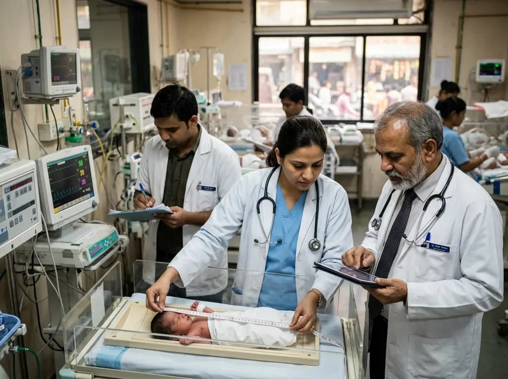
Scandinavian register evidence reveals that neonatal birth length strongly predicts adult health and mortality, whereas birth weight dictates cognitive capacity. Symmetric growth restriction causes far more severe long-term deficits than asymmetric restriction, underscoring the need for comprehensive anthropometry.
Today
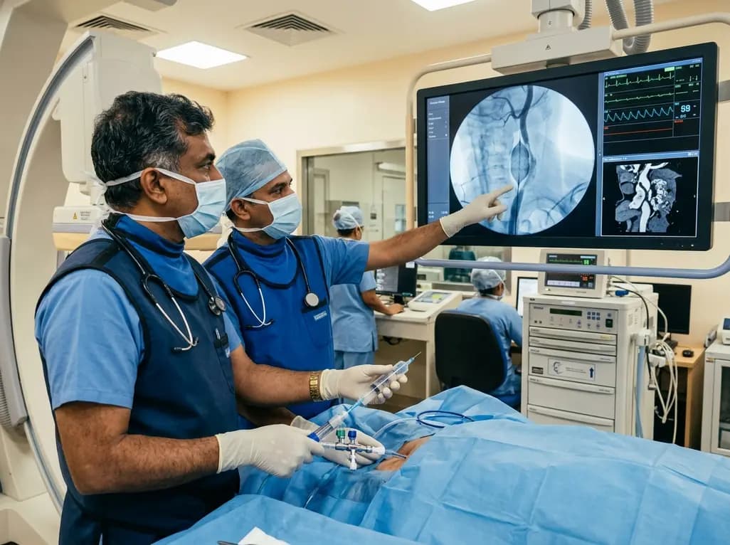
Discover how rescue cutting balloon angioplasty successfully modifies heavily calcified carotid stenosis, enabling stent delivery without baroreceptor instability and ensuring favorable long-term neurologic outcomes.
Today

Data from the 2026 Workforce Health Index reveals that 38.7% of urban employees and dependents exhibit abnormal glycemic control. Emerging at increasingly younger ages, this metabolic shift intersects with severe vitamin deficiencies, highlighting the urgent need for early workplace screenings and targeted clinical care.
Today
