.webp&w=384&q=75)

Loading, please wait...
Loading, please wait...
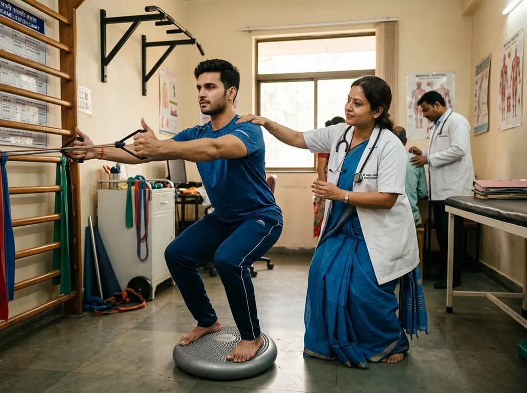
Shoulder dysfunction frequently stems from altered scapular mechanics and muscular imbalances across the periscapular stabilizers. Clinicians often observe hyperactivation of the upper trapezius accompanied by insufficient activation of the middle and lower trapezius muscles. Consequently, this dysfunctional recruitment pattern promotes abnormal scapulohumeral rhythm, subacromial space narrowing, and rotator cuff pathology. Restoring optimal trapezius muscle balance represents a cornerstone in contemporary orthopedic and sports rehabilitation. Recent therapeutic approaches emphasize functional movement patterns rather than isolated joint training. Specifically, therapists incorporate the kinetic chain, which links lower extremity and core movements to distal shoulder mechanics. Bilateral scapular retraction with shoulder external rotation serves as an effective corrective exercise. However, clinicians actively seek the most effective postural modifications to maximize lower trapezius recruitment while dampening excessive upper trapezius dominance. By manipulating postural stances and surface stability, practitioners can systematically alter neuromuscular recruitment strategies. Understanding how kinetic chain integration alters trapezius activation provides clinicians with valuable tools to tailor exercise prescriptions. Ultimately, optimizing these mechanical parameters allows rehabilitation specialists to design precise protocols that improve scapular stability, reduce chronic shoulder pain, and prevent reinjury across diverse patient populations.
A recent clinical investigation evaluated how kinetic chain variations and surface stability influence periscapular activation during bilateral scapular retraction exercises. The researchers enrolled twenty asymptomatic participants, comprising ten males and ten females, to establish baseline neuromuscular recruitment patterns. Investigators systematically measured surface electromyography across three distinct subdivisions: the upper trapezius, middle trapezius, and lower trapezius. Participants performed the standard exercise protocol across three progressive postural conditions: standing, squatting, and lunging. In addition, each exercise condition was executed on both a rigid, stable floor and a compliant, unstable surface. Surface electromyography signals were meticulously recorded, amplified, and normalized to maximal voluntary isometric contractions. This structured experimental design enabled precise comparisons across postural variations and environmental stability conditions. By analyzing both individual muscle activation levels and neuromuscular co-activation ratios, the researchers isolated the specific mechanical effects of trunk and lower limb engagement. Moreover, the study maintained strict kinematic monitoring to ensure uniform execution across all test trials. Consequently, the resulting data offer objective insights into how lower extremity posture and postural perturbation reshape scapulothoracic muscle dynamics.
The electromyographic findings demonstrated clear and statistically significant differences among the various postural and surface conditions. Specifically, the squat position generated substantially higher middle trapezius activation compared to the lunge position. Furthermore, lower trapezius activation peaked markedly during the squat position, exceeding activation levels observed in both standing and lunge postures. Regarding surface stability, lower trapezius muscle recruitment was significantly higher on the stable surface compared to the unstable surface. In contrast, upper trapezius activation during the squat posture decreased significantly when participants performed the movement on an unstable surface. Most importantly, the upper trapezius to middle trapezius ratio was significantly lower in the squat position compared to standing and lunging. Similarly, the upper trapezius to lower trapezius ratio on an unstable surface was lowest during the squat position. Therefore, executing the exercise in a squat posture consistently promoted favorable muscular recruitment ratios by reducing upper trapezius dominance while selectively stimulating the middle and lower trapezius fibers. These electromyographic findings strongly support the strategic selection of squat-based kinetic chain variations to restore periscapular muscular equilibrium.
Understanding the biomechanical pathways underlying these activation shifts provides valuable rationale for clinical practice. When an individual adopts a bilateral squat position, the nervous system engages extensive posterior chain musculature, including the gluteals, hamstrings, and thoracolumbar erectors. Consequently, this proximal co-activation facilitates reciprocal muscular firing along the posterior myofascial kinetic chain. The co-contraction of the core and pelvis establishes a rigid, stable base of support for scapulothoracic stabilization. Because the lower trapezius and middle trapezius anatomically anchor along the lower thoracic spine and fascial planes, lower body loading naturally reinforces their recruitment. Furthermore, introducing an unstable surface challenges balance and stimulates peripheral mechanoreceptors. In response to sudden postural perturbations, the central nervous system shifts neuromuscular control toward joint stabilization and spinal alignment. Interestingly, this dynamic adjustment inhibits compensatory superficial muscle overactivity, specifically down-regulating the upper trapezius. Therefore, combining lower extremity kinetic engagement with sensorimotor challenges effectively reprograms motor unit recruitment. This multi-joint coordination prevents the elevation and anterior tilting typically driven by upper trapezius overactivity.
These findings carry substantial clinical relevance for physical therapists, orthopedic specialists, and sports medicine practitioners. Scapular dyskinesis frequently manifests as excessive scapular elevation and anterior tilting, primarily driven by hyperactive upper trapezius fibers and weak lower stabilizers. Traditional rehabilitation often relies on isolated, non-weight-bearing exercises that fail to translate into functional movement patterns. However, integrating squat-based retraction protocols directly targets the underlying muscle imbalance without reinforcing compensatory habits. Clinicians should progressively introduce the squat posture on stable ground during early-to-intermediate rehabilitation to build robust middle and lower trapezius activation. Subsequently, progressing patients to an unstable surface can actively suppress excessive upper trapezius firing and refine neuromuscular motor control. Moreover, this kinetic chain approach improves dynamic trunk control, enhances athletic performance, and minimizes subacromial impingement risks. Overhead athletes, occupational workers, and general orthopedic patients can all benefit from these targeted postural modifications. By adopting evidence-based kinetic chain variations, clinicians optimize functional recovery and accelerate safe return to full daily and athletic activities.
Rehabilitation specialists must carefully sequence kinetic chain progressions to maximize therapeutic outcomes and restore durable trapezius muscle balance. In the initial phase, practitioners should evaluate the patient's baseline lower limb strength and postural stability. If a patient exhibits significant scapular dyskinesis, starting with bilateral scapular retraction in a stable squat posture provides immediate structural support while encouraging lower trapezius recruitment. As movement quality and endurance improve, therapists can introduce compliant foam pads or balance boards to stimulate proprioceptive feedback. This unstable environment effectively discourages upper trapezius compensation and promotes a balanced scapulothoracic force couple. In addition, practitioners must educate patients regarding proper lumbo-pelvic alignment and avoid excessive spinal extension during the retraction phase. Combining these biomechanical adjustments ensures consistent motor learning and long-term neuromuscular adaptation. Consequently, structured progression from stable bilateral squats to unstable multi-planar challenges creates a robust foundation for functional shoulder recovery.
Maintaining trapezius muscle balance is critical because the upper, middle, and lower trapezius fibers coordinate scapular upward rotation, posterior tilt, and retraction. When the upper trapezius becomes hyperactive and overpowers the lower stabilizers, the scapula tilts anteriorly and elevates excessively. Consequently, this faulty alignment reduces subacromial clearance, compressive loads on the rotator cuff increase, and chronic shoulder impingement develops. Restoring balanced recruitment ensures proper joint arthrokinematics and long-term functional recovery.
Performing scapular retraction in a squat posture engages the lower extremity and core kinetic chain, including the gluteal and paraspinal musculature. This proximal tension facilitates neural drive to the middle and lower trapezius muscles through fascial and neuromuscular connections. Furthermore, the squat stance stabilizes the spine and rib cage, which naturally reduces compensatory upper trapezius shrugging. Consequently, patients achieve higher middle and lower trapezius recruitment and favorable activation ratios.
Therapists should introduce an unstable surface once a patient demonstrates adequate lower limb strength, stable core control, and proper exercise kinematics on firm ground. Performing retraction exercises on compliant surfaces challenges dynamic balance and mechanoreceptors, which actively down-regulates upper trapezius overactivity. However, if introduced prematurely, surface instability might compromise exercise form and provoke unwanted compensations. Therefore, clinicians must ensure foundational stability before advancing to unstable rehabilitation variations.
Disclaimer: This content is for informational and educational purposes only and does not constitute medical advice, diagnosis, or treatment. Healthcare professionals must exercise independent clinical judgment. Patients should consult a qualified physician regarding any medical condition. Refer to the latest local and national guidelines for clinical practice.
References

Read summarized clinical updates, watch expert medical content, and earn CME certifications right from your smartphone.
Open your smartphone camera to scan and install.





Discover how integrating squat postures and unstable surfaces during scapular retraction with external rotation enhances middle and lower trapezius activation while reducing upper trapezius dominance to optimize shoulder rehabilitation outcomes.
last month

With post-monsoon dengue surges threatening South Asian health systems, physicians must refine triage and management. This guide covers vital warning signs, plasma leakage detection, crystalloid titration, and vector containment strategies to prevent mortality during seasonal arboviral epidemics.
Today

Findings from Project Viva indicate that sustained postpartum weight retention, rather than excessive gestational weight gain alone, directly worsens women's mid-life cardiovascular health, degrading blood pressure and glycemic parameters decades after delivery.
Today

A large UK Biobank study reveals that plasma proteomic signatures capture preclinical organ damage and improve multiorgan risk prediction across early CKM syndrome stages 0 to 2 beyond traditional PREVENT clinical models.
Today

A retrospective CBCT study reveals that superior TMJ space asymmetry correlates moderately with occipital condyle-atlas-axis asymmetry in unilateral TMJ osteoarthritis, highlighting an imaging correlate between jaw biomechanics and upper cervical alignment rather than a standalone diagnostic marker.
Today

A Bayesian multilevel meta-analysis reveals that aerobic training combined with moderate carbohydrate restriction modestly lowers HbA1c in type 2 diabetes. However, sparse data and very low certainty leave incremental benefits over exercise or diet alone unproven, highlighting the need for individualized care.
Today
