.webp&w=384&q=75)

Loading, please wait...
Loading, please wait...
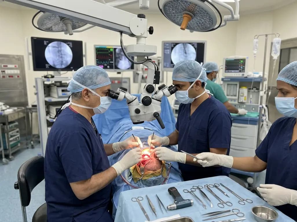
Surgical management of complex orbital and cranio-orbital lesions represents a formidable challenge in modern neurosurgery and skull base practice. Because lesions often abut crucial neural pathways and vascular structures, mastering transcranial orbital approaches is vital for achieving safe, radical resection. Selecting the proper surgical trajectory depends entirely on the precise anatomical compartment involved, requiring neurosurgeons and ophthalmic surgeons to possess comprehensive knowledge of both intraorbital and intracranial anatomy.
The orbit represents a compact, cone-shaped anatomical corridor surrounded by bony walls and densely packed neurovascular elements. Consequently, surgical pathology here demands unmatched anatomical clarity. Tumors such as spheno-orbital meningiomas, schwannomas, cavernous hemangiomas, and optic nerve gliomas frequently involve multiple orbital compartments. Furthermore, these lesions often breach the orbital roof or superior orbital fissure, directly extending into the anterior or middle cranial fossa.
Historically, surgeons used classical transorbital routes to reach superficial or anterior lesions. However, transcranial orbital corridors provide superior illumination, broad multi-angle visibility, and unmatched surgical control for pathologies seated in the orbital apex, optic canal, or superior compartments. By elevating a frontotemporal or pterional bone flap, the surgeon gains circumferential control over the cranial base. This superior vantage allows early bony unroofing and direct identification of the frontal nerve, trochlear nerve, and superior ophthalmic vein before manipulating deeper intraconal soft tissue structures.
Following a standard pterional craniotomy and orbitotomy, surgeons can tailor their intraorbital dissection through three fundamental trajectories: medial, central, and lateral. Each vector offers distinct access to specific intraorbital compartments. Therefore, choosing the correct corridor prevents unnecessary traction on sensitive ocular structures.
Specifically, the central transcranial approach proceeds directly between the levator palpebrae superioris and the superior rectus muscle. This route offers direct access to superior intraconal tumors, although surgeons must exercise extreme caution to prevent postoperative blepharoptosis or superior rectus paresis. Conversely, the medial corridor traverses the space between the superior oblique muscle and the medial rectus muscle. This path provides ideal visual access to lesions situated along the superomedial orbital wall, the optic canal, and the ethmoidal roof.
Meanwhile, the lateral transcranial corridor navigates between the superior rectus and lateral rectus muscles. It represents the workhorse trajectory for extensive lesions occupying the superior orbital fissure, the lateral intraconal space, and the middle cranial fossa.
Although standard lateral transcranial routes provide favorable visualization, deep inferolateral and apical exposure often remains constrained by bony overhangs. Recent anatomical investigations demonstrate that incorporating a temporary resection of the zygomatic arch dramatically expands the accessible surgical corridor. Consequently, this simple modification improves the surgeon's working angle significantly.
Removing the zygomatic arch allows surgeons to mobilize and displace the temporalis muscle inferolaterally. This maneuver completely clears the subtemporal corridor, flattening the angle of approach to the orbital floor and lateral orbital wall. As a result, the surgeon operates parallel to the skull base rather than peering downward over a steep bony ledge. This expanded exposure substantially improves illumination of the inferior orbital fissure, cavernous sinus interface, and infratemporal fossa without imposing excessive brain retraction. Ultimately, rigid miniplate fixation reliably restores cosmetic contours during closure.
Lesions situated at the orbital apex and within the central muscle cone remain notoriously difficult to resect. The confluence of extraocular muscle origins at the annular tendon of Zinn creates a tight anatomical bottleneck. Within this compact zone, inadvertent manipulation of the oculomotor, abducens, or optic nerves can lead to permanent visual deficit or severe ophthalmoplegia.
Transcranial corridors provide a substantial mechanical advantage when addressing these apex lesions. By unroofing the optic canal and incising the falciform ligament extradurally, the surgeon achieves early optic nerve decompression before opening the periorbita. Furthermore, expanding the lateral orbital corridor via zygomatic arch resection allows surgeons to access lateral intraconal pathology beneath the lateral rectus muscle. Thus, surgical teams can resect deep vascular malformations and neural sheath tumors with minimal manipulation of the ciliary ganglion and central retinal artery.
Preserving visual acuity, pupillary dynamics, and ocular motility requires meticulous microsurgical technique during every step of orbital entry. Intracranial brain relaxation through careful CSF drainage eliminates the need for aggressive frontal lobe retraction. Additionally, preserving the delicate periorbital membrane until clear bony landmarks are identified prevents premature herniation of intraorbital fat pads.
Moreover, modern skull base surgery relies heavily on multi-modality intraoperative neuromonitoring. Continuous visual evoked potentials and cranial nerve electromyography help identify neural structures within dense tumor capsules. When opening the muscular cone, surgeons must perform sharp, delicate dissections parallel to muscle fibers to avoid avascular necrosis or denervation. Following successful tumor resection, reconstructing the orbital roof with autologous bone grafts, titanium mesh, or biocompatible implants prevents postoperative pulsatile enophthalmos and orbital encephaloceles.
Mastering complex cranio-orbital procedures requires comprehensive collaboration between neurosurgeons, oculoplastic surgeons, otolaryngologists, and neuroradiologists. High-resolution pre-operative MRI and CT reconstructions allow precise 3D trajectory mapping, enabling the surgical team to select the ideal corridor well before entering the operating room.
Furthermore, while purely endoscopic endonasal techniques continue to expand for inferomedial pathologies, open transcranial routes remain the definitive gold standard for extensive, multi-compartmental, or highly vascular tumors crossing the superior and lateral skull base. Incorporating anatomical modifications, such as zygomatic osteotomy, enhances surgical versatility and promotes complete lesion excision. Consequently, systematic surgical training on human cadaveric specimens remains indispensable for refining these complex techniques and ensuring outstanding clinical results for patients undergoing cranio-orbital interventions.
Surgeons primarily employ transcranial orbital approaches for expansive lesions involving the orbital apex, optic canal, superior orbital fissure, or superior intraconal spaces. These routes also provide optimal exposure for tumors extending concurrently across both the orbital cavity and intracranial compartments, such as spheno-orbital meningiomas and extensive schwannomas.
Temporary zygomatic arch removal enables the surgical team to mobilize the temporalis muscle downward, significantly flattening the operative trajectory. This bone resection expands surgical exposure, improves illumination of deep inferolateral and apical structures, and minimizes the need for excessive frontal or temporal lobe retraction during tumor dissection.
Surgeons reconstruct the orbital roof using autologous bone grafts, titanium mesh, or biocompatible resorbable implants secured with miniplates. Creating a stable barrier between the anterior cranial fossa and intraorbital contents is essential to prevent severe long-term complications, such as pulsatile enophthalmos, diplopia, and orbital meningoencephaloceles.
Disclaimer: This content is for informational and educational purposes only. It is not intended to be a substitute for professional medical advice, diagnosis, or treatment. Always seek the advice of your physician or other qualified health provider with any questions you may have regarding a medical condition or procedure. Refer to the latest local and national guidelines for clinical practice.
References

Read summarized clinical updates, watch expert medical content, and earn CME certifications right from your smartphone.
Open your smartphone camera to scan and install.





An in-depth anatomical review of transcranial orbital approaches highlights how selecting central, medial, or modified lateral corridors—including zygomatic arch removal—maximizes surgical exposure to deep intraconal and orbital apex pathologies while protecting critical neurovascular structures.
last month
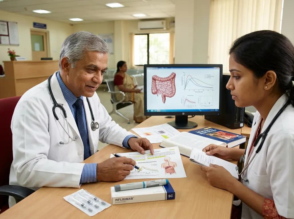
Subcutaneous infliximab provides stable steady-state pharmacokinetics and reduces immunogenicity in inflammatory bowel disease. This clinical review explores pivotal trial evidence, therapeutic drug monitoring recalibration, immunomodulator de-escalation, and real-world switching strategies.
Today
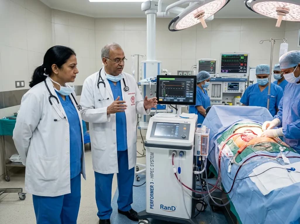
AIIMS Bathinda has inaugurated a specialized peritoneal oncology service featuring the RanD Performer 3 HIPEC platform. The facility offers cytoreductive surgery alongside hyperthermic intraperitoneal chemotherapy, transforming regional management for advanced ovarian, colorectal, and appendiceal malignancies.
Today

A cross-sectional study reveals that hypertension knowledge, illness perceptions, depression, and substance use significantly predict self-care behaviors. Addressing mental health and cognitive beliefs in primary care is crucial for optimal blood pressure control and cardiovascular disease prevention.
Today
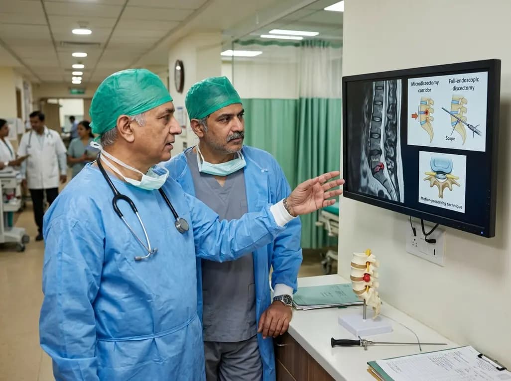
This clinical overview examines recent advancements in lumbar disc herniation surgery, comparing microscopic and full-endoscopic discectomy while highlighting biportal techniques and emerging motion-preserving innovations for spine specialists.
Today

A breakthrough study reveals that loss of SELENOT in hypothalamic POMC neurons triggers premature cellular senescence, ATF6α stress activation, and ER calcium depletion, linking neuroendocrine decay to metabolic disease.
Today
