

Loading, please wait...
Loading, please wait...

Exercise intolerance often puzzles clinicians when standard resting cardiovascular evaluations reveal structurally preserved myocardium and normal ventricular hemodynamics. In post-inflammatory neurological recovery, central chronotropic incompetence represents a severe yet frequently overlooked driver of persistent exertional dyspnea. Specifically, patients recovering from brainstem injury may suffer from profound autonomic failure despite resolving motor deficits. Therefore, understanding central autonomic regulation is critical when routine cardiac testing fails to explain lingering functional impairment after central nervous system inflammation.
The brainstem tightly coordinates cardiac chronotropy through continuous sympathetic and parasympathetic interplay. Specifically, the medulla oblongata houses crucial autonomic control centers, including the rostral ventrolateral medulla and the nucleus ambiguus. Sympathetic premotor neurons located within the ventrolateral medulla project directly to the intermediolateral cell column in the thoracic spinal cord. Consequently, these pathways stimulate cardiac postganglionic sympathetic fibers, which accelerate sinoatrial node firing during physical exertion. However, focal neuroinflammatory destruction can severely disrupt these descending sympathetic pathways. When necrotizing encephalitis damages medullary vasomotor centers, the heart loses its essential upstream accelerator drive. The sinoatrial node may retain intrinsic viability, yet it fails to receive obligatory efferent neural triggers. As a result, heart rate remains blunted when metabolic demand surges during exercise. Clinicians frequently misdiagnose this blunted response because resting electrocardiograms and resting echocardiography display normal findings. Moreover, postganglionic sympathetic reserve cannot compensate for destroyed upstream brainstem premotor pacemakers. Thus, structural medullary damage uncouples the heart-brain axis, producing rigid chronotropic failure despite preserved cardiac contractility and pulmonary airflow.
Rhombencephalitis represents a severe, inflammatory syndrome targeting the pons, cerebellum, and medulla oblongata. Etiologies encompass Listeria monocytogenes infection, enterovirus infections, paraneoplastic syndromes, and idiopathic autoimmune encephalitides. Although acute immunomodulatory therapy often halts tissue necrosis, chronic architectural injury frequently persists within autonomic nuclei. Patients frequently present months after apparent recovery complaining of profound, disabling exertional fatigue and breathlessness. Furthermore, physical examination in the post-acute clinic rarely reveals obvious cardiac pathology. Resting heart rates typically appear normal or slightly bradycardic, and standard laboratory biomarkers remain unrevealing. Physicians routinely perform transthoracic echocardiography, which demonstrates preserved ejection fraction without valvular abnormalities or pulmonary hypertension. Similarly, pulmonary function testing confirms normal spirometry and adequate gas diffusion capacity. Consequently, healthcare providers frequently attribute ongoing symptoms to deconditioning, functional neurological disorder, or post-viral asthenia. Such diagnostic delays can demoralize patients who struggle with everyday physical tasks. Therefore, clinicians must maintain high suspicion for autonomic pacemaking disruption whenever patients report persistent exertional intolerance following documented brainstem encephalitis.
Under physiologic conditions, physical exertion triggers immediate vagal withdrawal followed rapidly by robust sympathetic activation. This central autonomic orchestration elevates heart rate and enhances myocardial inotropy, thereby augmenting systemic cardiac output. However, inflammatory necrosis within the medullary tegmentum severs this dynamic autonomic dialogue. When medullary premotor sympathetic neurons suffer irreversible structural loss, the brain loses the capacity to accelerate cardiac rhythm proportionally. Notably, this central dysautonomia produces a striking functional paradox during sustained physical activity. Because peripheral vascular tone and intrinsic myocardial contractility may remain intact, stroke volume increases during initial exertion. Nevertheless, stroke volume reaches an anatomical plateau quickly. At higher metabolic workloads, cardiac output depends almost entirely on heart rate escalation. When heart rate cannot rise past an abnormally low ceiling, systemic oxygen delivery falters dramatically. Patients experience abrupt, overwhelming exhaustion, dyspnea, and lightheadedness at modest workloads. In addition, peripheral tissues incur rapid lactic acidosis because circulatory delivery cannot match peripheral oxygen demand. Thus, persistent heart-brain axis uncoupling creates a rigid physiological ceiling that mimics severe primary heart failure.
Standard resting evaluations cannot capture exercise-induced hemodynamic failure. Instead, clinicians require cardiopulmonary exercise testing to unmask isolated chronotropic dysfunction. Cardiopulmonary exercise testing continuously quantifies minute ventilation, oxygen consumption, carbon dioxide production, and continuous twelve-lead electrocardiography under incremental workloads. In patients with central autonomic failure, this modality demonstrates a preserved breathing reserve alongside a severely blunted peak heart rate. Specifically, diagnostic criteria for chronotropic incompetence include the failure to achieve eighty percent of the age-predicted maximal heart rate. Alternatively, clinicians evaluate the chronotropic index, where values below 0.8 reflect marked chronotropic deficiency. During testing, affected patients display an abnormally flat heart rate slope relative to metabolic expenditure. Furthermore, the oxygen pulse often shows an exaggerated early rise, reflecting compensatory stroke volume expansion. Once stroke volume reaches its physical limit, the oxygen pulse plateaus prematurely while heart rate remains fixed. Consequently, peak oxygen uptake falls short of predicted normal values despite maximal voluntary effort. Therefore, cardiopulmonary exercise testing delivers objective, indisputable evidence differentiating neurogenic chronotropic incompetence from intrinsic pulmonary disease, muscular weakness, or psychogenic dyspnea.
Managing central chronotropic incompetence demands collaborative care among neurologists, cardiologists, and exercise physiologists. First, clinicians must thoroughly review the patient's pharmacotherapy. They should eliminate rate-slowing agents such as beta-blockers, non-dihydropyridine calcium channel antagonists, and centrally acting alpha-agonists. In addition, secondary neuroinflammation warrants consideration, although repeat immunosuppression rarely restores dead medullary neuronal architecture. When functional limitation remains severe, permanent pacemaker implantation offers an effective therapeutic avenue. Specifically, modern dual-chamber pacemakers equipped with closed-loop stimulation or accelerometer-based rate-responsive algorithms can substitute for damaged medullary premotor drive. These intelligent pacing devices sense mechanical movement or intracardiac impedance changes, thereby elevating ventricular pacing rates dynamically during physical effort. Clinical studies demonstrate that rate-adaptive pacing significantly enhances peak oxygen consumption and functional capacity in chronotropically limited individuals. Concurrently, tailored cardiac rehabilitation programs provide supervised aerobic conditioning. Endurance training optimizes peripheral oxygen extraction across skeletal muscles and enhances endothelial vasodilation. Moreover, rehabilitation specialists guide patients on pacing strategies to maximize daily activity without triggering autonomic exhaustion. Thus, integrating advanced technological pacing with structured rehabilitation restores independence and physical vigor in recovering patients.
Encountering persistent exertional dyspnea following neuroinflammatory diseases challenges conventional clinical paradigms. In general practice and specialty clinics across India, viral and autoimmune central nervous system infections occur with notable frequency. When survivors present with protracted weakness and exercise intolerance, physicians often order standard resting tests that show completely normal organ structure. Consequently, clinicians must expand their diagnostic perspective beyond intrinsic cardiac and pulmonary parenchymal disorders. Furthermore, physicians must recognize that subjective dyspnea often reflects inadequate cardiac output during metabolic challenge rather than ventilatory restriction. A carefully calibrated exercise stress test or formal cardiopulmonary exercise test unmasks rigid chronotropic ceilings that resting examinations completely overlook. In addition, early collaboration between neurology and cardiology prevents prolonged diagnostic odysseys and unnecessary invasive testing. Clinicians should educate patients on physiological pacing while pursuing definitive autonomic testing. If cardiopulmonary testing confirms severe neurogenic rate limitation, timely electrophysiological consultation can determine candidacy for rate-responsive pacing. Ultimately, maintaining acute clinical awareness of central autonomic failure ensures accurate diagnosis, prevents therapeutic nihilism, and substantially improves long-term functional outcomes for survivors of brainstem injury.
Central chronotropic incompetence arises when inflammatory or necrotizing processes damage autonomic regulatory centers within the medulla oblongata. Specifically, the loss of medullary sympathetic premotor neurons disrupts descending signals to the spinal intermediolateral cell column. Although the sinoatrial node and intrinsic myocardial architecture remain completely healthy, the heart fails to receive necessary sympathetic acceleration during physical exercise. Consequently, patients experience rigid rate limitations and exercise intolerance.
Cardiopulmonary exercise testing objectively evaluates cardiopulmonary performance by measuring gas exchange and continuous electrocardiography under graded exercise. During testing, patients with central autonomic failure demonstrate an inability to achieve eighty percent of their age-predicted maximal heart rate. Furthermore, testing reveals an abnormally flat chronotropic response slope alongside preserved ventilatory reserve and normal gas exchange. This distinct physiologic pattern successfully differentiates central chronotropic impairment from primary pulmonary limitations or deconditioning.
Management begins by withdrawing any rate-limiting medications, including beta-blockers and calcium channel antagonists. For persistent, severe functional impairment, implantation of a permanent rate-responsive pacemaker serves as a highly effective intervention. Modern rate-adaptive algorithms detect physical motion or minute ventilation changes to artificially elevate pacing rates during exertion. Additionally, supervised cardiac rehabilitation helps optimize peripheral oxygen extraction, improving exercise endurance and daily functional capacity for affected patients.
Disclaimer: This content is for informational and educational purposes only... Refer to the latest local and national guidelines for clinical practice.
References

Read summarized clinical updates, watch expert medical content, and earn CME certifications right from your smartphone.
Open your smartphone camera to scan and install.





Central chronotropic incompetence is an underrecognized sequela of rhombencephalitis causing exertional dyspnea despite normal resting cardiac function. Cardiopulmonary exercise testing unmasks this heart-brain uncoupling, guiding rate-responsive pacing and targeted rehabilitation.
Today
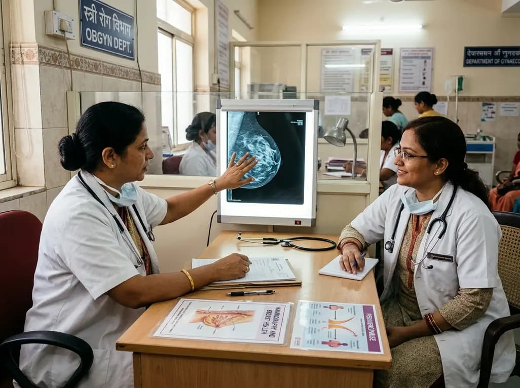
Perimenopause brings erratic hormonal surges and metabolic shifts that significantly affect breast tissue. This comprehensive clinical review examines benign breast conditions, cancer risk stratification, mammographic density challenges, and endocrine therapy transitions in midlife women.
Today

Understand the causal role of Lipoprotein(a) in cardiovascular disease, why international guidelines recommend once-in-a-lifetime molar testing, and the clinical implications of recent phase 3 RNA outcome trials.
Today
A multi-center study reveals that plain radiographs demonstrate poor sensitivity for detecting anterolateral tibial plafond involvement in pronation-external rotation (PER) ankle fractures. Routine preoperative CT imaging ensures accurate identification of articular impaction and guides surgical reduction.
Today

This review examines the pharmacogenomics of antidiabetic agents, highlighting how genetic variations in SLC2A2, SLC47A1, CYP2C9, ABCC8, and GLP1R influence drug transport, therapeutic efficacy, and hypoglycemia risk to enable tailored precision medicine in type 2 diabetes.
Today
