.webp&w=384&q=75)

Loading, please wait...
Loading, please wait...

Temporomandibular disorders present widespread diagnostic challenges in dental, maxillofacial, and pain management clinics across the globe. Among various radiological evaluation tools, magnetic resonance imaging provides unmatched soft-tissue visualization of the craniomandibular articulation. Clinicians regularly detect temporomandibular joint effusion on fluid-sensitive sequences such as T2-weighted and proton density scans. This intra-articular fluid accumulation typically reflects active synovial inflammation, capsular irritation, and biochemical disturbance. Historically, many practitioners assumed a direct linear relationship between excessive joint fluid and structural osteoarthritic degradation. However, published clinical studies frequently yielded conflicting conclusions regarding whether synovial fluid directly drives osseous damage or merely accompanies chronic joint friction. Therefore, clarifying this pathological interface remains critical for accurate diagnostic classification and therapeutic planning. When patients present with severe preauricular pain, acoustic joint sounds, or functional movement limitations, precise structural evaluation helps differentiate reversible inflammatory conditions from progressive degenerative disease. Furthermore, recognizing how fluid accumulation relates to condylar remodeling prevents unnecessary and premature invasive surgical interventions. Consequently, modern radiological interpretation requires a nuanced understanding of biological joint turnover. Clinicians must distinguish incidental physiological fluid from marked inflammatory exsudation that correlates with irreversible bone remodeling. Through targeted clinical and radiological assessments, dental specialists can establish reliable management protocols tailored to individual structural phenotypes.
To resolve enduring diagnostic controversies, researchers executed an exhaustive systematic review and meta-analysis synthesizing international observational datasets. The investigators systematically searched PubMed, Scopus, Embase, Cochrane Library, and Web of Science up to May 2026. Ultimately, eighteen rigorous observational studies comprising 7001 temporomandibular joints fulfilled the strict inclusion criteria for qualitative review. Fourteen observational cohorts contributed quantitative data to the primary meta-analysis. The pooled statistical calculations revealed a significant positive association between MRI-detected joint effusion and temporomandibular joint degenerative joint disease, yielding an odds ratio of 1.41. Heterogeneity across the included cohorts was substantial, registering an I-squared statistic of 70.1 percent. Therefore, the investigators performed a post hoc sensitivity analysis excluding outlier data from Zheng and colleagues. This refined calculation yielded an even stronger odds ratio of 1.51 with reduced statistical heterogeneity of 63.4 percent. Most remarkably, marked effusion exhibited an exceptional correlation with structural destruction, demonstrating an odds ratio of 2.63. In contrast, mild or physiological fluid accumulations failed to reach similar pathological thresholds. Thus, robust quantitative synthesis confirms that abundant intra-articular fluid strongly signals degenerative joint activity.
Degenerative joint disease does not manifest as a uniform pathological entity across every patient presenting with masticatory dysfunction. Instead, condylar remodeling exhibits several distinct morphological phenotypes on diagnostic scans. The meta-analysis categorized osseous transformations into specific subtypes to uncover selective associations with inflammatory effusion. Interestingly, the researchers demonstrated a statistically significant positive association with condylar flattening, which produced a pooled odds ratio of 1.49. Similarly, osteophyte formation demonstrated a pronounced link to fluid presence, recording an odds ratio of 1.61. Osteophytes represent advanced adaptive bone proliferations that respond to altered mechanical stress distributions and chronic articular irritation. In contrast, structural findings for condylar erosion and condylar atrophy remained inconclusive across the pooled data. This discrepancy likely reflects differing stages of biological disease progression. For instance, active subchondral erosions may emerge during aggressive acute catabolic phases without lingering synovial expansion. Conversely, extensive osteophytosis and flattening indicate long-standing mechanical overload paired with persistent inflammatory microenvironments. Consequently, clinicians must recognize that fluid accumulation preferentially aligns with chronic hypertrophic remodeling rather than isolated condylar volume loss.
Magnetic resonance imaging serves as the premier diagnostic modality for assessing intra-articular soft-tissue architecture within the masticatory apparatus. Because high-resolution T2-weighted and proton density sequences highlight liquid accumulation, radiologists can reliably quantify intra-articular exudates. However, clinicians must interpret these radiological signals with measured diagnostic restraint. The systematic review underscores that temporomandibular joint effusion cannot function as an isolated, standalone biomarker for osteoarthritis. Indeed, trace volumes of synovial fluid represent normal lubricating physiological reservoirs that facilitate smooth condylar translation. Furthermore, isolated internal disc derangement without osseous degeneration often produces reactive fluid spikes. Therefore, practitioners should regard marked effusion as an adjunctive imaging marker that heightens clinical suspicion of underlying structural wear. When severe effusion coincides with clinical crepitus, restricted translation, or regional arthralgia, clinicians should examine the bony margins with extreme care. In complex clinical scenarios, combining MRI with cone-beam computed tomography provides optimal diagnostic clarity. While MRI captures active capsular inflammation and disc morphology, computed tomography details fine cortical margins and microscopic osteophytic remodeling.
Integrating these radiological insights into day-to-day dental and surgical practice substantially improves patient management strategies. Because marked effusion correlates directly with degenerative bone remodeling, its radiological presence highlights active intra-articular pathology requiring timely intervention. Initially, clinicians should prioritize conservative modalities to control acute inflammation and unload stressed articular structures. Nonsteroidal anti-inflammatory drugs, stabilization splints, and targeted physical rehabilitation often alleviate mechanical strain. Furthermore, when persistent severe effusion accompanies intractable arthralgia, minimally invasive interventions offer substantial clinical benefits. Specifically, temporomandibular joint arthrocentesis or diagnostic arthroscopy effectively flushes out inflammatory cytokines and matrix metalloproteinases from the synovial cavity. Lavage therapy relieves intra-articular hydrostatic pressure, thereby halting the catabolic cascade before irreversible osteophytic expansion occurs. Additionally, clinicians should educate patients regarding parafunctional habit cessation, such as daytime clenching and nocturnal bruxism. In summary, identifying marked joint effusion shifts the therapeutic focus toward anti-inflammatory and joint-sparing protocols. By combining precise radiological interpretation with patient-centered conservative care, clinicians optimize long-term jaw function and prevent progressive degenerative joint disease.
No, joint effusion does not definitively confirm temporomandibular osteoarthritis when observed in isolation. While meta-analytic evidence demonstrates a positive association with degenerative osseous changes, fluid accumulation also occurs in isolated disc displacement and acute capsulitis. Furthermore, minor fluid collections exist physiologically to lubricate the craniomandibular joint. Therefore, clinicians must treat effusion as an adjunctive imaging indicator, corroborating imaging findings with clinical palpation, mandibular functional range, and cortical bony architecture.
Marked effusion correlates strongly with condylar remodeling because abundant intra-articular fluid reflects intense, persistent synovial inflammation. Elevated levels of pro-inflammatory cytokines, including interleukin-1 beta and tumor necrosis factor-alpha, stimulate catabolic enzymes that degrade articular fibrocartilage. Over time, this chronic biological environment impairs natural tissue repair and accelerates subchondral bone adaptations. Consequently, joints with marked fluid collections experience sustained biomechanical stress, which directly promotes cortical flattening and osteophyte growth.
When MRI identifies marked joint effusion alongside degenerative changes, clinicians should implement structured anti-inflammatory protocols. Initial non-invasive interventions include occlusal splint therapy, nonsteroidal anti-inflammatory medications, and physical therapy to relieve biomechanical loading. If conservative strategies fail to alleviate severe joint pain or dysfunction, clinicians may recommend minimally invasive joint arthrocentesis. Arthrocentesis flushes out inflammatory mediators, reduces intra-articular pressure, and improves mandibular mobility, thereby halting further degenerative osseous remodeling.
Disclaimer: This content is for informational and educational purposes only and should not be considered medical advice. Always consult a qualified healthcare professional regarding any medical condition or treatment. Refer to the latest local and national guidelines for clinical practice.
References

Read summarized clinical updates, watch expert medical content, and earn CME certifications right from your smartphone.
Open your smartphone camera to scan and install.





A systematic review and meta-analysis shows that MRI-detected temporomandibular joint effusion is positively linked to degenerative osseous changes, particularly marked fluid, flattening, and osteophytes, serving as an adjunctive imaging marker.
Today
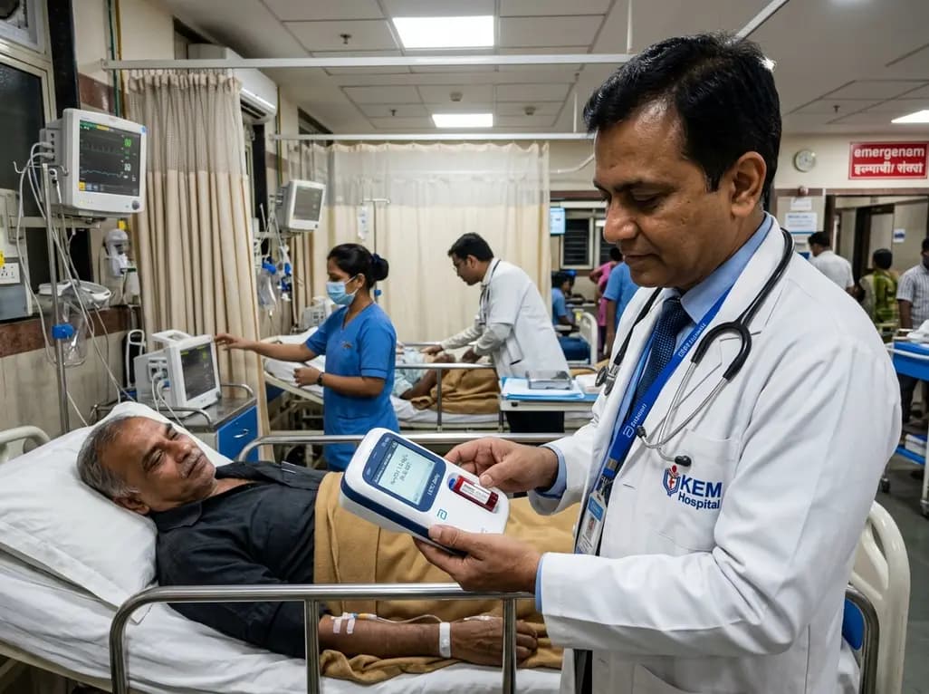
A real-world emergency department evaluation demonstrates that the Abbott i-STAT Alinity point-of-care high-sensitivity troponin I assay achieves excellent analytical precision and safely rules out myocardial infarction without false negatives, offering rapid bedside results to decongest acute care pathways.
Today

Integrating adult genetic services into reproductive and pregnancy care substantially alters clinical management. A study reveals that diagnostic genetic evaluations directly inform maternal, fetal, and neonatal care in nearly 57% of tested patients, highlighting the value of preconception and antenatal genomics.
Today

A cross-sectional study in Nepal evaluates cardiovascular risk factors among adults working above 4,000 meters. The findings reveal unexpected rates of diabetes, hypertension, and mountain sickness, underscoring the urgent need for targeted cardiometabolic screening in high-altitude occupational cohorts.
Today
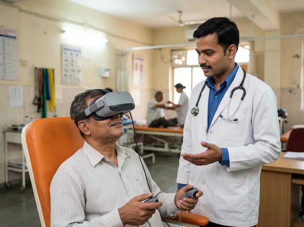
A recent scoping review highlights the efficacy, usability, and patient satisfaction of virtual reality pain management for chronic musculoskeletal pain, emphasizing psychotherapy-based VR protocols.
Today

Preclinical findings demonstrate that dietary flavonoid hyperoside ameliorates high-fat diet-induced type 2 diabetes by stimulating brown adipose tissue thermogenesis, promoting white fat browning, dampening adipose macrophage inflammation, and restoring gut microbiota homeostasis.
Today
