.webp&w=384&q=75)

Loading, please wait...
Loading, please wait...
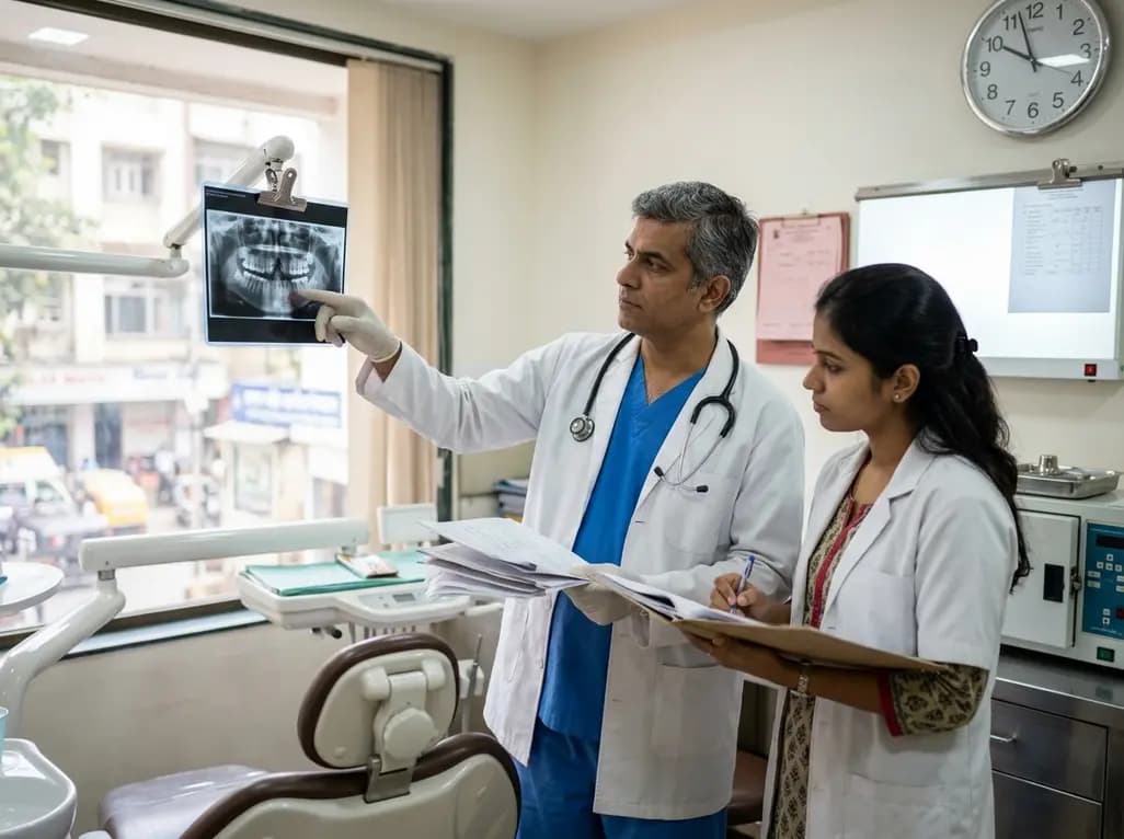
Managing dental anomalies in children with cleft lip and palate requires precise surgical timing and coordinated orthodontic planning. Clinicians frequently encounter supernumerary, malformed, or severely displaced dentition directly adjacent to the alveolar defect. Therefore, determining the exact surgical schedule for removing these compromised units remains a subject of ongoing debate among craniofacial teams. Secondary alveolar bone grafting serves as the gold standard reconstructive approach to restore maxillary arch integrity and facilitate canine eruption. However, surgeons often disagree on whether to extract cleft-adjacent teeth weeks before the graft or during the primary reconstructive operation. Recent comprehensive scoping reviews provide critical clarity regarding how extraction timing influences osseous healing, mucosal closure, and graft survival.
Secondary alveolar bone grafting remains an essential milestone in the comprehensive rehabilitation of pediatric cleft lip and palate patients. The primary surgical goals include stabilizing the maxillary dental arch, closing residual oronasal fistulas, and providing adequate osseous support for erupting permanent dentition. Typically, surgical teams execute this intervention during mixed dentition when the permanent canine root achieves approximately one-half to two-thirds of its development. However, teeth situated immediately adjacent to the bony defect frequently present severe hypoplasia, ectopic eruption paths, or significant periodontal compromise. Consequently, surgeons must decide whether extracting these compromised units prior to grafting provides a superior biological bed compared to concurrent intraoperative removal. Historically, many protocols recommended early extraction to permit complete soft tissue healing before bone placement. Proponents argue that healed keratinized mucosa minimizes the risk of oral contamination and flap dehiscence during graft incorporation. Conversely, other specialists emphasize that delaying extractions until the main surgical procedure preserves alveolar height and limits the overall number of general anesthesia episodes for the child. Understanding the physiological balance between soft tissue readiness and bony preservation helps clinicians formulate standardized surgical protocols.
Recent systematic evaluations have scrutinized whether pre-graft dental removal genuinely improves clinical outcomes compared to concurrent intraoperative extraction. Interestingly, initial univariate analyses across clinical studies frequently suggest that extracting cleft-adjacent teeth weeks before grafting enhances bone consolidation. However, when researchers apply rigorous multivariate statistical models, this apparent advantage often disappears completely. Confounding variables, such as defect dimensions, patient age, baseline oral hygiene, and surgical flap design, exert a substantially stronger influence on overall graft survival. Furthermore, secondary evaluations comparing intraoperative extractions directly against procedures without any extractions reveal highly inconsistent associations regarding bone graft failure. In fact, many successful reconstructive cases occur concurrently with dental extractions without increasing post-operative infectious complications or graft resorption. Therefore, current empirical literature demonstrates that extraction timing alone is not an independent determinant of surgical success. Clinicians should not feel bound to rigid dogmatic timelines that force children into staged interventions. Instead, teams must interpret the available evidence critically, recognizing that meticulous surgical technique and anatomical defect geometry play far more pivotal roles in determining graft incorporation than the specific extraction schedule.
The biological environment surrounding the alveolar cleft requires careful manipulation to avoid compromise of adjacent healthy periodontal structures. When surgeons perform concurrent extractions, they must achieve tension-free, watertight mucosal closure over the freshly evacuated socket and the placed cancellous bone. Consequently, improper soft tissue handling during concurrent procedures might elevate the risk of wound dehiscence and bacterial penetration. However, modern mucoperiosteal advancement techniques and lateral releasing incisions reliably ensure complete coverage, mitigating these historical concerns. Furthermore, leaving severely compromised or non-restorable primary teeth in situ can harbor persistent local inflammation, which impairs osteoblast function during graft revascularization. Therefore, eliminating sources of chronic dental sepsis represents an absolute clinical necessity before or during bone augmentation. Periodontal health around the remaining permanent incisors and canines also dictates long-term arch stability. Preserving interdental bone septa during cleft-adjacent tooth removal prevents secondary marginal bone loss around vital permanent teeth. Ultimately, surgical success relies on minimizing soft tissue trauma, ensuring robust vascularity of mucoperiosteal flaps, and establishing rigid containment of the graft material regardless of the chosen extraction timeframe.
Beyond anatomical considerations, clinical decision-making must integrate patient safety, logistical feasibility, and healthcare resource stewardship. Performing dental extractions weeks in advance of the grafting procedure often necessitates a separate surgical session. For young pediatric patients, this approach frequently requires an additional episode of general anesthesia or deep conscious sedation. Repeated anesthetic exposure in early childhood carries recognized neurodevelopmental and physiological considerations that clinicians must weigh carefully. In addition, staging these procedures increases the cumulative burden on families, requiring extra clinic visits, time off school, and parental leave from employment. Consequently, intraoperative extraction during the definitive bone grafting procedure offers profound practical advantages by consolidating surgical interventions into a single hospital admission. This streamlined pathway reduces overall healthcare expenditures, optimizes operating room utilization, and improves patient adherence. However, in scenarios where a patient exhibits profound pre-existing dental infection or severe soft tissue deficiency, staged extractions under local anesthesia remain justified. Therefore, surgical teams must evaluate institutional capabilities, anesthetic safety profiles, and patient socioeconomic factors alongside purely anatomical metrics.
Optimizing outcomes in alveolar cleft reconstruction demands robust multidisciplinary collaboration among pediatric dentists, orthodontists, and oral and maxillofacial surgeons. Orthodontic preparation frequently involves maxillary expansion to correct crossbites and align cleft segments before surgical intervention. During this preparatory phase, orthodontists and pediatric dentists accurately assess the restorability, periodontal prognosis, and spatial orientation of cleft-adjacent dentition. Advanced three-dimensional cone-beam computed tomography provides precise visualization of root morphology and surrounding alveolar bone volume. Therefore, the team can determine whether a specific tooth will assist in preserving bone or act as an obstacle to proper graft placement. Furthermore, clear communication between dental specialists and the operating surgeon prevents unnecessary extractions of restorable permanent units. Establishing individualized care pathways ensures that each child receives treatment tailored to their specific dental stage, anatomic defect, and psychological tolerance. Ultimately, collaborative planning bridges the gap between scientific evidence and clinical reality, delivering predictable functional and aesthetic outcomes for pediatric patients.
Surgeons perform secondary alveolar bone grafting during mixed dentition, typically between ages eight and eleven, to synchronize with permanent canine root development. Grafting during this window establishes adequate maxillary bone volume, allowing the permanent canine or lateral incisor to erupt naturally through the graft. Furthermore, this timing stabilizes the maxillary dental arch, facilitates continuous orthodontic tooth movement, and supports periodontal health while successfully closing residual oronasal fistulas.
Current scientific evidence indicates that extracting cleft-adjacent teeth in advance does not independently improve bone graft success rates. Although initial univariate analyses suggested potential benefits, multivariate studies demonstrate that extraction timing is not a primary determinant of osseous healing. Instead, factors such as defect size, precise surgical flap management, tension-free closure, and meticulous oral hygiene exert a much greater impact on long-term clinical graft survival.
Clinicians determine the extraction timing by evaluating local periodontal health, the presence of active infection, soft tissue adequacy, and overall treatment burden. If a severely infected tooth threatens graft sterility, advance extraction is warranted. However, if the soft tissue is healthy, concurrent intraoperative extraction is often preferred. This approach prevents repeated general anesthesia exposures for pediatric patients and significantly reduces healthcare costs and family burden.
Disclaimer: This content is for informational and educational purposes only and should not be considered medical advice. Always consult a qualified healthcare professional regarding any medical condition or treatment. Refer to the latest local and national guidelines for clinical practice.
References

Read summarized clinical updates, watch expert medical content, and earn CME certifications right from your smartphone.
Open your smartphone camera to scan and install.





A scoping review reveals that timing the extraction of erupted cleft-adjacent teeth does not independently dictate secondary alveolar bone grafting success. Treatment decisions must balance surgical technique, patient safety, and resources.
last month

New evidence from the J-ECOH study demonstrates that exclusive and dual use of heated tobacco products significantly increases sleep latency and restless sleep among workers. These findings refute harm reduction claims, highlighting critical sleep health risks relevant to clinical counseling in India.
Today

A multiparametric MRI approach combining quantitative T2 shading, apparent diffusion coefficient (ADC) measurements, and morphological signs provides superior diagnostic accuracy over subjective evaluation to differentiate ovarian endometriomas from benign hemorrhagic cysts.
Today

Discover a novel laparoscopic approach for iatrogenic diaphragmatic hernia repair following pedicled omentoplasty. This modified Sugarbaker technique integrates dorsal pedicle lateralization and round ligament reinforcement to secure the diaphragmatic defect while preserving vital omental vascularity.
Yesterday
A new randomized crossover trial reveals that a hinged ulnar gutter orthosis significantly improves hand function and weight-bearing tolerance compared to standard volar splints in patients with degenerative triangular fibrocartilage complex lesions, supporting functionally permissive orthotic designs.
Today

New research reveals that M1 macrophage-derived exosomes aggravate diabetic nephropathy by transferring WTAP to stabilize S1PR2 mRNA. Silencing WTAP in these vesicles attenuates endothelial injury and renal fibrosis, pointing toward innovative nanomedicine therapies.
Today
