.webp&w=384&q=75)

Loading, please wait...
Loading, please wait...

Reconstructive surgeons frequently utilize pedicled omental flaps to treat severe deep sternal wound infections and mediastinitis following major cardiothoracic operations. However, mobilizing this vascularized tissue into the anterior mediastinum requires creating a surgical defect through the anterior diaphragm. Consequently, this anatomic breach predisposes patients to delayed visceral herniation into the thoracic cavity. Performing an effective diaphragmatic hernia repair in this specific setting presents formidable clinical and technical hurdles. Surgeons must successfully reduce herniated abdominal viscera while maintaining adequate tension-free closure across the diaphragmatic defect. Furthermore, the operating team must avoid any compression or mechanical injury to the omental vascular pedicle that protects thoracic structures. Because standard primary suture closure frequently fails under respiratory motion, tension-free prosthetic repair remains essential. Traditional intraperitoneal mesh repair techniques can constrict the vascular pedicle, thereby causing flap necrosis or mediastinal sepsis. Therefore, surgical teams require innovative reconstructive strategies that ensure diaphragmatic integrity while completely preserving vascular inflow.
Mobilization of the greater omentum relies primarily on the right or left gastroepiploic arterial arch to maintain reliable tissue perfusion. Surgeons typically tunnel this pedicle through the diaphragm adjacent to the retroxiphoid space to reach the anterior mediastinum. Consequently, the constant transdiaphragmatic pressure gradient exerts substantial mechanical force on the surrounding muscular tissue over time. Negative intrathoracic pressure during inspiration constantly pulls intra-abdominal contents upward through this non-physiologic aperture. Meanwhile, elevated positive intra-abdominal pressure pushes mobile hollow organs, such as the transverse colon and stomach, toward the low-pressure thoracic cavity. Incarceration or strangulation of these organs can rapidly lead to respiratory compromise, bowel ischemia, or secondary mediastinitis. Therefore, surgeons must address the hernia defect promptly upon radiological confirmation. However, simple prosthetic coverage risks strangulating the feeding omental vessels against rigid diaphragmatic edges. Hence, surgical planning demands an approach that lateralizes and isolates the vascular conduit from direct prosthetic compression.
To overcome the limitations of conventional keyhole mesh repairs, clinicians adapted the principles of the Sugarbaker parastomal hernia repair for this unique diaphragmatic application. During this totally laparoscopic procedure, the surgical team first reduces the herniated transverse colon back into the abdominal cavity using atraumatic graspers. Next, the surgeon carefully mobilizes and lateralizes the omental vascular pedicle toward the dorsal margin of the defect. This dorsal orientation prevents the conduit from undergoing anterior tethering or acute angulation during normal diaphragmatic excursion. Subsequently, the surgical team approximates the central diaphragmatic defect using continuous barbed sutures to establish an initial fascial closure. The surgeon then positions a composite prosthetic mesh over the defect with broad parietal overlap. Rather than slitting the mesh to create a keyhole opening, the operator secures the composite prosthesis over the lateralized vascular pedicle. Consequently, the vascular structure courses smoothly beneath the mesh lateral edge into the mediastinum without constrictive ring formation.
Although the modified Sugarbaker mesh configuration protects vascular patency, a potential weak point persists along the mesh periphery where the pedicle traverses the diaphragm. To address this vulnerability, the surgical team can harvest and mobilize the round ligament of the liver. The falciform and round ligaments provide robust, well-vascularized autologous tissue readily accessible in the upper abdomen. By mobilizing this tissue on its vascular base, the operator overlays the ligament directly across the peri-pedicle hiatus. Furthermore, anchoring the round ligament between the lateral edge of the composite mesh and the diaphragmatic border creates an effective biological gasket. This biological buffer seals potential gaps that could otherwise allow recurrent herniation alongside the moving pedicle. Moreover, the autologous tissue cushion shields the gastroepiploic vessels from abrasive friction against the prosthetic material. Thus, this dual-layer strategy combines durable synthetic mesh reinforcement with protective biological coverage.
Long-term clinical follow-up demonstrates excellent durability for this novel reconstructive approach without radiological or symptomatic recurrence. Patients experience rapid post-procedure recovery because the totally laparoscopic approach avoids the significant morbidity of repeat sternotomy or open laparotomy. Additionally, avoiding rigid prosthetic constriction preserves uninterrupted omental perfusion, which maintains mediastinal graft protection and prevents delayed sternal complications. Surgeons should always maintain low pneumoperitoneum pressures when dissecting dense mediastinal adhesions to avoid sudden capnothorax or hemodynamic instability. Barbed non-absorbable sutures facilitate rapid defect reapproximation under continuous tension, which simplifies mesh placement against the dynamic diaphragm. Furthermore, surgeons must select an anti-adhesive composite mesh to prevent subsequent visceral adhesion formation to the visceral bowel surface. Ultimately, combining a modified Sugarbaker mesh orientation with autologous ligamentous reinforcement represents a safe, reproducible technique for managing complex post-omentoplasty diaphragmatic defects.
Keyhole mesh designs require cutting a central aperture for the vascular pedicle, which introduces significant mechanical failure points. Over time, respiratory motion causes keyhole margins to pull apart, leading to high recurrence rates. Furthermore, scarring and contraction around the keyhole aperture frequently constrict the delicate gastroepiploic vessels. This mechanical constriction can cause pedicle thrombosis, resulting in secondary necrosis of the omental flap within the reconstructed anterior mediastinum.
The round ligament of the liver provides an immediately accessible, vascularized autologous tissue flap within the upper abdomen. Its flexible connective tissue structure conforms easily to complex diaphragmatic contours without requiring separate donor incisions. Consequently, placing the ligament over the peri-pedicle area cushions the vascular bundle against the prosthetic mesh edge. This biological barrier effectively closes peripheral gaps and prevents direct abrasive contact between synthetic materials and vessels.
Because laparoscopic surgery avoids painful re-sternotomy or large subcostal incisions, postoperative recovery proceeds rapidly. Most patients mobilize comfortably on the first postoperative day and resume enteral nutrition immediately. However, clinicians usually advise restricting strenuous physical exertion and heavy lifting for approximately six to eight weeks. This protective period allows durable fibrovascular ingrowth into the composite mesh and ensures solid integration between autologous tissues and the diaphragm.
Disclaimer: This content is for informational and educational purposes only. It is not intended to provide medical advice or to be a substitute for professional medical advice, diagnosis, or treatment. Always seek the advice of your physician or other qualified health provider with any questions you may have regarding a medical condition. Refer to the latest local and national guidelines for clinical practice.
References
Kawate Y et al. Laparoscopic repair of iatrogenic diaphragmatic hernia following pedicled omentoplasty using a modified Sugarbaker technique with round ligament reinforcement. J Surg Case Rep. 2026 Sep undefined. doi: 10.1093/jscr/rjag278. PMID: 42798326.
Donck B, Cappelle M, Decaluwé H, Depypere L. Repair of an iatrogenic posterolateral diaphragmatic hernia after pedicled omentoplasty for chronic empyema in an immunocompromised patient: taking down the omentum or not?—A case report. Curr Chall Thorac Surg. 2021;3:34. doi: 10.21037/ccts-20-156.
Sugarbaker PH. Peritoneal parastomal hernia repair: a new technique using synthetic mesh. Dis Colon Rectum. 1985;28(5):359-362. doi: 10.1007/BF02560359.

Read summarized clinical updates, watch expert medical content, and earn CME certifications right from your smartphone.
Open your smartphone camera to scan and install.





Discover a novel laparoscopic approach for iatrogenic diaphragmatic hernia repair following pedicled omentoplasty. This modified Sugarbaker technique integrates dorsal pedicle lateralization and round ligament reinforcement to secure the diaphragmatic defect while preserving vital omental vascularity.
Today

High-dose anakinra provides rapid, life-saving remission in refractory SLE-associated haemophagocytic lymphohistiocytosis. Escalating interleukin-1 blockade halts hyperinflammation without cytotoxic marrow suppression, marking a key paradigm shift in managing secondary macrophage activation.
Today
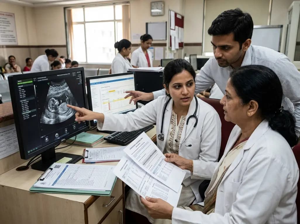
Prenatal evaluation of fetal growth restriction requires an integrated diagnostic approach. While chromosomal microarray remains fundamental, exome sequencing offers significant diagnostic yield in isolated and syndromic cases, especially when ultrasound shows skeletal anomalies or normal placental function.
Today
High-grade trochlear dysplasia disrupts patellofemoral stability. Although the patellotrochlear index measures cartilage overlap on MRI, cartilage contact does not ensure osseous containment. Relying solely on two-dimensional metrics may lead surgeons to overlook necessary tibial tubercle osteotomy procedures.
Today
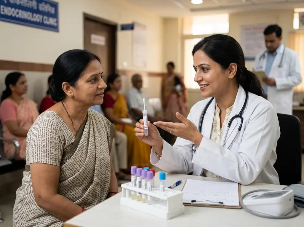
A study reveals that salivary cortisone correlates more strongly with serum cortisol than salivary cortisol during dynamic adrenal testing, while salivary dexamethasone accurately confirms compliance. This provides a non-invasive, precise alternative to venipuncture for diagnosing adrenal disorders.
Today
