

Loading, please wait...
Loading, please wait...
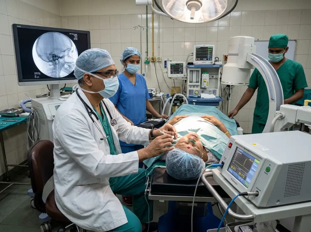
Percutaneous trigeminal radiofrequency rhizotomy remains an indispensable interventional treatment for medically refractory trigeminal neuralgia. Clinicians historically navigate the cannula through the foramen ovale into the Gasserian ganglion using fluoroscopy and physiological stimulation. However, conventional electrophysiological testing requires an alert, cooperative patient to report paresthesias in specific trigeminal divisions. Consequently, general anesthesia or deep sedation impairs classic verbal feedback, increasing technical uncertainty. Temperature-based interrogation has therefore emerged as an attractive physiological localization strategy. In particular, this technique allows interventionalists to evaluate probe position before triggering permanent thermal ablation.
Nevertheless, clinicians frequently worry about unintended neural tissue damage during preliminary thermal interrogation. Specifically, uncontrolled thermal spread can cause devastating complications, including corneal anesthesia, masseter weakness, or dysesthesia dolorosa. Therefore, establishing a definitive biophysical safety margin is paramount for wider clinical adoption. Recently, a groundbreaking investigation led by Dr. Michael D. Staudt and colleagues evaluated this exact risk. Their team meticulously quantified the precise thermal exposures generated during multicontact electrode mapping. Consequently, their biophysical data provide reassuring evidence for surgical teams seeking high anatomical precision without premature neurodestructive injury.
To determine whether temperature mapping damages neural tissue, researchers must quantify cumulative thermal exposure rather than transient peak temperatures. Crucially, thermal injury follows the Arrhenius damage integral, where cumulative damage depends exponentially on absolute temperature and linearly on exposure duration. When the Arrhenius damage integral equals or exceeds 1.0, irreversible coagulative tissue necrosis occurs. Therefore, the investigators analyzed high-resolution thermocouple logs from twelve patients undergoing rhizotomy with cylindrical platinum band electrodes. These electrodes featured a 0.8-millimeter diameter and a 2.0-millimeter contact length, capturing 54 mapping runs and 19 definitive lesioning episodes.
In addition, the authors constructed a sophisticated reduced-order effective-medium model representing the Gasserian ganglion. This computational model accounted for complex tissue factors, including local cerebrospinal fluid fraction and distributed microvascular heat exchange. The investigators calibrated their computational model using intraoperative burn data before applying it directly to mapping sequences. Furthermore, they conducted rigorous sensitivity analyses incorporating two distinct sets of neural Arrhenius parameters and simulated a plus-or-minus two-degree Celsius thermocouple bias. Consequently, this rigorous computational framework ensured that variations in probe calibration or tissue perfusion would not alter the fundamental safety conclusions.
Importantly, the experimental findings demonstrated an overwhelming safety buffer separating mapping interrogation from therapeutic tissue destruction. Under neural-relevant biophysical parameters, the median Arrhenius damage integral during mapping was merely 0.000373, with an interquartile range from 0.0000416 to 0.00165. Furthermore, the absolute maximum damage integral recorded across all mapping episodes reached only 0.00992. In striking contrast, definitive therapeutic burn episodes produced a median damage integral of 1850, with values regularly exceeding 3000. Therefore, thermal exposure during mapping remained several orders of magnitude below the critical threshold of 1.0 required for cellular destruction.
Similarly, the computational models revealed an unmistakable divergence in predicted anatomical lesion dimensions. Definite therapeutic burn episodes produced predictable destructive lesions, yielding computed radial margins between 1.8 and 2.2 millimeters from the electrode. Conversely, the predicted tissue damage radius for temperature mapping was exactly zero millimeters across every single evaluation. Even when investigators introduced a conservative plus-or-minus two-degree Celsius measurement artifact, mapping values stayed strictly subthreshold. Thus, mapping produces transient thermal elevations that dissipate harmlessly into surrounding tissues without triggering axonal degeneration or structural protein denaturation within the trigeminal ganglion.
Traditional radiofrequency systems utilize single active monopolar tips that generate uniform, non-directional spherical heat clouds. However, multicontact cylindrical platinum band electrodes permit discrete, segmental thermal mapping along distinct anatomical vectors within Meckel's cave. Consequently, surgeons can selectively interrogate specific neural rootlets that correlate with V1, V2, or V3 dermatomal pain. Because the mapping step incurs zero thermal injury dose, operators can reposition the probe safely without risking cumulative tissue trauma. Furthermore, this incremental verification process substantially reduces the likelihood of mistargeting adjacent cranial structures or motor fibers of the mandibular division.
In addition, temperature mapping provides objective biophysical feedback that diminishes reliance on subjective patient cooperation during painful trans-facial cannulation. Many modern centers perform percutaneous rhizotomy under monitored anesthesia care with intermittent boluses of propofol or remifentanil. However, waking patients repeatedly for sensory verification often induces disorientation, hypertension, and involuntary head movement. By establishing that temperature mapping maintains an exceptionally wide safety margin, interventionalists can systematically map target segments under continuous sedation. Consequently, this advance optimizes procedural workflow, mitigates patient distress, and reinforces operating room safety during needle manipulation.
These biophysical insights carry direct relevance for interventional pain specialists, neurosurgeons, and neurologists managing drug-resistant trigeminal neuralgia. Although carbamazepine and oxcarbazepine provide reliable initial symptom control, many patients eventually develop intolerable pharmacological adverse effects or treatment resistance. Percutaneous radiofrequency rhizotomy offers durable, cost-effective relief, particularly for elderly individuals or patients unfit for posterior fossa microvascular decompression. However, fears of corneal ulceration and persistent sensory dysesthesia historically restricted aggressive lesioning. By clarifying that preliminary thermal interrogation causes no permanent injury, this study reassures practitioners when refining electrode trajectories prior to irreversible ablation.
Furthermore, standardized temperature interrogation protocols could help harmonize procedural training across specialized neurosurgical centers in developing and developed nations. Indian tertiary healthcare institutions routinely handle heavy caseloads of typical and atypical facial pain where minimally invasive techniques are essential. Therefore, integrating computational safety boundaries into clinical cannulation protocols ensures maximum treatment efficacy with minimal morbidity. As multicontact electrode technology becomes more widely available, clinicians can confidently employ temperature mapping as an evidence-based localization tool. Ultimately, this approach bridges advanced computational bioengineering with patient-centered bedside care.
The Arrhenius damage integral serves as a biophysical mathematical formula that calculates cumulative thermal injury based on absolute temperature and exposure duration. In radiofrequency ablation, an integral value of 1.0 or greater signifies irreversible cellular protein denaturation and tissue necrosis. In this study, temperature mapping produced a median value of only 0.000373. This confirmed that mapping exposures remain several orders of magnitude below thresholds that cause permanent neural damage.
Temperature mapping enables clinicians to verify probe contact position before triggering permanent neurodestructive lesions. Traditional techniques require conscious patient cooperation to report tingling sensations during electrical stimulation. However, temperature interrogation works effectively under sedation, sparing patients distressing intraprocedural discomfort. Furthermore, by evaluating localized thermal dynamics using multicontact electrodes, surgical teams can achieve precise anatomical localization. This targeted approach significantly minimizes the risk of off-target complications such as inadvertent corneal numbness or masticatory motor weakness.
No, the biophysical modeling confirmed that temperature mapping produced an injury radius of exactly zero millimeters across all test episodes. In comparison, definitive therapeutic lesions produced thermal ablation radii between 1.8 and 2.2 millimeters from the electrode band. Even when researchers evaluated potential calibration errors with a plus-or-minus two-degree Celsius bias, mapping remained completely subthreshold. Therefore, practitioners can perform repetitive mapping without generating cumulative structural damage to the Gasserian ganglion.
Disclaimer: This content is for informational and educational purposes only... Refer to the latest local and national guidelines for clinical practice.
References

Read summarized clinical updates, watch expert medical content, and earn CME certifications right from your smartphone.
Open your smartphone camera to scan and install.





A biophysical study confirms that temperature mapping during trigeminal radiofrequency rhizotomy produces thermal doses orders of magnitude below lesion thresholds, demonstrating a 0-mm injury radius and substantial procedural safety for trigeminal neuralgia treatment.
Today

A randomized crossover trial evaluated fifth-year medical students interacting with an AI standardized patient versus a human standardized patient. The study revealed key differences in Mini-CEX scores, active engagement, and interview dynamics.
Today

A comparative evaluation of INNO-LiPA, Cobas 4800, and Amplicor highlights significant assay-dependent variation in high-risk HPV detection, emphasizing the clinical distinction between analytical sensitivity and screening specificity.
Today

A Canadian nutrition study reveals that higher diet costs predict lower dietary energy density and higher fruit intake, but also increase sodium and processed meat consumption.
Today
Idiopathic spinal cord herniation is a rare pathology presenting with progressive thoracic myelopathy. Early MRI identification and meticulous microsurgical reduction with dual bolstered dural defect repair prevent irreversible neurological deterioration.
Today
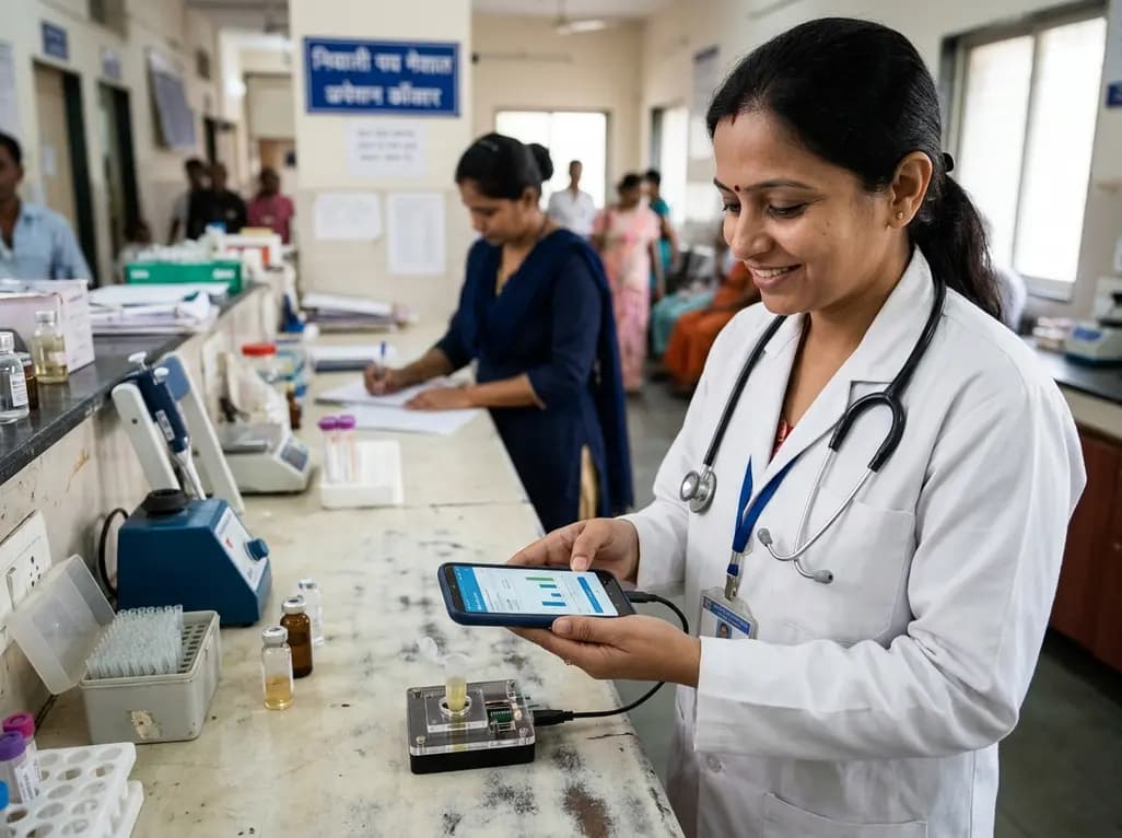
A novel smartphone-assisted biosensor utilizing molecularly imprinted polymers and catalytic nanozymes offers antibody-free, enzyme-free urinary microalbumin testing, enabling rapid, low-cost point-of-care screening for early chronic kidney disease.
Today
