.webp&w=384&q=75)

Loading, please wait...
Loading, please wait...
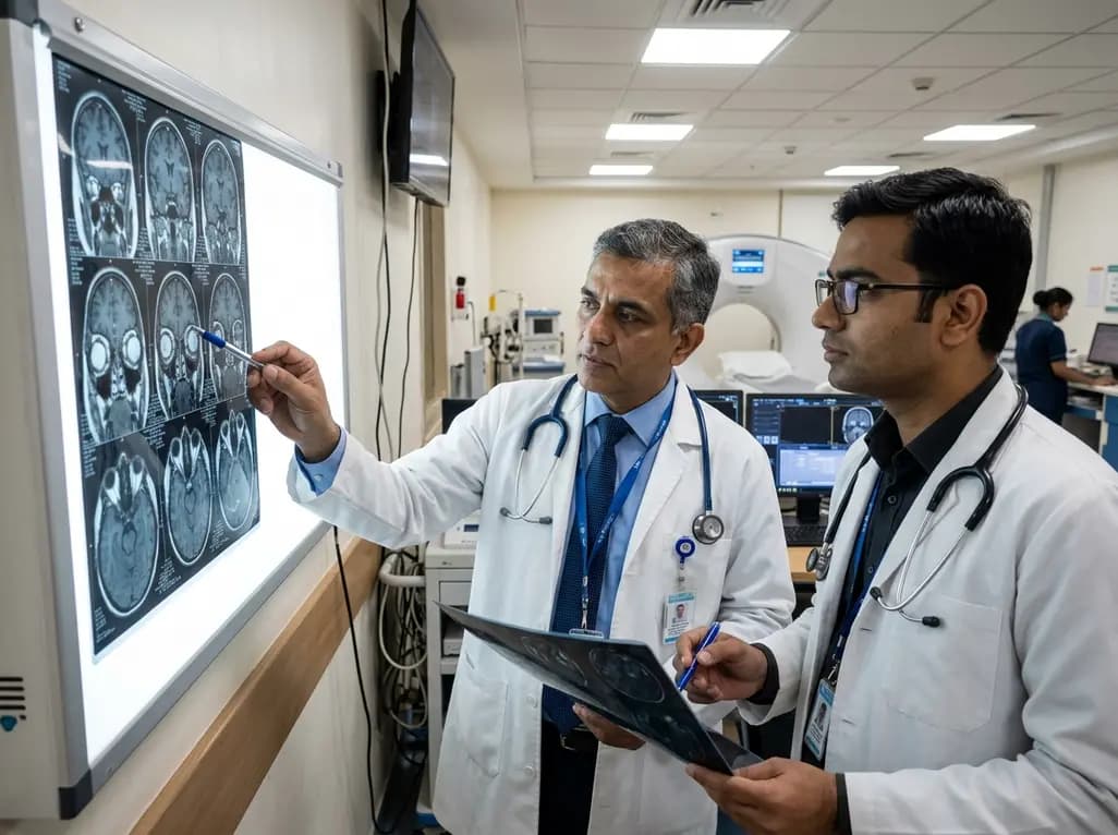
Thyroid eye disease causes distressing orbital inflammation, tissue remodeling, and significant proptosis that impairs visual function and patient quality of life. Recent clinical developments involving targeted biologic therapies have revolutionized management strategies for active moderate-to-severe disease. Specifically, teprotumumab thyroid eye disease management offers clinicians a non-surgical option to target underlying autoimmune pathophysiology. Evaluating objective radiologic changes provides essential insights into how this insulin-like growth factor-1 receptor inhibitor reduces orbital soft tissue enlargement and stabilizes clinical presentation.
Active thyroid eye disease involves an autoimmune cascade where autoantibodies stimulate orbital fibroblasts. These activated fibroblasts express high levels of insulin-like growth factor-1 receptors along with thyroid-stimulating hormone receptors. Consequently, fibroblast activation stimulates excessive glycosaminoglycan accumulation, inflammatory cytokine secretion, and tissue expansion behind the eye. This pathological remodeling leads directly to extraocular muscle enlargement, orbital fat expansion, elevated intraorbital pressure, severe proptosis, and troublesome diplopia.
Historically, clinicians relied heavily on high-dose intravenous corticosteroid therapy or orbital radiotherapy to suppress active inflammation. However, these conventional interventions frequently failed to yield structural regression of retrobulbar tissue expansion. Teprotumumab operates by selectively blocking the insulin-like growth factor-1 receptor on orbital fibroblasts. By suppressing downstream signaling pathways, the drug effectively stops hyaluronic acid synthesis and adipogenesis. Consequently, blocking this molecular pathways suppresses active inflammation while promoting tissue volume reduction. Understanding these cellular mechanisms helps clinicians recognize why targeted biologic therapy modifies actual disease anatomy rather than simply providing transient symptomatic relief.
The post hoc exploratory substudy from the OPTIC-J trial provides valuable magnetic resonance imaging evidence regarding retrobulbar tissue changes. Japanese investigators examined orbital scans from patients treated with teprotumumab compared against placebo control subjects over twenty-four weeks. Quantitative volumetric analysis demonstrated a marked thirty-one percent reduction in total extraocular muscle volume among teprotumumab-treated study eyes. Conversely, placebo-treated control eyes showed minimal change, confirming the selective structural impact of active therapy.
In addition to muscle volume reduction, orbital fat volume decreased significantly by six percent in the teprotumumab cohort. Furthermore, placebo controls demonstrated a four percent increase in orbital fat volume during the same observation period. Statistical comparison confirmed that reductions in both extraocular muscle volume and orbital fat volume were significantly greater with targeted biologic therapy. Additionally, signal intensity ratio measurements on magnetic resonance imaging showed a thirty-one percent reduction in muscle edema and inflammation. These objective radiologic improvements demonstrate that teprotumumab directly reverses retrobulbar soft tissue expansion in active ophthalmic disease.
Radiologic reductions in retrobulbar tissue directly correlate with meaningful clinical improvements in affected patients. In the OPTIC-J MRI substudy, eighty-three percent of patients receiving teprotumumab achieved significant proptosis reduction within twenty-four weeks. Decreasing orbital tissue volume creates space inside the rigid bony orbit, directly allowing the eyeball to settle back into its natural position. Consequently, relieving mechanical pressure on retrobulbar structures significantly mitigates optic nerve compression risks.
Furthermore, sixty-seven percent of patients receiving active treatment experienced notable improvements in diplopia. Reduction of extraocular muscle swelling restores normal extraocular muscle motility, thereby alleviating debilitating double vision. Additionally, fifty percent of teprotumumab-treated patients achieved complete resolution of inflammatory disease activity, as measured by the Clinical Activity Score. These concurrent improvements demonstrate a strong relationship between imaging findings and clinical outcomes. Decreasing tissue swelling and retrobulbar volume translates directly into functional visual recovery and enhanced patient comfort. Clinicians can confidently utilize orbital magnetic resonance imaging as an objective biomarker during patient monitoring.
Magnetic resonance imaging provides an exceptional non-invasive tool for assessing active orbital inflammation and structural remodeling. Standard clinical exams often rely on subjective scoring systems that may underrepresent deep retrobulbar inflammation. In contrast, T2-weighted MRI sequences calculate exact signal intensity ratios relative to cerebral white matter, yielding objective measurements of muscle edema. Similarly, ordinal grading scales effectively quantify inflammation within orbital fat compartments.
Utilizing quantitative imaging helps clinicians differentiate active inflammatory edema from chronic, burnt-out fibrotic changes. This distinction is critical because biologic therapies targeting receptor pathways demonstrate optimal efficacy during active inflammatory phases. Furthermore, sequential volumetric tracking enables clinicians to monitor therapeutic response accurately over a standard twenty-four-week treatment course. Identifying distinct changes in individual extraocular muscle groups and fat pads aids in personalizing long-term treatment strategies. Consequently, integrating magnetic resonance imaging into routine clinical protocols elevates diagnostic accuracy and refines patient management in complex orbitopathy cases. Ultimately, objective radiologic assessment supports evidence-based therapeutic decisions.
The objective radiologic proof of orbital tissue volume reduction reinforces the importance of early intervention in active disease. Endocrinologists and ophthalmologists must collaborate closely to identify candidates who will benefit most from targeted therapy. Early initiation of receptor-targeted treatment during the active phase prevents irreversible fibrotic remodeling and persistent double vision. Moreover, understanding that both extraocular muscle swelling and orbital fat expansion respond to therapy helps tailor patient expectations regarding aesthetic and functional recovery.
While clinical trial results are highly promising, real-world practice requires careful patient selection and comprehensive safety monitoring. Clinicians must screen for potential adverse effects, including hyperglycemia, auditory disturbances, and gastrointestinal symptoms. Additionally, establishing standardized orbital imaging protocols across clinical centers will optimize monitoring accuracy and facilitate comparative research. As post-marketing data continue to accumulate across diverse ethnic populations, targeted biological therapies will further redefine standard care algorithms. Collaborative multidisciplinary care models remain essential for delivering comprehensive, personalized management to patients with active disease.
Teprotumumab is a human monoclonal antibody that selectively blocks the insulin-like growth factor-1 receptor on orbital fibroblasts. By inhibiting this key signaling receptor, the drug stops hyaluronic acid synthesis, reduces inflammatory cytokine release, and inhibits adipogenesis. Consequently, this molecular blockade suppresses retrobulbar inflammation, leading to significant volume reduction in both enlarged extraocular muscles and expanded orbital fat tissue within twenty-four weeks of active treatment.
Magnetic resonance imaging with T2-weighted sequences is ideal for evaluating treatment response in thyroid eye disease. T2-weighted signal intensity ratios accurately quantify muscle edema relative to cerebral white matter, while volumetric software measures exact changes in extraocular muscle and orbital fat volumes. These quantitative radiologic metrics provide objective, non-invasive evidence of reduced soft tissue inflammation, helping clinicians distinguish active inflammatory disease from chronic fibrotic tissue changes.
Reductions in extraocular muscle and orbital fat volumes directly correlate with clinical improvements in proptosis and diplopia. Shrinking retrobulbar tissue relieves intraorbital pressure, allowing the eye to recede and reducing exophthalmos. Furthermore, decreasing muscle swelling restores normal extraocular muscle motility, significantly relieving double vision. Additionally, reduced retrobulbar inflammation promotes resolution of active disease, as demonstrated by lower Clinical Activity Scores in treated patients.
Disclaimer: This content is for informational and educational purposes only and does not constitute medical advice, diagnosis, or treatment. Refer to the latest local and national guidelines for clinical practice.
References

Read summarized clinical updates, watch expert medical content, and earn CME certifications right from your smartphone.
Open your smartphone camera to scan and install.





An exploratory MRI analysis from the OPTIC-J trial demonstrates that teprotumumab significantly reduces extraocular muscle (-31%) and orbital fat (-6%) volumes in Japanese patients with active thyroid eye disease, correlating with clinical improvements in proptosis, diplopia, and disease activity score.
last month

The JAKPPPOT feasibility trial evaluated oral upadacitinib in moderate-to-severe palmoplantar pustulosis. Meeting all feasibility goals with 95% adherence, upadacitinib demonstrated a substantial baseline-adjusted reduction in disease severity compared to historic placebo, supporting future randomised trials.
Today

Prenatal evaluation of fetal growth restriction requires an integrated diagnostic approach. While chromosomal microarray remains fundamental, exome sequencing offers significant diagnostic yield in isolated and syndromic cases, especially when ultrasound shows skeletal anomalies or normal placental function.
Today

Evaluating disagreement among Friedewald, Martin-Hopkins, and Sampson LDL-C equations reveals diagnostic uncertainty at statin cutoffs. An interpretable regime-aware calibration model rescues patient misclassifications without extra testing costs.
Today

A randomized controlled trial compared ultrasound-guided intra-articular injections of PRP, hydrolyzed type I collagen, and hyaluronic acid in knee osteoarthritis. All therapies demonstrated significant improvements in WOMAC and pain scores at six months, supporting hydrolyzed collagen as a viable biological option.
Today

A study reveals that salivary cortisone correlates more strongly with serum cortisol than salivary cortisol during dynamic adrenal testing, while salivary dexamethasone accurately confirms compliance. This provides a non-invasive, precise alternative to venipuncture for diagnosing adrenal disorders.
Today
