

Loading, please wait...
Loading, please wait...

Temporal lobe epilepsy represents the most prevalent form of drug-resistant focal epilepsy encountered in adult clinical neurology. While conventional neuroimaging frequently reveals heterogeneous cortical atrophy patterns across individual patients, the precise mechanisms uniting these dispersed structural lesions into a cohesive clinical syndrome have remained poorly understood. A landmark study published in Neurobiology of Disease provides compelling evidence that individualized structural alterations systematically converge into shared functional networks.
Neurologists have long observed that patients with temporal lobe epilepsy exhibit diverse structural abnormalities on volumetric magnetic resonance imaging. Although hippocampal sclerosis remains the classic pathological hallmark, extrahippocampal cortical thinning often appears across wide anatomical areas, including prefrontal, insular, and parietal cortices. Consequently, this pronounced patient-specific variability has historically complicated attempts to establish universal structural biomarkers for diagnosis and prognosis. To overcome this diagnostic hurdle, investigators leveraged advanced normative modeling of regional gray matter volume. Normative modeling functions analogously to pediatric growth charts, mapping each individual patient's regional brain volume deviations against an extensive normative reference dataset. Therefore, this statistical framework accurately detects subtle, patient-specific cortical atrophy that traditional group-level comparisons routinely overlook. Furthermore, the researchers mapped these dispersed atrophic foci onto normative human connectome data to evaluate their broader architectural impact. Rather than acting as isolated structural defects, these distinct lesions interact dynamically through intrinsic neural pathways. Ultimately, this paradigm shift illuminates how anatomically inconsistent structural lesions produce remarkably consistent electroclinical features across diverse patient cohorts.
Although individual patients demonstrated unique distributions of cortical thinning, network connectivity mapping uncovered a striking organizational phenomenon. Specifically, the analysis revealed that spatially dispersed atrophic foci systematically converge onto a shared temporal-limbic functional network. This shared circuitry prominently encompasses the mesial temporal structures, amygdala, entorhinal cortex, insula, and interconnected paralimbic regions. Because these anatomical hubs form the core architecture governing seizure propagation, their functional convergence explains the uniform clinical semiology seen in focal seizures. Additionally, this convergence demonstrates that temporal lobe epilepsy operates fundamentally as a large-scale network disorder rather than a localized focal defect. When discrete cortical regions degenerate, they appear to propagate downstream functional disruptions across shared synaptic pathways. As a result, structurally distinct injuries across disparate neocortical sites repeatedly target the same vulnerable temporal-limbic pathways. Consequently, this common network convergence reconciles the longstanding discrepancy between heterogeneous structural neuroimaging findings and stereotypical clinical presentations. Neurologists can therefore conceptualize individualized structural injuries as distinct entry points into a singular pathogenic network.
To decipher the biological substrates linking structural atrophy to functional circuitry, investigators integrated whole-brain transcriptomic profiles from the Allen Human Brain Atlas. Remarkably, spatial enrichment analyses demonstrated that the identified temporal-limbic convergence network strongly correlates with specific glial-immune gene expression profiles. Rather than aligning predominantly with pure neuronal markers, the vulnerable network exhibited profound enrichment for astrocytic molecular pathways. Furthermore, these transcriptomic signatures implicated active neuroinflammatory processes, cytokine signaling cascades, and blood-brain barrier dysfunction in circuit degeneration. Astrocytes play essential roles in maintaining extracellular potassium buffering, glutamate clearance, and local neurovascular coupling. Therefore, chronic astrocytic dysfunction and reactive neuroinflammation can severely impair synaptic homeostasis, precipitating both neuronal hyperexcitability and progressive tissue loss. In addition, these findings suggest that chronic neuroinflammatory signaling perpetuates circuit-level vulnerability throughout the epileptic temporal-limbic axis. Consequently, targeting glial-mediated neuroinflammation may represent a promising disease-modifying therapeutic avenue alongside traditional ion channel-modulating therapies. Integrating spatial transcriptomics with connectomics thus bridges the critical gap between microscopic cellular mechanisms and macroscopic neuroimaging alterations.
Temporal lobe epilepsy frequently exhibits a progressive trajectory characterized by escalating cognitive complaints and worsening seizure control over time. In this study, cross-sectional analyses stratified by disease duration revealed clear patterns of functional reorganization across the brain. Specifically, as disease duration increased, functional coupling systematically shifted away from higher-order transmodal networks toward unimodal primary cortices. In the early stages of disease, network disturbances concentrated heavily within associative and limbic structures. However, prolonged illness duration correlated with progressive decoupling in heteromodal hubs and compensatory hyperconnectivity across primary sensory and motor cortices. Furthermore, this chronicity-related topological reorganization mirrors the cumulative cognitive decline and sensory processing deficits commonly documented in long-standing epilepsy. Therefore, these dynamic alterations emphasize that neurodegeneration in epilepsy is not a static initial insult. Instead, ongoing seizure activity, subclinical epileptiform discharges, and chronic neuroinflammation drive continuous network remodeling over decades. Clinicians must recognize that early therapeutic intervention remains vital to prevent irreversible global network destabilization and secondary neocortical decline.
Achieving seizure freedom remains a substantial challenge, as approximately one-third of patients develop pharmacoresistance despite modern pharmacotherapy. Crucially, the researchers evaluated whether intrinsic functional network metrics could predict therapeutic outcomes in patients receiving anti-seizure medication. The investigators identified fronto-parietal network coupling strength as a powerful, non-invasive biomarker of anti-seizure medication responsiveness. Specifically, patients exhibiting preserved functional coupling within the fronto-parietal control network demonstrated significantly superior responses to pharmacological therapy. Conversely, patients with marked fronto-parietal decoupling experienced higher rates of treatment failure and drug resistance. The fronto-parietal network governs executive control, adaptive cognitive processing, and top-down regulation across distributed cortical modules. Consequently, severe disruption of this regulatory network may impair intrinsic inhibitory controls, rendering conventional anti-seizure medications less effective. In clinical practice, this network-based stratification could enable early identification of individuals unlikely to benefit from repeated drug trials. As a result, clinicians could expedite comprehensive presurgical evaluations or initiate neuromodulatory therapies much earlier in the disease course.
Network convergence proves that variable, patient-specific cortical lesions coalesce into a shared temporal-limbic circuit. Rather than viewing temporal lobe epilepsy as a purely isolated anatomical focal lesion, clinicians can now understand it as a distributed circuit disorder. This explains why patients with diverse structural atrophy patterns exhibit remarkably similar clinical seizure presentations.
Transcriptomic analyses demonstrate that temporal-limbic network vulnerability correlates strongly with glial-immune and astrocytic gene expression pathways. Astrocytes regulate extracellular potassium clearance, glutamate homeostasis, and neurovascular coupling. Impaired astrocytic function and sustained neuroinflammation provoke neuronal hyperexcitability, promoting circuit degradation, progressive cortical atrophy, and persistent seizure activity across connected limbic networks.
Fronto-parietal network coupling strength serves as an objective biomarker for predicting anti-seizure medication responsiveness. Preserved connectivity correlates with favorable drug response, whereas disrupted coupling signals pharmacoresistance. Identifying this decoupling early allows clinicians to minimize ineffective medication trials and accelerate timely evaluations for resective surgery or advanced neuromodulation therapies.
Disclaimer: This content is for informational and educational purposes only and should not be construed as medical advice. Always consult qualified healthcare professionals for diagnosis and treatment. Refer to the latest local and national guidelines for clinical practice.
References

Read summarized clinical updates, watch expert medical content, and earn CME certifications right from your smartphone.
Open your smartphone camera to scan and install.





New research reveals that spatially dispersed cortical atrophy in temporal lobe epilepsy converges into a shared temporal-limbic network enriched in glial-immune transcriptomic signatures. Furthermore, fronto-parietal network coupling predicts anti-seizure medication response, offering a key stratification biomarker.
Today

The DOMINO study protocol presents a multidisciplinary implementation mapping approach to overcome provider, patient, and payer barriers to antiobesity medications, offering vital insights for Indian clinicians managing the growing diabesity burden.
Today

An intracranial EWSR1::CREM fusion sarcoma in a 31-year-old female manifested as a paraneoplastic inflammatory syndrome with anemia and secondary infertility. The dural mass mimicked meningioma, but gross-total resection resolved her systemic inflammation and restored natural fertility within one month.
Today

A real-world cohort study of 6,537 patients from the FOUNTAIN platform demonstrates that initiating finerenone significantly reduces albuminuria by 40.7% with minimal eGFR decline and manageable hyperkalemia risk, confirming its practical effectiveness and safety in diabetic kidney disease.
Yesterday

A comprehensive analysis of MAP therapy resistance in osteosarcoma, examining canonical and non-canonical mechanisms across damage avoidance, enhanced DNA repair, and replication stress tolerance to identify novel vulnerabilities.
Today
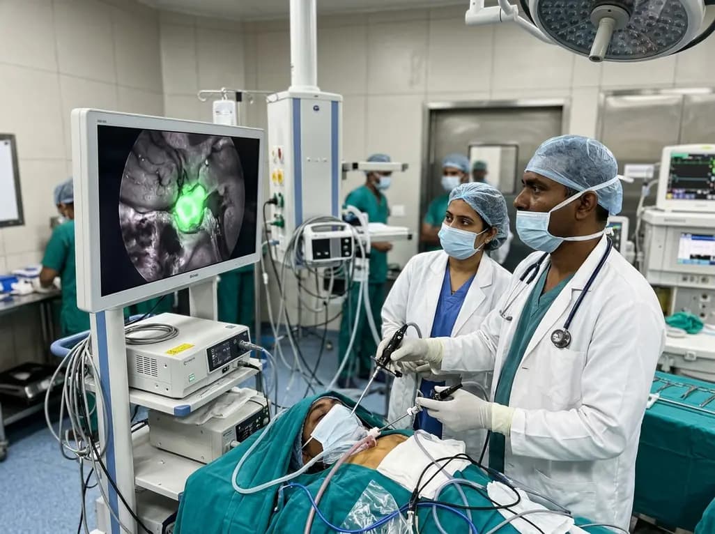
Near-infrared autofluorescence provides real-time label-free optical guidance during endoscopic transsphenoidal pituitary surgery. By highlighting stark signal contrasts between normal pituitary gland and adenomas, this technique aids gross-total tumor resection while safeguarding critical endocrine function.
Today
