
Loading, please wait...
Loading, please wait...

Neuroimaging researchers are increasingly utilizing T2 relaxometry in gliomas to refine the distinction between peritumoral edema and infiltrative malignancy. Standard magnetic resonance imaging (MRI) often fails to separate these two entities effectively. Both appearing as hyperintense signals on T2-weighted and FLAIR sequences, they look nearly identical to the human eye. Consequently, neuro-oncologists face significant challenges when defining surgical margins or radiation fields. However, recent findings suggest that quantitative analysis via multi-echo T2 relaxometry offers a more precise solution for tissue characterization.
Recent studies evaluated the efficacy of this technique using specialized multi-echo sequences and analysis software. Specifically, researchers compared high-grade gliomas (HGGs) with non-infiltrative lesions like meningiomas and metastases. These non-glioma tumors typically present with pure vasogenic edema. In contrast, HGGs exhibit a complex mixture of edema and active tumor infiltration. Furthermore, the data showed significant statistical differences in T2 relaxation values between these groups. Therefore, this quantitative approach provides a clearer picture of the tumor microenvironment than conventional scans.
Notably, the mean T2 relaxation times for pure vasogenic edema in metastases were significantly lower than the values observed in HGG infiltrated zones. Moreover, central tumor areas showed distinct relaxation patterns compared to peripheral zones. Additionally, these peripheral zones often shared characteristics with pure edema, suggesting a gradient of infiltration. This distinction is vital for accurate preoperative assessment. Consequently, clinicians can better predict the extent of tumor resection and optimize postoperative outcomes for their patients.
Advanced software allows for the mapping of T2 relaxation curves across different regions of interest. By analyzing these curves, radiologists can identify subtle changes in water mobility and tissue structure. Consequently, this method improves the detection of infiltrative zones that standard FLAIR imaging might overlook. Furthermore, the use of quantitative metrics reduces inter-observer variability among radiologists. Therefore, T2 relaxometry serves as a valuable adjunct to traditional neuroimaging protocols in oncology.
Conventional MRI sequences like T2-weighted and FLAIR imaging show both vasogenic edema and tumor infiltration as bright hyperintense areas. Because they share similar water content characteristics, standard imaging cannot reliably separate healthy reactive fluid from migrating cancer cells.
T2 relaxometry provides quantitative values that differentiate pure fluid from infiltrated tissue. This data helps surgeons identify high-risk zones of infiltration beyond the main tumor mass, allowing for more precise surgical margins and better-targeted radiation therapy.
Disclaimer: This content is for informational and educational purposes only and does not constitute medical advice. It is not intended to be a substitute for professional medical advice, diagnosis, or treatment. Always seek the advice of your physician or other qualified healthcare provider with any questions you may have regarding a medical condition. Refer to the latest local and national guidelines for clinical practice.
References
1. Mansano de Souza L et al. T2 relaxometry in high-grade gliomas: Preliminary analysis of peritumoral tissue characteristics. Neuroradiol J. 2026 May 11. doi: 10.1177/19714009261450379. PMID: 42109220.
2. Oh J et al. Quantitative apparent diffusion coefficients and T2 relaxation times in characterizing contrast enhancing brain tumors and regions of peritumoral edema. J Magn Reson Imaging. 2005;21(6):701-708.
3. Ellingson BM et al. Quantification of edema reduction using differential quantitative T2 (DQT2) relaxometry mapping in recurrent glioblastoma treated with bevacizumab. J Neurooncol. 2012;106(1):111-119.
"
Read summarized clinical updates, watch expert medical content, and earn CME certifications right from your smartphone.
Open your smartphone camera to scan and install.


T2 relaxometry shows a significant difference between pure vasogenic edema and tumor-infiltrated tissue in high-grade gliomas, aiding precise diagnostics....
2 months ago
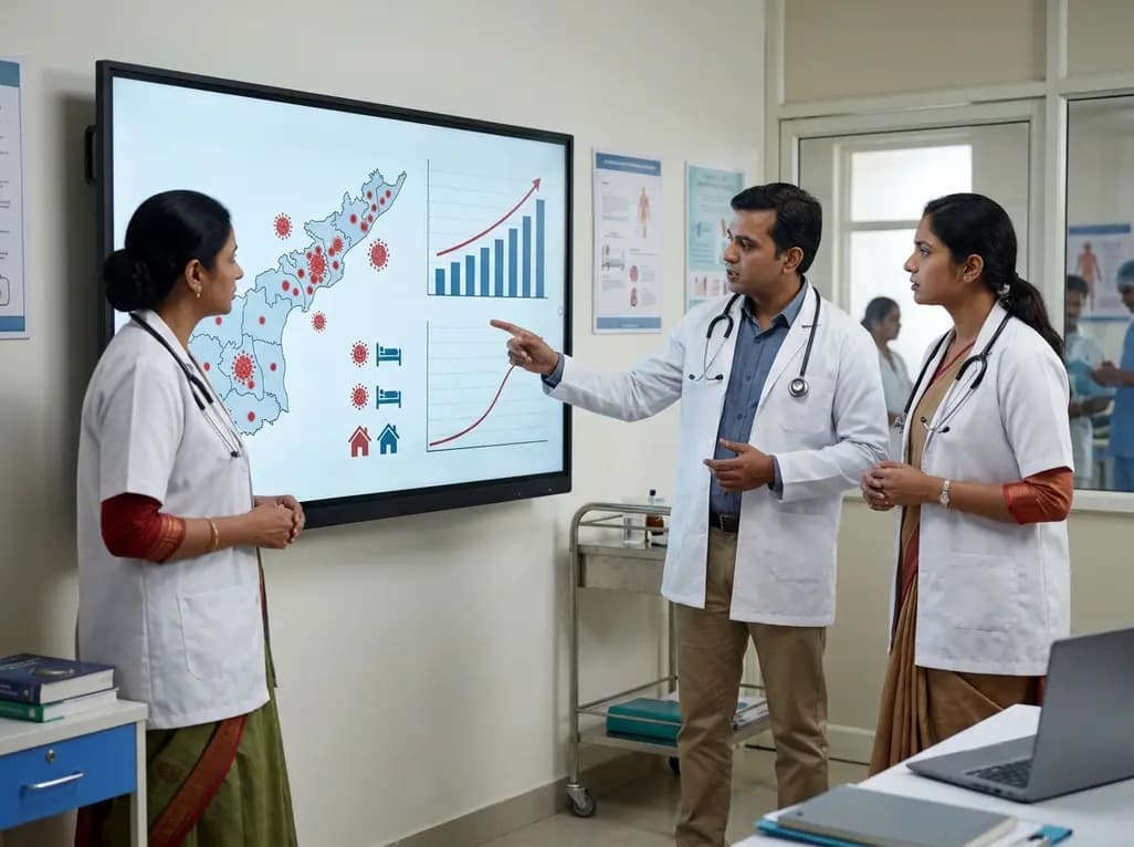
Andhra Pradesh reported 10 new Covid-19 cases, taking the state tally to 49 while deaths remain at four. With 24 patients hospitalized and 16 under home isolation, the Health Department has intensified monitoring. Medical professionals should review regional distribution, diagnostic protocols, and management plans.
Today

An 11-year Swedish registry study of 618 uterine sarcoma patients found that minimally invasive surgery yielded survival comparable to open surgery in early stages. However, adjuvant chemotherapy conferred no survival benefit in localized or advanced disease, highlighting stage and histology as key outcomes.
3 days back
A cross-sectional study evaluates post-intensive care syndrome in cardiac patients 2-4 weeks post-ICU discharge, highlighting cognitive, psychological, and functional impairments and the need for structured multidisciplinary rehabilitation.
3 days back
Anterior cruciate ligament reconstruction failure lacks uniform definition. A narrative review proposes an integrative framework incorporating objective and subjective instability, persistent pain, restricted motion, graft rupture, and secondary meniscal injury to standardize clinical reporting.
3 days back

With World Obesity Atlas data warning that over 41 million Indian children are overweight or obese, ICMR and NIN have unveiled a 10-point policy roadmap. The initiative calls for mandatory front-of-pack labeling, HFSS taxes, strict marketing bans, and healthier school environments to curb non-communicable diseases.
Today
