

Loading, please wait...
Loading, please wait...

Paediatric portal hypertension represents a challenging clinical condition that leads to severe gastrointestinal complications, progressive splenomegaly, and liver decompensation. Clinicians historically viewed this syndrome through a hemodynamic lens, focusing primarily on vascular resistance and collateral blood flow. However, emerging translational research reveals that profound immune dysregulation actively influences disease progression and systemic complications in affected children. A landmark pilot study conducted by Si and colleagues prospectively investigated these immunological changes in paediatric cohorts. The researchers enrolled forty-four children with portal hypertension, separating them into chronic liver disease and portal vein thrombosis cohorts, alongside fifteen healthy pediatric controls. By collecting paired peripheral and portal venous blood, the investigators characterized cellular phenotypes, cytokine cascades, and immune checkpoints using multiparameter flow cytometry and enzyme-linked immunosorbent assays. Consequently, the study demonstrated that children with portal hypertension experience a marked depletion of circulating T cells. In addition, these cells exhibit defective effector functions and altered phenotypic properties. Therefore, evaluating cellular immunity provides critical insights into underlying pathobiology, transforming our understanding of vascular and parenchymal hepatic disorders.
The underlying aetiology significantly dictates the precise pattern of immune disruption in affected pediatric patients. Specifically, children presenting with chronic liver disease-related portal hypertension demonstrate a pronounced systemic pro-inflammatory cascade. Laboratory profiling identified dramatic elevations in interferon-gamma, tumour necrosis factor-alpha, and matrix metalloproteinase-7 among these patients. Furthermore, these children display substantial elevations of soluble programmed death-ligand 1 alongside increased expression of CD39 and PD-1 on circulating lymphocytes. These specific surface markers define a classical phenotype of immune exhaustion. When persistent antigenic stimulation and ongoing parenchymal injury barrage the liver, T cells upregulate inhibitory receptors to restrain unbridled immune damage. Although this mechanism limits local tissue destruction, it simultaneously dampens functional cell-mediated immune responses. Consequently, exhausted T cells lose their capacity to mount effective antimicrobial responses, increasing host vulnerability to secondary systemic infections. In addition, the systemic surge of soluble PD-L1 highlights widespread compensatory checkpoint activation. Thus, in chronic liver disease, chronic inflammation directly intertwines with progressive immunological senescence, creating an environment that fuels both vascular remodeling and systemic immune incompetence.
In sharp contrast to chronic parenchymal liver disease, portal vein thrombosis generates a fundamentally different immune signature in paediatric patients. Children suffering from extrahepatic portal vein thrombosis maintain relatively preserved liver synthetic function and display significantly milder systemic inflammation. However, multiparameter flow cytometry reveals that these patients experience unique disturbances in T cell homing and migratory trafficking. Specifically, circulating T lymphocytes exhibit marked upregulation of CC chemokine receptor 4 alongside a notable downregulation of CXC chemokine receptor 3. Chemokine receptors direct the geographic navigation of immune cells throughout lymphoid tissues and visceral vascular beds. Elevated circulating levels of CXCL9, a principal ligand for CXCR3, significantly correlate with surface receptor expression on these lymphocytes. This inverse chemokine receptor pattern indicates aberrant homing signals that divert lymphocytes away from normal hepatic surveillance routes. Furthermore, non-cirrhotic portal hypertension induces mechanical shear stress and altered portal hemodynamics, which may actively reshape endothelial interactions. Therefore, rather than driving profound immune exhaustion, portal vein thrombosis primarily alters chemokine responsiveness and lymphocyte positioning. Recognizing this distinct trafficking defect enables pediatricians to differentiate vascular pathology from true parenchymal failure.
A compelling discovery from paired blood analyses is the profound immunological difference between peripheral blood and portal venous blood. Regardless of primary disease aetiology, T cells residing within the portal circulation demonstrate significantly decreased cytokine production and attenuated cytotoxic activity. The portal vein delivers blood rich in microbial antigens and lipopolysaccharides derived directly from the gastrointestinal microbiome. Consequently, the liver microenvironment must maintain an intricate balance between pathogen defence and immune tolerance. If hepatic immune cells responded aggressively to every portal antigen, lethal systemic inflammation would quickly ensue. Hepatic sinusoidal endothelial cells, Kupffer cells, and resident dendritic cells actively condition incoming lymphocytes toward functional tolerance. Thus, the observed reduction in portal T cell effector capacity reflects an essential biological adaptation. However, this physiologic dampening can become detrimental during advanced liver disease. As portal pressures rise, intestinal barrier integrity breaks down, flooding the hepatic sinusoids with heavy microbial burdens. When portal T cells remain functionally suppressed, bacterial clearance falters, facilitating spontaneous bacterial peritonitis and systemic endotoxemia. Understanding this regional tolerance helps hepatologists appreciate why children with elevated portal pressures develop unique infectious complications despite heightened peripheral inflammation.
Beyond mapping immunological mechanisms, evaluating immune signatures delivers meaningful prognostic value for pediatric gastroenterologists. Notably, the study revealed strong positive correlations between liver stiffness measurements and circulating levels of pro-inflammatory cytokines, including IFN-gamma and TNF-alpha. In addition, elevated concentrations of regulatory checkpoint markers, such as soluble PD-L1 and CD39, closely tracked structural hepatic fibrogenesis. Non-invasive assessment of hepatic fibrosis via transient elastography remains a cornerstone of pediatric hepatology. However, integrating soluble immune markers can significantly enhance clinical risk stratification. Clinicians can identify patients experiencing silent disease acceleration before overt decompensation occurs. Furthermore, profiling circulating chemokine receptors and cytokines may help predict variceal development and gastrointestinal bleeding risks. Because children with severe portal hypertension frequently face life-threatening hemorrhage, non-invasive risk scores could refine the timing of endoscopic screening and prophylactic band ligation. Future management algorithms might also incorporate immune checkpoint modulators or anti-cytokine therapies to arrest fibrotic remodeling. Ultimately, prospective validation across larger multicentre cohorts will determine whether these candidate biomarkers can guide personalized therapeutic interventions in paediatric hepatology.
Children with chronic liver disease exhibit prominent systemic inflammation and T cell exhaustion, marked by elevated IFN-gamma, TNF-alpha, MMP7, soluble PD-L1, CD39, and PD-1 expression. In contrast, children with portal vein thrombosis maintain relatively preserved liver function and demonstrate milder systemic inflammation. However, they display distinct alterations in lymphocyte trafficking, featuring increased CCR4 expression alongside significantly reduced CXCR3 levels. Consequently, each clinical aetiology manifests a distinct immunological profile that drives pathology.
T cells isolated from the portal circulation demonstrate significantly decreased cytokine production and attenuated cytotoxic responses compared to peripheral lymphocytes. This phenotypic shift represents a functional adaptation within the distinct hepatic microenvironment. Chronic antigenic stimulation and constant exposure to gut-derived endotoxins prompt local immune down-regulation to prevent overwhelming tissue damage. Therefore, this local immunosuppressive response protects hepatic architecture, although it simultaneously compromises cellular immune defence against prospective local infections.
Elevated circulating levels of pro-inflammatory cytokines, specifically IFN-gamma and TNF-alpha, exhibit a direct positive correlation with shear-wave liver stiffness measurements in paediatric patients. Furthermore, immune checkpoint molecules such as soluble PD-L1 and cellular CD39 parallel fibrotic remodeling and structural organ rigidity. These biomarkers directly reflect progressive hepatic parenchymal inflammation and active extracellular matrix expansion. As a result, monitoring these soluble mediators offers clinicians non-invasive immunological indicators to gauge disease progression and parenchymal deterioration.
Disclaimer: This content is for informational and educational purposes only... Refer to the latest local and national guidelines for clinical practice.
References
Si T et al. Altered T cell immune profile and its prognostic implications in children with portal hypertension: A pilot study. J Pediatr Gastroenterol Nutr. 2026 Oct 18. doi: 10.1002/jpn3.70613. PMID: 42893940.
Didato F, et al. The immunological profile of children with portal hypertension. Dig Liver Dis. 2025; doi: 10.1016/j.dld.2025.11.025. PMID: 41423404.
Grammatikopoulos T, McKiernan PJ, Dhawan A. Portal hypertension and its management in children. Arch Dis Child. 2018;103(2):186-191. doi: 10.1136/archdischild-2015-310022. PMID: 28814421.

Read summarized clinical updates, watch expert medical content, and earn CME certifications right from your smartphone.
Open your smartphone camera to scan and install.





A recent pilot study reveals that paediatric portal hypertension features distinct immune dysregulation based on aetiology: chronic liver disease triggers T cell exhaustion and chronic inflammation, whereas portal vein thrombosis alters chemokine trafficking, with key implications for prognosis.
Today
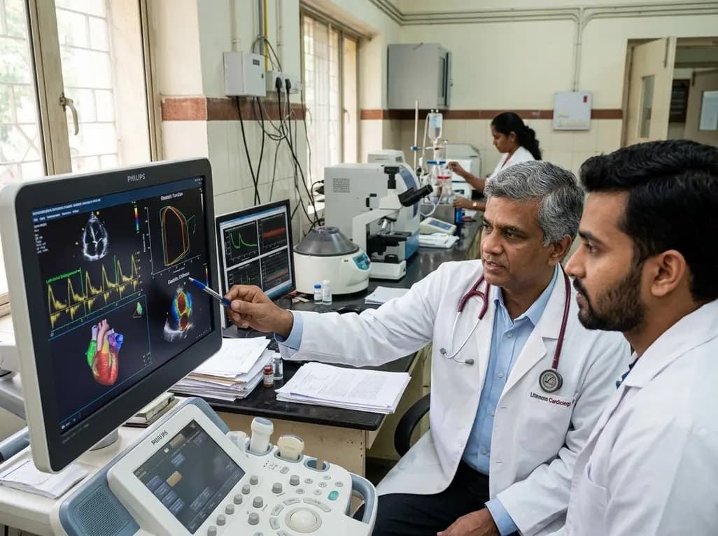
A study evaluates a cardiometabolic HFpEF model combining metabolic stress with chronic mineralocorticoid excess. The findings reveal profound diastolic stiffness, endothelial dysfunction, and pulmonary hypertension, illuminating key pathophysiological mechanisms for targeted cardiopulmonary therapies.
Today

The Human Reproduction and Development Database (HRD) consolidates bulk and single-cell transcriptomics, epigenomics, metabolomics, proteomics, and clinical trial records into a unified platform to advance reproductive health research.
Today

A study reveals that the MYLK c.4079G>A, p.(Gly1360Asp) variant is linked to high-penetrance heritable thoracic aortic disease and early-onset aortic dissection, showing a founder effect in Finnish cohorts. Findings emphasize early cascade screening and vigilant surveillance.
Yesterday

A single-cell transcriptomic atlas reveals how peripheral nerve injuries cause dorsal root ganglion remodeling. By evaluating proximal nerve crush and resection, researchers identify distinct glial and immune programs that resolve neuropathic pain and promote regeneration.
Today
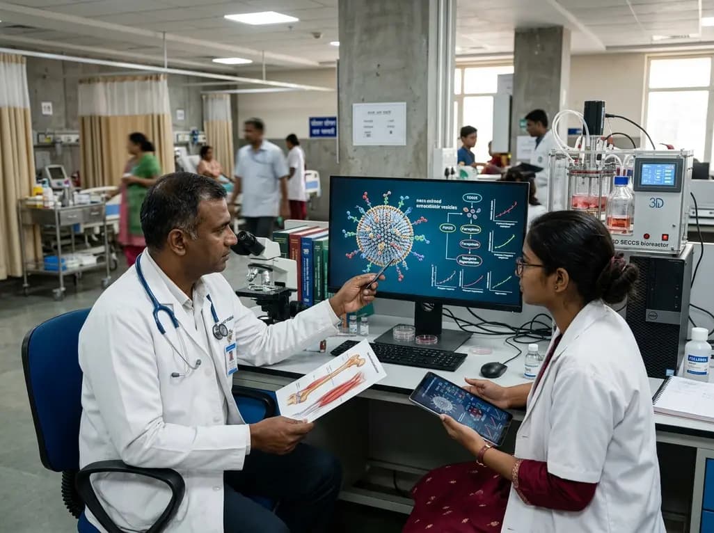
A breakthrough study demonstrates that fullerol-primed extracellular vesicles generated in a 3D SECCS bioreactor attenuate ferroptosis and restore myogenic regeneration in diabetes via the FOXO3 pathway, offering a cell-free therapeutic strategy for diabetic musculoskeletal degeneration.
Today
