.webp&w=384&q=75)

Loading, please wait...
Loading, please wait...
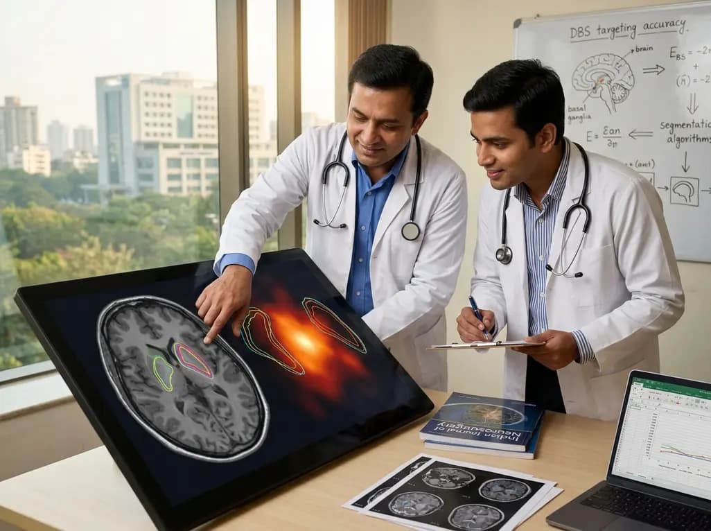
Deep brain stimulation (DBS) represents a cornerstone surgical therapy for advanced Parkinson's disease. However, achieving optimal motor symptom relief while minimizing adverse effects requires millimeter-level precision. Consequently, accurate subthalamic nucleus segmentation has emerged as a fundamental prerequisite for successful preoperative planning and postoperative programming. Clinicians increasingly rely on automated algorithms to define complex midbrain nuclei. Nevertheless, neurosurgeons must understand how these anatomical segmentations correspond spatially to the actual volume of tissue activated (VTA) during chronic therapeutic stimulation.
The subthalamic nucleus (STN) is an exceptionally small, biconvex structure situated deep within the midbrain. Because the motor, associative, and limbic territories lie in tight proximity within the STN, electrode placement must target the dorsolateral motor zone with high fidelity. When stimulation fields inadvertently spread into adjacent limbic areas or internal capsule fibers, patients frequently develop debilitating psychiatric or motor side effects. Therefore, advanced preoperative neuroimaging and high-resolution delineation remain paramount for optimal electrode placement.
Historically, neurosurgeons relied on direct visual targeting on magnetic resonance imaging (MRI) combined with standardized stereotactic coordinates. However, significant inter-individual anatomical variation and age-related brain shifts can compromise the accuracy of rigid atlas coordinates. Modern stereotactic workflows now incorporate algorithmic tools that automatically delineate the nucleus directly from patient-specific T1-weighted and T2-weighted MRI sequences. As a result, automated subthalamic nucleus segmentation enables individualized surgical trajectory planning, thereby enhancing anatomical confidence before entering the operating suite.
To evaluate the spatial reliability of automated tools, researchers recently compared commercial image-guided surgery algorithms against an external anatomical standard. Specifically, the study evaluated automated segmentations generated by Brainlab Elements against a multi-atlas segmentation (MAS) library derived from twenty manually segmented midbrain nuclei. The investigative team analyzed imaging and electrophysiological data from 40 patients with Parkinson's disease who underwent chronic bilateral STN-DBS.
Multi-atlas segmentation combines multiple expert-delineated reference datasets to register anatomical boundaries through non-linear spatial transformations. Although MAS offers robust anatomical generalizability, automated commercial pipelines rely on dedicated local intensity algorithms and shape priors. The researchers investigated which method provides closer anatomical concordance to actual functional stimulation volumes. Consequently, they analyzed effective VTAs determined after comprehensive monopolar contact reviews to examine the clinical relevance of each segmentation technique.
The study utilized quantitative volumetric and geometric metrics, including Dice similarity coefficients, Jaccard indices, and Euclidean centroid distances, to measure spatial concordance. The findings revealed that automated segmentations derived from Brainlab Elements exhibited significantly superior spatial overlap with therapeutic VTAs compared to the multi-atlas approach. In particular, the automated platform demonstrated significantly smaller Euclidean distances between the segmentation centroids and the centers of the active stimulation fields.
Furthermore, the active VTA centroids demonstrated consistent spatial alignment with the dorsolateral motor zone of the STN. This close proximity confirms that automated software accurately captures the true functional sweet spot of subthalamic stimulation. Because effective stimulation fields closely mirror the algorithmic boundaries, clinicians can use these models to anticipate electrical spread and verify electrode positioning during postoperative follow-up.
Precise anatomical segmentation directly influences long-term motor outcomes and battery efficiency in neurostimulation. When clinicians accurately visualize the relationship between active lead contacts and subthalamic boundaries, they can program electrical fields more rationally. For instance, directional leads allow neurosurgeons to steer current toward the motor STN while steering away from adjacent capsular tracts, effectively widening the therapeutic window.
Additionally, knowing that automated segmentation tools closely correlate with functional stimulation volumes reduces the time required for empiric postoperative programming. Rather than testing every contact through exhaustive clinical trial and error, neurologists can use patient-specific VTA modeling relative to the segmented STN as an initial roadmap. Thus, reliable imaging models streamline clinical workflows, reduce patient fatigue during programming sessions, and ensure consistent motor improvement across motor fluctuations.
These findings provide strong reassurance regarding the anatomical validity of automated commercial segmentation pipelines in routine clinical practice. As stereotactic neurosurgery transitions toward integrated, automated platforms, rigorous validation against physiological and clinical endpoints remains indispensable. However, surgical teams must continue exercising expert clinical oversight, because severe motion artifacts, vascular pathology, or magnetic susceptibility variations can occasionally degrade automated boundary detection.
Moving forward, the integration of artificial intelligence and automated subthalamic nucleus segmentation with closed-loop adaptive DBS systems holds substantial promise. In the future, real-time computational models may dynamically optimize stimulation parameters as patient symptoms fluctuate throughout daily activities. By combining reliable anatomical delineations with sophisticated electrophysiological feedback, multidisciplinary teams can further personalize neurosurgical interventions and maximize quality of life for individuals with Parkinson's disease.
Subthalamic nucleus segmentation provides precise anatomical boundaries for preoperative surgical planning and postoperative DBS programming. By accurately delineating the nucleus on MRI, neurosurgeons can position electrodes directly within the dorsolateral motor territory. Consequently, this precision maximizes motor symptom relief and minimizes stimulation-induced side effects in patients with Parkinson's disease.
Clinical studies show that automated software segmentations achieve superior spatial correspondence with effective therapeutic volumes of tissue activated compared to multi-atlas techniques. Specifically, automated segmentations demonstrate significantly smaller Euclidean centroid distances and greater spatial overlap with active stimulation fields, confirming high anatomical and functional accuracy in clinical practice.
Yes, reliable automated STN segmentations provide a visual anatomical roadmap for postoperative programming. By visualizing lead contacts relative to segmented nuclear boundaries and modeled activation volumes, clinicians can predict optimal stimulation contacts faster. Therefore, this approach significantly shortens programming sessions and reduces patient fatigue during monopolar reviews.
Disclaimer: This content is for informational and educational purposes only. It is not intended to be a substitute for professional medical advice, diagnosis, or treatment. Always seek the advice of your physician or other qualified health provider with any questions you may have regarding a medical condition. Refer to the latest local and national guidelines for clinical practice.
References

Read summarized clinical updates, watch expert medical content, and earn CME certifications right from your smartphone.
Open your smartphone camera to scan and install.





Precise targeting in deep brain stimulation relies on anatomical accuracy. A new clinical study compares automated subthalamic nucleus segmentation tools against multi-atlas references, demonstrating superior spatial correspondence with therapeutic volumes of tissue activated in Parkinson's disease.
last month

Discover how to identify superimposed entrapment neuropathy in diabetic patients. This review details clinical evaluation, surgical decompression evidence, and a structured algorithm for primary care referrals to prevent ulcers and preserve limb function.
Today
A retrospective cohort study evaluated 1,000 low-risk singleton births to assess maternal and neonatal outcomes during office-hours versus off-hours. Off-hours deliveries were significantly associated with higher odds of intrapartum cesarean sections and episiotomies, highlighting critical obstetric staffing considerations.
Today
A comprehensive systematic review reveals that CT-based epicardial adipose tissue radiomics combined with machine learning holds significant promise for predicting atrial fibrillation occurrence and recurrence. However, technical heterogeneity and validation gaps currently limit immediate clinical adoption.
Today

Researchers have engineered an artificial sensory-pain integrated receptor using a multi-threshold organic synaptic transistor. The system distinguishes benign touch from noxious stimuli, enabling safer, more responsive prosthetic limbs with built-in memory and simplified hardware architecture.
Today

Medical robotics is transitioning beyond operative suites into diagnostics, cardiology, and endocrinology. Explore how innovations in tele-robotics, AI integration, and nanomedicine are overcoming geographical barriers and transforming precision healthcare delivery across India's evolving disease landscape.
Today
