

Loading, please wait...
Loading, please wait...

Trauma teams frequently encounter complex pelvic and spinal column disruptions in polytrauma settings. However, occult complications can emerge unexpectedly during hospitalisation. Intracranial subarachnoid fat deposition represents an exceptionally uncommon sequela of pelvic and lower spinal injuries. In this condition, retroperitoneal or bone marrow adipose tissue traverses traumatic dural breaches. It then ascends through the cerebrospinal fluid pathways into basal cisterns and ventricles. Clinicians often associate lipid dissemination with ruptured intracranial dermoid cysts. Yet, blunt mechanical force can yield a similar imaging picture. Recognizing this unusual manifestation prevents erroneous diagnoses, unwarranted neurosurgical interventions, and excessive diagnostic testing in critically ill patients.
High-energy trauma, such as falls from significant heights or vehicular collisions, imparts severe compressive and shear forces across the axial skeleton. Patients typically sustain burst fractures of the thoracolumbar junction alongside comminuted or displaced sacral fractures. In many cases, initial cranial computed tomography examinations demonstrate entirely normal intracranial compartments. However, clinicians may notice subtle intracranial alterations on subsequent imaging performed days later. Remarkably, affected individuals often remain completely asymptomatic from a neurological standpoint. They do not demonstrate focal deficits, signs of meningismus, or impaired consciousness. Consequently, bedside clinicians may overlook these lesions unless routine follow-up or protocolized repeat imaging occurs. Orthopedic and neurosurgical teams must therefore maintain heightened clinical suspicion when managing combined axial and pelvic ring disruptions.
The mechanistic cascade leading to subarachnoid fat deposition requires two distinct mechanical events. First, the fracture must disrupt cancellous bone spaces or the retroperitoneal fat pad. This trauma liberates free lipid globules directly into surrounding tissue compartments. Second, sharp fracture fragments or torsional stress must create an occult laceration in the adjacent thecal sac. Once this dural opening develops, buoyant fat globules enter the thecal space. Physical forces then drive these particles cephalad. Because lipid has lower density than cerebrospinal fluid, it floats upward within the subarachnoid spaces. Furthermore, patient posture, respiratory fluctuations, and fluid pulsations actively accelerate this cranial migration. As a result, fat droplets travel past the spinal cord and lodge within the basilar cisterns, Sylvian fissures, and ventricular cavities.
Radiologists identify intracranial fat primarily by analyzing attenuation values on unenhanced computed tomography scans. Adipose tissue exhibits characteristic negative Hounsfield units, typically ranging from minus fifty to minus one hundred forty. In contrast, free air produces values closer to minus one thousand, while cerebrospinal fluid reads between zero and fifteen. Magnetic resonance imaging provides clear confirmation of lipid architecture. On magnetic resonance sequences, the deposits exhibit hyperintensity on T1-weighted images and show variable signal on T2-weighted acquisitions. Crucially, frequency-selective fat-suppression techniques extinguish these hyperintense signals entirely, thereby confirming lipid identity. Clinicians must distinguish subarachnoid fat from subarachnoid hemorrhage, pneumocephalus, and artifactual imaging phenomena to prevent inappropriate therapeutic administration or surgical exploration.
Spinal dural lacerations frequently precipitate occult cerebrospinal fluid leaks that lower intrathecal hydrostatic pressure. Magnetic resonance imaging of the neuroaxis may subsequently reveal classic signs of intracranial hypotension. These features include diffuse pachymeningeal thickening, prominent venous engorgement, and bilateral thin subdural fluid collections. Notably, cerebrospinal fluid leakage decreases buoyant support of the brain, predisposing fragile bridging veins to microvascular tearing. Interestingly, standard computed tomography myelography may fail to locate a definitive fistulous tract if the dural rent is small or intermittently sealed. In addition, migrating fat particles can theoretically trigger chemical meningitis if free fatty acids provoke arachnoid inflammation. Fortunately, isolated mechanical fat migration rarely incites severe inflammatory cascades, allowing patients to stay clinically stable without aggressive debridement.
Managing post-traumatic intracranial fat requires a vigilant, conservative approach rather than urgent neurosurgical exploration. Unless patients develop progressive acute hydrocephalus from mechanical ventricular outflow obstruction, supportive care remains the standard protocol. Clinicians should prioritize anatomical stabilization of unstable lumbar and pelvic fractures to prevent continued tissue shearing. Bed rest, adequate hydration, and close neurological observations help resolve transient low-pressure symptoms. Furthermore, serial neuroimaging plays an indispensable role throughout the recovery phase. Follow-up magnetic resonance studies performed weeks to months post-injury typically reveal persistent yet stable intracisternal fat droplets without progressive inflammatory changes. Therefore, multidisciplinary communication among orthopedic surgeons, neurosurgeons, and radiologists guarantees rational treatment, preventing unnecessary interventional procedures in trauma wards.
Subarachnoid fat deposition is the abnormal presence of free marrow or retroperitoneal adipose droplets within the cerebrospinal fluid spaces. It occurs when traumatic pelvic or spinal injuries lacerate the thecal sac, allowing buoyant lipid globules to migrate upward through spinal pathways into the intracranial cisterns and cerebral sulci.
Radiologists differentiate fat from air by measuring Hounsfield unit attenuation values. Fat consistently measures between minus fifty and minus one hundred forty Hounsfield units. In contrast, intracranial air or pneumocephalus displays substantially lower density, typically registering near minus one thousand Hounsfield units on standard non-contrast cranial computed tomography scans.
Most cases of traumatic subarachnoid fat migration require conservative management without direct neurosurgical intervention. Unless migrating droplets obstruct ventricular pathways to induce acute hydrocephalus or produce severe chemical arachnoiditis, treatment focuses primarily on stabilizing underlying skeletal fractures, managing intracranial hypotension, and maintaining careful neurological surveillance.
Disclaimer: This content is for informational and educational purposes only and is not intended as medical advice. It should not replace professional clinical judgment or direct patient evaluation. Clinical decisions must always be individualized according to the patient’s specific condition, diagnostic findings, and current standard care protocols. Refer to the latest local and national guidelines for clinical practice.
References
Suzuki K et al. Subarachnoid Fat Deposition After Lumber and Sacral Fractures: Case Imaging. World Neurosurg. 2025 Jul. doi: 10.1016/j.wneu.2025.124071. PMID: 40368095.
Woo JH, et al. Intracranial transthecal subarachnoid fat emboli and subarachnoid haemorrhage arising from a sacral fracture and dural tear. Br J Radiol. 2010;83(986):e18-e21.
Schucht P, et al. When Fat Hits the Brain: Intraventricular and Subarachnoid Fat Migration Secondary to a Complex Sacropelvic Fracture—Diagnosis and Treatment. J Neurol Surg A Cent Eur Neurosurg. 2022;83(3):290-293.

Read summarized clinical updates, watch expert medical content, and earn CME certifications right from your smartphone.
Open your smartphone camera to scan and install.





Explore the rare phenomenon of subarachnoid fat deposition following lumbar and sacral fractures. This clinical overview details the pathophysiology, neuroimaging hallmarks, intracranial hypotension links, and conservative monitoring protocols vital for trauma teams.
Today

A randomized crossover trial evaluated fifth-year medical students interacting with an AI standardized patient versus a human standardized patient. The study revealed key differences in Mini-CEX scores, active engagement, and interview dynamics.
Today

A comparative evaluation of INNO-LiPA, Cobas 4800, and Amplicor highlights significant assay-dependent variation in high-risk HPV detection, emphasizing the clinical distinction between analytical sensitivity and screening specificity.
Today

A Canadian nutrition study reveals that higher diet costs predict lower dietary energy density and higher fruit intake, but also increase sodium and processed meat consumption.
Today
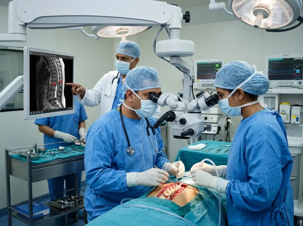
Idiopathic spinal cord herniation is a rare pathology presenting with progressive thoracic myelopathy. Early MRI identification and meticulous microsurgical reduction with dual bolstered dural defect repair prevent irreversible neurological deterioration.
Today

A novel smartphone-assisted biosensor utilizing molecularly imprinted polymers and catalytic nanozymes offers antibody-free, enzyme-free urinary microalbumin testing, enabling rapid, low-cost point-of-care screening for early chronic kidney disease.
Today
