.webp&w=384&q=75)

Loading, please wait...
Loading, please wait...
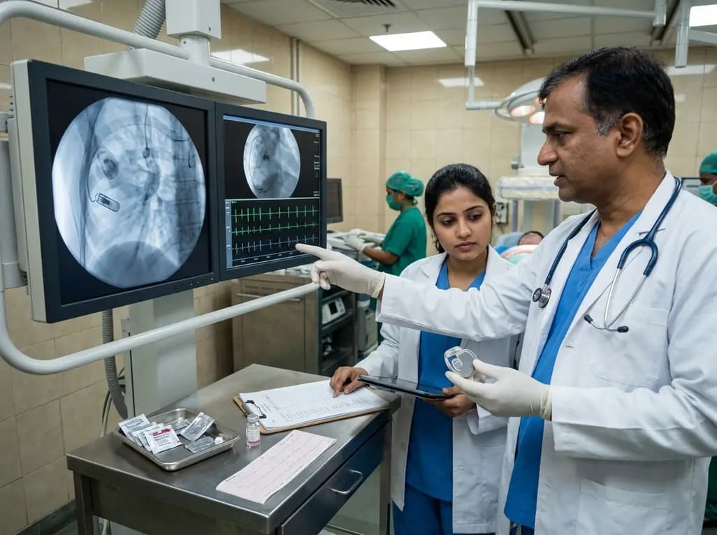
The atrial leadless pacemaker has emerged as a groundbreaking advancement in modern cardiac electrophysiology, offering physiological pacing without transvenous leads or subcutaneous generator pockets. Historically, conventional transvenous pacing systems carried notable clinical risks, including lead dislodgement, venous thrombosis, tricuspid valve regurgitation, and pocket hematomas or infections. Consequently, leadless systems significantly reduce these long-term hardware morbidities. However, anchoring a leadless capsule within the thin right atrial wall requires specialized active-fixation screw helix technology. Because atrial myocardium is thin and delicate, localized mechanical tissue trauma and an acute current of injury naturally occur upon helix deployment. Electrophysiologists commonly observe elevated pacing thresholds immediately after device fixation. Fortunately, these acute capture thresholds typically decline within twenty-four hours as acute myocardial trauma stabilizes. Nevertheless, unexpected sub-acute electrical instability can occasionally manifest after hospital discharge. Understanding these delayed threshold shifts is essential for cardiologists who manage modern leadless systems. Systematic post-implantation monitoring ensures clinicians accurately differentiate transient inflammatory responses from catastrophic mechanical dislodgement, avoiding unnecessary invasive interventions.
A notable clinical case recently highlighted the diagnostic dilemmas of delayed electrical volatility following leadless device placement. An eighty-year-old female with symptomatic sinus bradycardia underwent successful implantation of an active-fixation atrial leadless device. During the index procedure, the final pacing threshold measured 4.0 V at 0.4 ms. However, subsequent testing showed rapid electrical improvement. By post-operative day one, the capture threshold dropped to 1.0 V at 0.4 ms, where it remained stable through discharge on day four. Surprisingly, the patient presented back to the hospital on day five due to new-onset atrial flutter. Clinicians administered an intravenous dose of 150 mg amiodarone for rhythm control. Following drug infusion, device interrogation revealed complete loss of atrial capture at maximum output. Serial telemetry over the next three days confirmed pacing thresholds exceeding 6.0 V at 1.5 ms. Because recurrent sinus asystole caused severe dizziness, physicians implanted a conventional DDD pacemaker for immediate backup pacing. This presentation demonstrates how rapidly stable early thresholds can deteriorate during sub-acute recovery.
The underlying mechanisms driving sub-acute pacing threshold spikes involve complex tissue dynamics at the electrode-myocardium interface. When the fixation helix engages the atrial myocardium, localized mechanical micro-trauma triggers microvascular bleeding and focal tissue edema. Consequently, local electrical resistance rises, temporarily increasing pacing thresholds. While this initial inflammation generally subsides within forty-eight hours, secondary inflammatory reactions can occasionally develop. Microscopic peri-electrode edema creates a physical barrier between the stimulating electrode and excitable atrial myocytes. Therefore, the delivered electrical impulse attenuates markedly before depolarizing viable myocardium. Furthermore, local cellular hypoxia and inflammatory cytokines alter membrane excitability, resulting in transient inexcitability. Although loss of capture raises immediate concerns regarding helix dislodgement, anatomical position often remains completely stable on imaging. Because myocardial healing and fibrous maturation require several weeks, these electrical disturbances frequently demonstrate complete reversibility over time. Recognizing this natural pathophysiological trajectory allows clinicians to avoid hasty, high-risk retrieval procedures.
Pharmacological agents can significantly influence cardiac conduction tissue and pacing thresholds. In the reported case, sudden loss of capture developed immediately after intravenous amiodarone administration. Amiodarone acts as a broad-spectrum antiarrhythmic drug with potent class I sodium channel blocking properties. By suppressing fast inward sodium channels, amiodarone reduces myocardial excitability and slows conduction velocity. Consequently, the energy required to achieve myocardial depolarization increases considerably. When newly injured, edematous atrial tissue encounters acute sodium channel blockade, the pacing threshold can elevate dramatically. In uninjured myocardium, this drug effect remains clinically silent; however, at an inflamed helix fixation site, it can induce complete pacing failure. Furthermore, rate-dependent threshold variations and phase 4 depolarization abnormalities can exacerbate pacing unresponsiveness during bradycardia episodes. Electrophysiologists must carefully weigh the risks when administering intravenous membrane-stabilizing agents to patients with recently implanted leadless devices. Recognizing drug-induced threshold spikes prevents misattributing temporary pharmacological capture failure to permanent device malfunction.
Managing severe sub-acute pacing threshold elevations requires balancing patient safety with conservative clinical restraint. If a patient remains hemodynamically stable without severe bradycardia, watchful waiting with continuous telemetry represents the optimal strategy. When profound sinus arrest or symptomatic asystole develops, temporary backup pacing provides vital safety. In this case, clinicians strategically implanted a transvenous DDD pacemaker as a temporary safety net while leaving the leadless device intact. Remarkably, at the one-month follow-up, the atrial leadless threshold spontaneously normalized to an optimal 0.5 V at 0.4 ms. Subsequent Holter monitoring confirmed robust, uninterrupted atrial capture after lowering the transvenous pacemaker base rate. At three months, pacing thresholds remained stable at 0.5 V. This outcome underscores that leadless threshold elevations may persist beyond one week but normalize within thirty days. Electrophysiologists should therefore favor conservative monitoring over immediate device extraction, ensuring patient safety while preserving leadless device functionality.
Pacing thresholds often elevate due to localized mechanical trauma, peri-electrode edema, and cellular inflammation caused by active helix fixation. When the helix enters the atrial myocardium, tissue injury creates electrical resistance. As local tissue swelling peaks, the electrical signal struggles to reach excitable myocytes. Consequently, capture thresholds rise temporarily before declining as the acute inflammatory cascade naturally resolves over subsequent weeks.
Amiodarone exerts potent sodium and potassium channel blocking properties that stabilize myocardial cell membranes and decrease tissue excitability. In the setting of fresh peri-electrode myocardial inflammation, these pharmacological effects can substantially elevate the stimulation energy required for capture. Consequently, intravenous amiodarone administration may trigger a severe, transient threshold spike, leading to complete loss of pacing capture in recently implanted leadless devices.
Clinicians should adopt a conservative approach with continuous hemodynamic monitoring if threshold elevation is suspected to stem from peri-electrode inflammation. If severe symptomatic bradycardia or asystole emerges, temporary transvenous backup pacing provides vital hemodynamic support. Electrophysiologists should avoid hasty leadless device retrieval, as capture thresholds frequently normalize to optimal levels within one month as myocardial tissue fully heals.
Disclaimer: This content is for informational and educational purposes only. It is not intended to be a substitute for professional medical advice, diagnosis, or treatment. Always seek the advice of your physician or other qualified health provider with any questions you may have regarding a medical condition. Refer to the latest local and national guidelines for clinical practice.
References

Read summarized clinical updates, watch expert medical content, and earn CME certifications right from your smartphone.
Open your smartphone camera to scan and install.





A clinical case illustrates sub-acute re-elevation of pacing thresholds after atrial leadless pacemaker implantation, triggered by peri-electrode inflammation and amiodarone, which spontaneously normalized within one month under conservative clinical management.
4 weeks back

With post-monsoon dengue surges threatening South Asian health systems, physicians must refine triage and management. This guide covers vital warning signs, plasma leakage detection, crystalloid titration, and vector containment strategies to prevent mortality during seasonal arboviral epidemics.
Today

Findings from Project Viva indicate that sustained postpartum weight retention, rather than excessive gestational weight gain alone, directly worsens women's mid-life cardiovascular health, degrading blood pressure and glycemic parameters decades after delivery.
Today

A large UK Biobank study reveals that plasma proteomic signatures capture preclinical organ damage and improve multiorgan risk prediction across early CKM syndrome stages 0 to 2 beyond traditional PREVENT clinical models.
Today

A retrospective CBCT study reveals that superior TMJ space asymmetry correlates moderately with occipital condyle-atlas-axis asymmetry in unilateral TMJ osteoarthritis, highlighting an imaging correlate between jaw biomechanics and upper cervical alignment rather than a standalone diagnostic marker.
Today

A Bayesian multilevel meta-analysis reveals that aerobic training combined with moderate carbohydrate restriction modestly lowers HbA1c in type 2 diabetes. However, sparse data and very low certainty leave incremental benefits over exercise or diet alone unproven, highlighting the need for individualized care.
Today
