.webp&w=384&q=75)

Loading, please wait...
Loading, please wait...
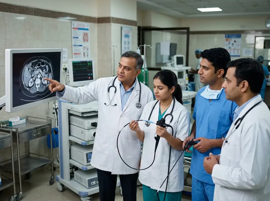
Obscure gastrointestinal bleeding presents a complex diagnostic hurdle for clinicians worldwide because the small intestine remains anatomically challenging to evaluate completely. Patients frequently present with melena or hematochezia, which indicates significant luminal hemorrhage. Standard endoscopic procedures, such as esophagogastroduodenoscopy and colonoscopy, often fail to identify the precise source of hemorrhage when massive blood pooling obscures visualization. Consequently, physicians must adopt a structured, systematic protocol to locate deep small bowel lesions effectively. A Meckel's diverticulum represents a classic yet easily missed etiology in young adults presenting with lower intestinal hemorrhage. This congenital anomaly arises from incomplete obliteration of the vitelline duct during embryonic development. Because ectopic gastric mucosa inside the diverticulum secretes acid, adjacent ileal tissue suffers ulceration and extensive bleeding. Therefore, timely localization becomes vital to prevent severe anemia, hemodynamic instability, and repeated emergency visits. Clinicians must quickly transition to non-invasive cross-sectional imaging when traditional upper and lower endoscopies remain non-diagnostic. By combining advanced imaging with endoscopic interventions, medical teams can rapidly identify hidden vascular or structural defects. Ultimately, establishing a clear diagnostic algorithm shortens patient suffering and prevents life-threatening complications.
When acute gastrointestinal hemorrhage persists without a clear source, abdominal computed tomography angiography serves as an essential preliminary diagnostic tool. This non-invasive modality quickly evaluates the abdominal vasculature and identifies specific structural abnormalities within the small intestine. In cases involving a bleeding Meckel's diverticulum, computed tomography angiography can demonstrate segmental hyper-enhancement or outline a distinct blind-ending diverticular pouch in the distal ileum. Moreover, high-resolution cross-sectional images provide clinicians with a reliable anatomical compass prior to invasive interventions. Consequently, gastroenterologists can determine whether an anterograde or retrograde endoscopic approach is appropriate based on precise lesion localization. Furthermore, multiplanar reconstructions allow radiologic teams to differentiate diverticular lesions from active vascular malformations, tumors, or inflammatory bowel disease. Identifying active contrast extravasation also helps confirm ongoing arterial bleeding in unstable patients. Thus, utilizing cross-sectional imaging as a primary screening tool minimizes diagnostic delay and reduces unnecessary exploratory operations. Furthermore, this roadmap guides subsequent endoscopic procedures with high accuracy, optimizing both patient safety and procedural efficiency.
Following preliminary localization via cross-sectional imaging, device-assisted enteroscopy provides direct visualization and therapeutic access to the small intestine. Specifically, retrograde double-balloon enteroscopy allows endoscopists to navigate the distal ileum and directly inspect suspicious mucosal abnormalities. In this clinical scenario, enteroscopic evaluation confirms the presence of the diverticulum alongside irregular, deep ulcerations showing recent stigmata of hemorrhage. Beyond visual confirmation, enteroscopy plays a crucial role in preoperative planning through precise lesion marking. Endoscopists can deploy titanium endoclips or inject mucosal dyes around the diverticular orifice to designate the exact lesion boundaries. Consequently, these endoscopic markers serve as visible targets for surgical teams during subsequent operative management. Furthermore, therapeutic maneuvers during enteroscopy, such as hemoclip application or epinephrine injection, can achieve temporary hemostasis in actively bleeding patients. This dual diagnostic and therapeutic capability stabilizes the patient prior to definitive surgical intervention. Consequently, combining enteroscopic evaluation with pre-surgical clip placement bridges the gap between non-invasive imaging and targeted surgical resection.
Surgical excision remains the definitive cure for symptomatic Meckel's diverticulum, particularly when complicated by recurrent ulceration and massive hemorrhage. Minimally invasive surgery offers significant advantages over conventional open laparotomy, including reduced postoperative pain, shorter hospital stays, and quicker recovery. However, identifying small bowel lesions during laparoscopy can prove challenging due to limited tactile feedback and extensive intestinal loops. Preoperative endoscopic marking with metallic clips effectively overcomes this obstacle by providing an unmistakable intraoperative target. Surgical teams can utilize intraoperative fluoroscopy or laparoscopic palpation to locate clips swiftly and accurately. As a result, surgeons can perform a targeted, tissue-sparing segmental small bowel resection rather than an extensive exploratory bowel mobilization. Preserving healthy small intestine reduces surgical trauma and lowers the risk of short-bowel syndrome or postoperative adhesion formation. Subsequently, primary end-to-end anastomosis restores intestinal continuity safely. Overall, combining enteroscopic clip localization with laparoscopic resection highlights the powerful synergy between gastroenterology and surgical discipline.
Postoperative management following minimally invasive small bowel resection heavily emphasizes rapid recovery and early restoration of physiological function. Implementing Enhanced Recovery After Surgery principles optimizes patient outcomes through standardized perioperative protocols. Specifically, multimodal analgesia minimizes opioid consumption, which in turn promotes early bowel motility and reduces postoperative ileus. Furthermore, clinicians encourage early enteral nutrition and mobilization to accelerate functional bowel recovery and preserve muscle mass. In patients treated for obscure gastrointestinal bleeding secondary to diverticular ulceration, successful surgical removal completely eliminates the risk of recurrent hemorrhage. Follow-up evaluations confirm steady hemoglobin recovery without the need for additional blood transfusions. Additionally, multidisciplinary collaboration between gastroenterologists, radiologists, and surgeons ensures seamless transition from emergency presentation to outpatient recovery. Integrating non-invasive imaging, device-assisted enteroscopy, and targeted laparoscopic surgery establishes a safe, reproducible standard of care. Consequently, this structured pathway provides an effective roadmap for managing complex small bowel vascular and structural abnormalities safely.
Bleeding in a Meckel's diverticulum primary occurs due to mucosal ulceration driven by ectopic gastric tissue. This congenital abnormality often contains acid-secreting parietal cells that produce gastric acid within the small intestine. Consequently, the adjacent ileal mucosa suffers chronic irritation, leading to deep ulcerations, erosion of local blood vessels, and significant gastrointestinal bleeding. Prompt diagnostic identification is necessary to prevent severe acute anemia and recurrent hemorrhagic episodes.
Computed tomography angiography acts as an crucial anatomical compass by identifying the precise location and vascular nature of obscure small bowel lesions. Because the small intestine is exceptionally long, performing enteroscopy without prior imaging can lead to prolonged procedural times and diagnostic failure. Cross-sectional imaging helps clinicians determine whether an anterograde or retrograde enteroscopic route is appropriate, thereby maximizing diagnostic yield and improving patient safety during invasive procedures.
Endoscopic clip placement provides precise intraoperative localization for surgical teams during minimally invasive laparoscopy. Because small bowel diverticula or flat mucosal ulcers can be difficult to identify visually from the serosal surface, clips serve as radio-opaque or tactile markers. Consequently, surgeons can quickly locate the exact lesion using intraoperative fluoroscopy or gentle manipulation, allowing targeted, tissue-sparing bowel resection while minimizing unnecessary tissue dissection and operative duration.
Disclaimer: This content is for informational and educational purposes only and does not constitute medical advice, diagnosis, or treatment. Always seek the advice of a qualified healthcare provider with any questions regarding a medical condition. Refer to the latest local and national guidelines for clinical practice.
References

Read summarized clinical updates, watch expert medical content, and earn CME certifications right from your smartphone.
Open your smartphone camera to scan and install.





A multidisciplinary CTA-DBE-Laparoscopy pathway enabled successful diagnostic localization and targeted laparoscopic resection of a bleeding Meckel's diverticulum in a young adult presenting with severe lower intestinal hemorrhage.
last month

A landmark study clarifies the chemical basis of human PAD4 citrullination, demonstrating strict substrate selectivity driven by noncovalent interactions and active site desolvation. These findings illuminate epigenetic histone regulation and provide vital structural blueprints for selective inhibitor design in RA.
Today

A school-based intervention using the Health Action Process Approach (HAPA) significantly enhanced adolescents' perceived benefits of consulting healthcare providers and promoted proactive menstrual health behaviors, offering key clinical insights for managing adolescent dysmenorrhea and endometriosis.
Today
A comprehensive analysis of the Translational Maturity Framework for nanomedicine-device combination products, evaluating regulatory complexity, manufacturing hurdles, and clinical readiness across oncology, cardiology, and orthopedics.
Today

A cross-sectional study reveals that sarcopenic obesity independently increases functional impairment and severe disc degeneration in patients with degenerative lumbar spinal stenosis, highlighting the need for body composition assessment.
Today
A landmark dose-response meta-analysis of 8.8 million adults reveals that high ultra-processed food intake significantly elevates risks for cardiovascular disease, cancer, diabetes, depression, and all-cause mortality, highlighting urgent needs for clinical lifestyle interventions.
Today
