.webp&w=384&q=75)

Loading, please wait...
Loading, please wait...
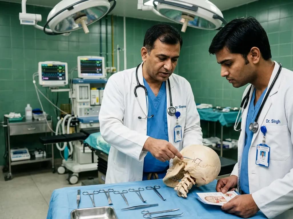
Surgical management of cerebellopontine angle pathology demands exceptional precision to protect critical neurovascular structures. The classical retrosigmoid route provides expansive visualization, yet traditional openings frequently incur significant soft tissue morbidity and prolonged operative duration. In recent years, minimally invasive skull base surgery has advanced rapidly. Consequently, the retrosigmoid keyhole craniotomy has emerged as an indispensable corridor for treating vestibular schwannomas, meningiomas, and microvascular compression syndromes. Nevertheless, surgeons have long faced challenges regarding precise external landmark localization and standardized osteotomy execution without risking venous sinus laceration. A landmark investigation by Liao and colleagues establishes an objective, reproducible protocol that refines keyhole access, bridging previous theoretical localization systems with dependable, everyday surgical execution.
The lateral suboccipital corridor offers direct line-of-sight visualization toward the internal acoustic meatus, petroclival region, and lower cranial nerves. However, performing an ultra-conservative keyhole opening requires uncompromising topographical accuracy. If the surgeon positions the bone window too far posteriorly, extensive cerebellar retraction becomes unavoidable, increasing the risk of contusion or cerebellar edema. Conversely, an excessively anterior burr hole directly imperils the transverse-sigmoid sinus junction. Although neuronavigation provides computerized orientation, anatomical shifts and registration inaccuracies can limit reliability during bony work. Therefore, neurosurgeons continuously seek surface bone landmarks that consistently correlate with intracranial vascular boundaries. Previous research delineated the "one point, two lines, two distances" localization concept to identify the burr hole target. However, translating this localization model into a standardized craniotomy remained an unaddressed surgical challenge until now.
To eliminate ambiguity during skull drilling, the researchers systematically analyzed cranial landmarks across cadaveric specimens and clinical cohorts. Specifically, the team focused on the superior point of the digastric sulcus as an anchor for orientation. Anatomical measurements confirmed that this distinct groove serves as a reliable external surrogate for the anterior margin of the sigmoid sinus. Because muscular attachments define this region, surgeons can readily palpate and visualize the landmark after elevating the suboccipital musculature. Furthermore, the relationship between the digastric sulcus and the transverse-sigmoid junction remains remarkably stable across diverse cranial morphologies. Consequently, integrating this skeletal reference into the initial surgical design removes guesswork. Operators no longer need to depend exclusively on inconsistent emissary foramen variants or ambiguous suture junctions such as the asterion, which frequently exhibits significant anatomical divergence from the underlying venous sinuses.
The standardized procedural technique begins with placing a precisely measured burr hole at the precalculated keypoint. Following dural separation, surgeons elevate the cranial bone flap sequentially in a controlled posterior-inferior-anterior trajectory. Importantly, the osteotomy cut traverses the superior point of the digastric sulcus, ensuring that the lateral margin precisely reaches the posterior border of the sigmoid sinus. The surgical team then addresses the bone surrounding the mastoid emissary vein. By utilizing a high-speed diamond burr under continuous cold irrigation, operators safely thin the bone around the emissary channel before completing hemostasis. Finally, clinicians resect residual bony ledges along the sinus margin using fine Kerrison rongeurs. This methodical workflow prevents uncontrolled dural tearing, minimizes unexpected venous hemorrhage, and maximizes the lateral working corridor toward the cerebellopontine angle cisterns.
The investigators rigorously validated this refined craniotomy protocol in both laboratory and clinical environments. Initially, researchers executed craniotomy simulations across 12 adult cadaveric skull specimens, consistently demonstrating complete exposure of the target dural margins without lacerating the venous sinuses. Subsequently, the surgical team applied this technique in 122 consecutive neurosurgical patients presenting with diverse cerebellopontine angle lesions. Every single operative case achieved a satisfactory, tailored bone window that delivered uncompromised microsurgical visualization. Most notably, the surgical team encountered zero intraoperative lacerations to the transverse sinus, sigmoid sinus, or surrounding critical vascular structures. In addition, meticulous perisinus bony resection eliminated the need for aggressive cerebellar retraction, fostering smoother postoperative recovery, reducing wound morbidity, and preventing excessive cerebrospinal fluid accumulation.
For modern skull base teams, this standardized technique provides a definitive roadmap for optimizing patient outcomes during cerebellopontine angle surgery. In tertiary centers where complex acoustic neuromas and microvascular decompressions occur routinely, procedural standardization significantly reduces operative anxiety and craniotomy time. Furthermore, establishing clear bone trajectory rules aids surgical education, allowing trainees to master safe posterior fossa access without arbitrary bone removal. By utilizing constant skeletal landmarks rather than relying solely on soft tissue signs, surgeons achieve consistent results regardless of anatomical variations. In summary, anchoring the keyhole osteotomy to the superior point of the digastric sulcus transforms retrosigmoid surgery into a truly reproducible, safe, and minimally invasive intervention.
The superior point of the digastric sulcus provides an exceptionally constant, palpable anatomical reference for the posterior margin of the sigmoid sinus. Unlike the asterion, which displays variable relationships with underlying sinuses, this landmark enables neurosurgeons to place osteotomy cuts safely along the sinus border without causing accidental vascular injury or relying solely on computerized neuronavigation.
By placing the bone window directly adjacent to the transverse-sigmoid sinus junction, the approach aligns the surgical corridor parallel to the petrous face. Consequently, releasing cerebrospinal fluid from the lateral cerebellomedullary cistern achieves rapid cerebellar relaxation. This dynamic exposure allows surgeons to manipulate deep microsurgical instruments along the skull base while avoiding fixed mechanical cerebellar retractors entirely.
Surgeons must avoid rapid or blind rongeuring near the mastoid emissary vein, which can tear the adjacent sigmoid sinus wall. Instead, operators should deploy a high-speed diamond burr with copious irrigation to thin the surrounding bone deliberately. Once exposed, the emissary vessel can be coagulated or sealed with bone wax before completing sinus margin decompression.
Disclaimer: This content is for informational and educational purposes only... Refer to the latest local and national guidelines for clinical practice.
References
Liao CC et al. Further Study of Craniotomy in Suboccipital Retrosigmoid Keyhole Approach. J Craniofac Surg. 2026 Sep 15. doi: 10.1097/SCS.0000000000013382. PMID: 42743543.
Jian ZH, Sheng MF, Li Z, Li Y, Weng W, Chen G. Precise Localization in Craniotomy With a Retrosigmoid Keyhole Approach: Microsurgical Anatomy and Clinical Study. Front Surg. 2022;9:809098. doi: 10.3389/fsurg.2022.809098.
Rhoton AL Jr. The cerebellopontine angle and posterior fossa cranial nerves by the retrosigmoid approach. Neurosurgery. 2000;47(3 Suppl):S93-S129. doi: 10.1097/00006123-200009001-00013.

Read summarized clinical updates, watch expert medical content, and earn CME certifications right from your smartphone.
Open your smartphone camera to scan and install.





A landmark study validates standardized retrosigmoid keyhole craniotomy anchored by the superior point of the digastric sulcus. Evaluating 12 cadaveric heads and 122 patients, researchers demonstrated safe bone flap elevation, excellent cerebellopontine angle exposure, and zero neurovascular complications.
Today
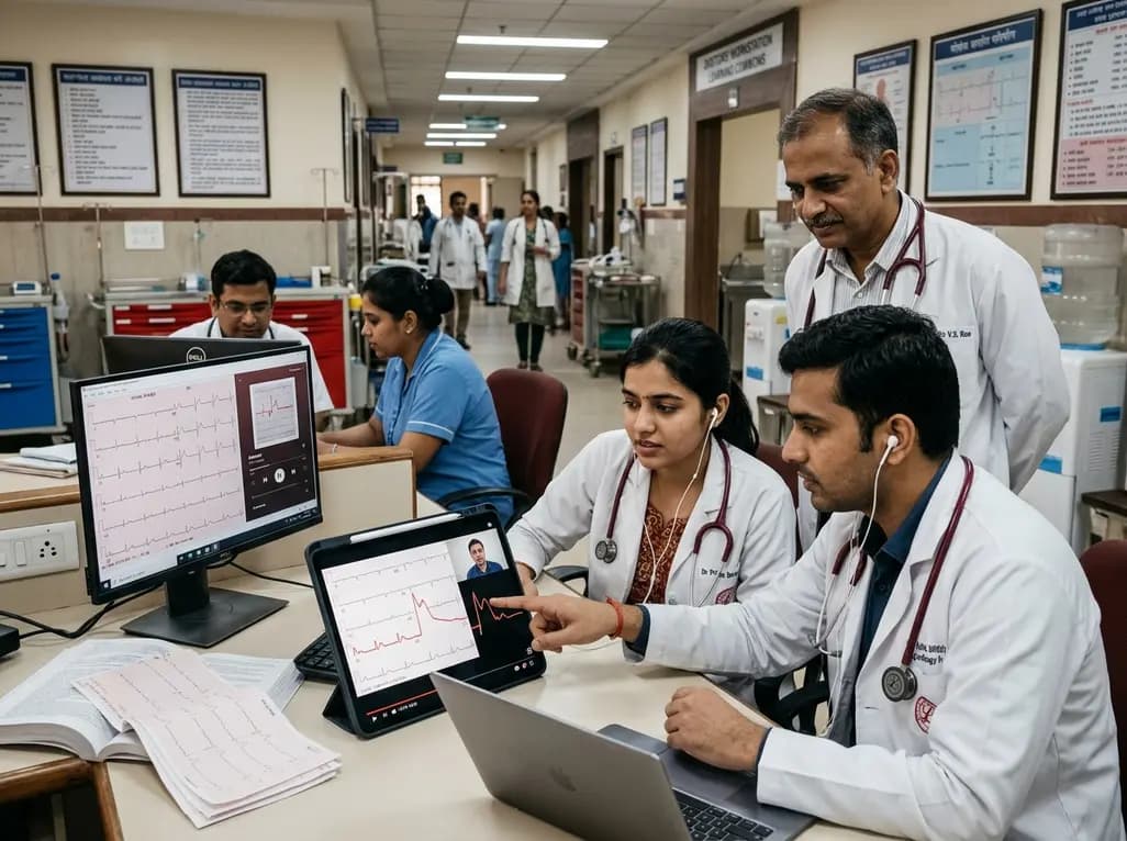
A randomized controlled trial demonstrates that curated FOAMed videos and podcasts produce superior gains in high-risk ECG interpretation and acute coronary syndrome clinical decision-making compared with print-based materials among interprofessional emergency care teams.
Today

Intimate partner violence during pregnancy significantly increases the risk of miscarriage, premature membrane rupture, and fetal growth restriction. This clinical review explores epidemiological insights, maternal depression, medico-legal documentation, and universal antenatal screening strategies.
Today

A Bayesian network meta-analysis evaluates exercise training in HFpEF, demonstrating that combined aerobic and resistance training, HIIT, and moderate continuous training significantly enhance peak VO2, while functional electrical stimulation uniquely improves quality of life without altering cardiac remodeling.
Today
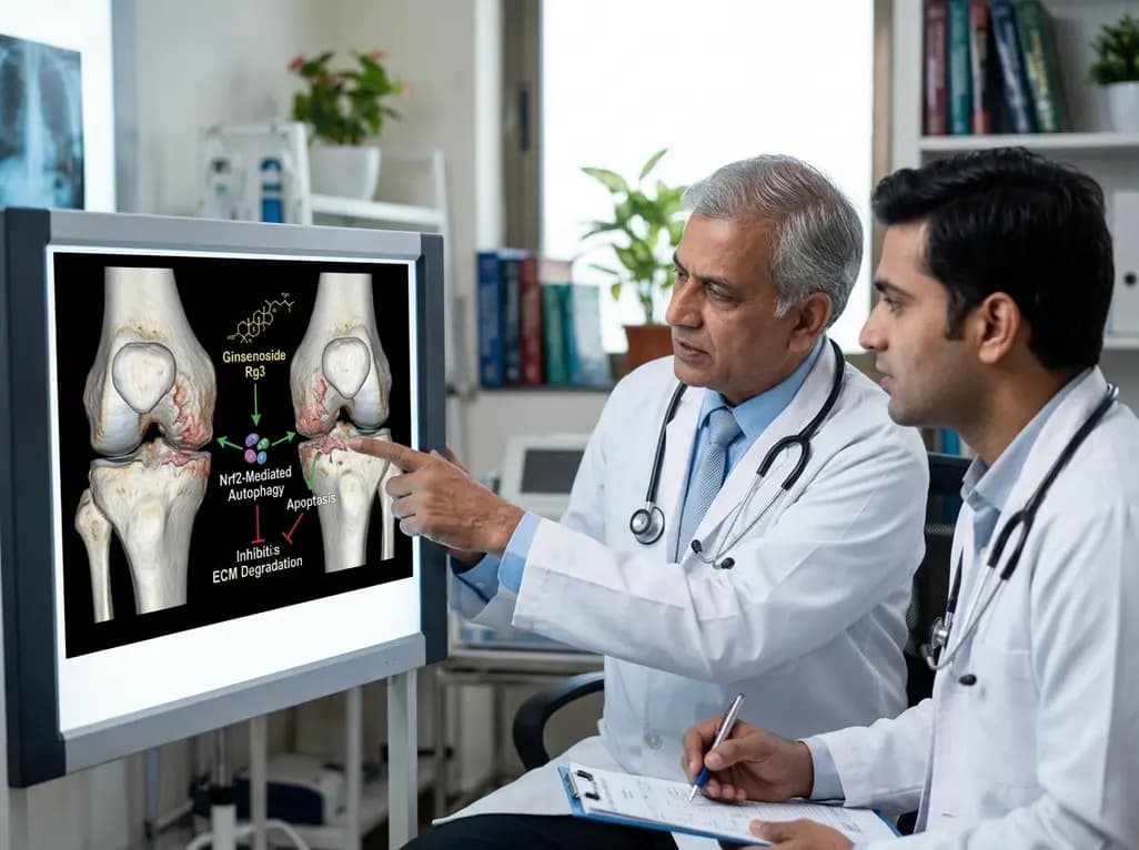
A study reveals that Ginsenoside Rg3 attenuates osteoarthritis progression by modulating the Nrf2-mediated autophagy pathway, reducing chondrocyte apoptosis, and curbing extracellular matrix degradation, offering promising chondroprotective potential.
Today

A cross-sectional study demonstrates that lower pediatric motor competence strongly correlates with adverse metabolic syndrome markers, including central adiposity, elevated triglycerides, and low HDL cholesterol, emphasizing early neuromuscular interventions in youth with overweight and obesity.
Today
