.webp&w=384&q=75)

Loading, please wait...
Loading, please wait...

Surgical resection of oral squamous cell carcinoma frequently necessitates bone conservation when oncologic margins allow. In these scenarios, marginal mandibulectomy reconstruction preserves mandibular continuity while achieving adequate disease clearance. Although microvascular free flaps represent the standard approach for composite oral defects, they carry prolonged operative times and substantial donor morbidity. Consequently, reconstructive surgeons increasingly evaluate simpler, tissue-sparing options for selected bone defects. Recent evidence indicates that split-thickness skin grafts placed directly onto cortical bone provide a durable, reliable epithelial barrier. This reconstructive strategy significantly shortens operative duration while avoiding complex microvascular interventions. Furthermore, it restores essential mucosal barriers and establishes a stable foundation for functional recovery.
Marginal mandibulectomy removes the involved alveolar cortex while preserving the inferior border of the mandible. Therefore, the structural continuity of the lower jaw remains completely intact, preventing facial collapse and functional deviation. However, resecting the alveolar margin leaves exposed cortical bone and adjacent soft tissue margins. Historically, surgeons believed that skin grafts required a vascularized periosteal bed to achieve adequate imbibition and neovascularization. Recent surgical series decisively challenge this dogma. Clinical teams now demonstrate that split-thickness skin grafts survive predictably when secured directly against properly prepared cortical bone. Cortical perforations and adjacent soft tissue margins supply robust vascular ingrowth, enabling rapid graft incorporation. In addition, avoiding bulky free tissue transfer preserves natural oral sulcus depth and tongue mobility. Consequently, patients retain superior articulation and oral competence. Surgeons can successfully employ this conservative approach in diverse anatomical sites, including the floor of mouth and the gingivobuccal sulcus.
Achieving successful graft take directly over bone demands meticulous surgical technique and rigid immobilization. First, the surgical team performs the oncologic resection with clear margins using sagittal saws or rotary instruments. Next, the operating surgeon smoothes sharp osseous ledges and creates fine decortication holes to promote punctate bleeding. The surgeon then harvests a split-thickness skin graft, typically from the anterolateral thigh, at a thickness of 0.012 to 0.015 inches. Subsequently, the surgical team insets the graft and secures peripheral edges with absorbable sutures to establish intimate contact. Rigid bolstering remains the cornerstone of successful integration. Clinicians utilize prefabricated acrylic partial dentures or intraoperatively shaped silicone and foam stents to exert continuous pressure. In addition, circummandibular retaining sutures maintain bolster stability against masticatory forces and salivary shearing. The surgical team typically leaves the bolster in place for five to seven days. This undisturbed stabilization supports undisturbed capillary ingrowth during the critical phases of revascularization.
Clinical data demonstrate exceptional reliability when utilizing split-thickness skin grafts for alveolar defects. In a recent five-year clinical series, all eleven treated patients achieved successful primary graft take without total graft loss. Minor complications occurred in four patients, predominantly manifesting as localized cortical bone exposure and mild donor site discomfort. Fortunately, clinicians managed all minor wound issues conservatively through chlorhexidine rinses and selective debridement without secondary surgery. Because the skin graft produces a thin, non-redundant epithelial lining, the reconstructed alveolus exhibits minimal redundancy. Therefore, the wound bed resists maceration and hyperkeratotic breakdown over extended surveillance periods. Furthermore, postoperative healing progresses rapidly, which substantially reduces initial hospital stays compared to microvascular tissue transfers. Patients resume protective oral hygiene within two weeks, ensuring clean intraoral conditions. Thus, this reconstructive technique provides reliable epithelial coverage while maintaining low morbidity profiles across diverse patient groups.
Preserving oral function constitutes a fundamental goal of head and neck reconstructive oncology. Bulky fasciocutaneous flaps often crowd the oral cavity, which impairs lingual excursion and alters articulation. In contrast, split-thickness skin grafts adhere tightly to the alveolar architecture, preventing obliteration of the buccal and lingual sulci. As a result, patients maintain physiologic tongue elevation, lateral movement, and dynamic swallowing mechanics. Nutritional intake resumes swiftly, enabling rapid transition from enteral tube feeding to an unrestricted oral diet. Moreover, the thin grafted surface establishes an ideal foundation for prosthetic dental rehabilitation. Patients desiring restoration can receive either removable tissue-borne dentures or osseointegrated dental implants. The attached, immobile graft tissue resists mechanical shearing from prostheses during mastication. Consequently, dental prosthodontists encounter favorable ridge contours for appliance retention, thereby enhancing long-term masticatory performance and overall quality of life.
Careful patient selection dictates long-term success in marginal mandibulectomy reconstruction. This technique suits patients presenting with superficial oral cavity tumors that abut the alveolar ridge without gross cortical invasion. In addition, medically compromised individuals who cannot tolerate lengthy general anesthesia derive profound benefits from this expedited approach. Advanced age, severe cardiopulmonary comorbidities, and peripheral vascular disease frequently increase microvascular flap failure risks. Under such circumstances, skin grafting provides an efficient, low-risk alternative that eliminates microsurgical stress. However, surgeons must evaluate previous radiotherapy carefully, as irradiated bone exhibits reduced osteogenic and capillary budding capacity. When extensive full-thickness soft tissue resections accompany the ostectomy, vascularized tissue flaps remain necessary to prevent orocutaneous fistulae. Nevertheless, for isolated alveolar ridge and sulcus defects, direct skin grafting represents an invaluable, tissue-sparing reconstructive modality.
The skin graft initially survives through plasma imbibition from wound margins and bleeding bony pores. Within several days, active neovascularization establishes capillary connections from punctate cortical perforations, surrounding periosteum, and adjacent vascularized mucosa. Rigid bolster stabilization prevents micromotion, thereby ensuring uninterrupted capillary budding into the dermal matrix until complete graft incorporation occurs.
Surgeons frequently use prefabricated acrylic dental splints, sterile foam bolsters, or elastomer stents lined with antiseptic gauze. These bolsters are secured firmly using circummandibular sutures or tie-over silk ligatures. This arrangement maintains continuous, uniform pressure against the osseous contour, which successfully prevents fluid pooling, hematoma formation, and graft shear during postoperative swallowing.
Yes, patients can successfully undergo dental rehabilitation with osseointegrated implants following complete healing of the skin graft. Because the graft provides a thin, immobile epithelial layer adhering firmly to underlying cortical bone, it mimics attached keratinized gingiva. This stable environment resists mechanical breakdown, allowing prosthodontists to place endosseous implants or construct stable removable dental prostheses.
Disclaimer: This content is for informational and educational purposes only... Refer to the latest local and national guidelines for clinical practice.
References

Read summarized clinical updates, watch expert medical content, and earn CME certifications right from your smartphone.
Open your smartphone camera to scan and install.





Split-thickness skin grafts placed directly on cortical bone offer an efficient, reliable alternative to free flaps for marginal mandibulectomy reconstruction, preserving speech and swallowing while minimizing donor morbidity.
Today
Explore evidence-based recommendations on enteral nutrition in dementia. Understand when tube feeding is appropriate, its risks in advanced neurocognitive disorders, and why individualized oral feeding and caregiver communication remain essential cornerstones of palliative nutritional care.
Today

The government of Maharashtra is establishing a novel state-level Living Lab to elevate clinical outcomes, bridge public-private service gaps, and test real-world health interventions. By integrating digital innovations and frontline feedback, the platform fosters sustainable, equitable care models across communities.
Today
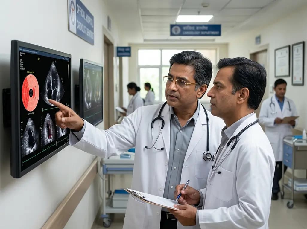
Cardiac involvement is the leading cause of mortality in EGPA. This report highlights an overlap phenotype of lymphocyte-predominant myocardial injury, Takotsubo-like cardiomyopathy, and left ventricular thrombus, emphasizing early multimodality diagnostics and aggressive immunosuppressive biologic therapy.
Today
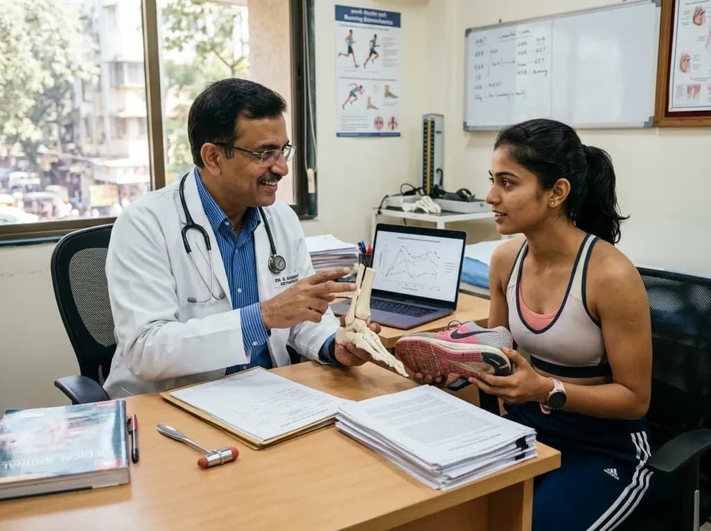
A landmark biomechanical study demonstrates that sports bra support and running shoe cushioning interact significantly to influence tibial acceleration, breast motion, and comfort in female runners, providing crucial insights for clinical counseling in sports medicine and women's health.
Yesterday
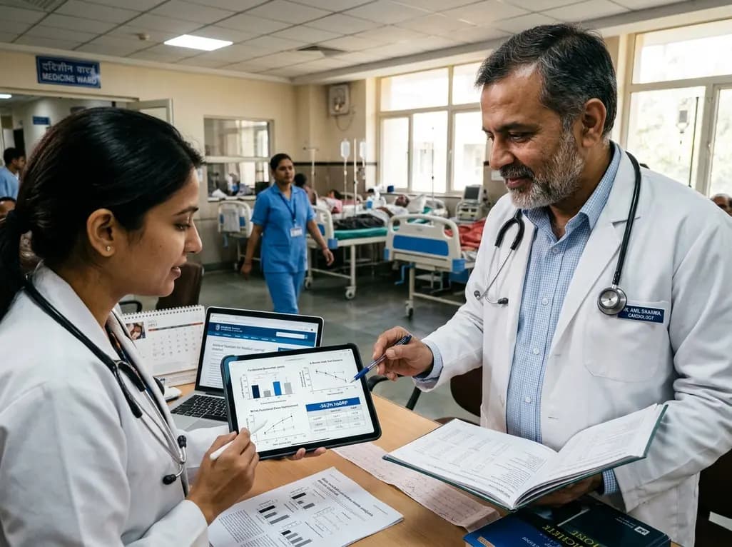
A systematic review and meta-analysis of 5,406 participants reveals that tirzepatide significantly reduces hsCRP by 34.3%, improves cardiorenal biomarkers, and enhances functional capacity and exercise endurance in patients with heart failure.
Today
