.webp&w=384&q=75)

Loading, please wait...
Loading, please wait...

Eosinophilic granulomatosis with polyangiitis represents a rare, systemic necrotizing vasculitis affecting small-to-medium vessels. Clinicians broadly recognize that cardiac manifestations of EGPA account for the primary source of patient mortality. Consequently, identifying myocardial involvement early determines overall survival. Although asthma, allergic rhinosinusitis, and peripheral blood hypereosinophilia traditionally herald the disease, cardiac involvement develops in substantial subsets of patients. In particular, antineutrophil cytoplasmic antibodies negative individuals demonstrate a predilection for profound myocardial disease. Classically, eosinophilic myocarditis drives this injury through the local degranulation of toxic eosinophil proteins. These cytotoxic mediators cause direct cardiomyocyte damage, microvascular thrombosis, and diffuse tissue necrosis. However, recent evidence suggests that the spectrum of myocardial disease is far more heterogeneous than previously recognized. Some patients present without typical necrotizing tissue lesions, exhibiting instead atypical inflammatory infiltrates. Therefore, clinicians must maintain high suspicion when evaluating cardiorespiratory complaints in patients with eosinophilic disorders. Recognizing these diverse clinical features prevents catastrophic progression toward irreversible fibrotic remodeling and refractory systolic heart failure. Furthermore, urgent multidisciplinary collaboration between cardiologists and rheumatologists remains essential during the early diagnostic phase to optimize clinical outcomes.
A newly documented clinical presentation illustrates an overlap phenotype uniting acute inflammatory injury, Takotsubo-like apical ballooning, and intracardiac thrombus. In this remarkable presentation, a middle-aged woman with systemic EGPA experienced severe, transient left ventricular systolic dysfunction. Echocardiography demonstrated regional wall-motion abnormalities mirroring classical stress cardiomyopathy. However, systemic inflammation accompanied the acute mechanical collapse, highlighting an intricate interaction between systemic stress and immune-mediated vascular injury. Moreover, clinicians identified an apical left ventricular thrombus, which added significant thromboembolic jeopardy to her acute instability. Severe apical hypokinesia frequently creates blood stasis, which accelerates platelet aggregation and fibrin deposition within the ventricle. In addition, localized endothelial damage from circulating inflammatory cytokines amplifies intracavitary hypercoagulability. Although Takotsubo syndrome typically presents after intense physical or emotional triggers, inflammatory cascades can equally provoke catecholaminergic myocardial toxicity. Consequently, systemic vasculitis directly induces microvascular spasm and profound sympathetic dysregulation. This overlap phenotype demonstrates that transient neurohormonal myocardial stunning and direct autoimmune cytotoxicity can coexist simultaneously. Therefore, clinicians should consider both inflammatory myocarditis and neurogenic stress patterns when evaluating acute hemodynamic failure in EGPA.
Endomyocardial biopsy provides essential definitive information when systemic vasculitis compromises cardiac performance. Historically, pathologists expect dense eosinophilic infiltrates with myocardial necrosis in such presentations. Surprisingly, tissue samples in this overlap phenotype revealed lymphocyte-predominant myocardial inflammation rather than classic eosinophilic dominance. This unexpected histologic finding reveals distinct cellular mechanisms underlying myocardial injury in systemic vasculitis. Specifically, activated T-lymphocytes can drive localized cytotoxic responses, cytokine production, and subsequent cellular necrosis independently of heavy eosinophil accumulation. Furthermore, rapid peripheral clearance or prior corticosteroid exposure might alter tissue cellularity before biopsy procurement. Consequently, the absence of prominent eosinophils on biopsy does not eliminate EGPA as the primary cause. This diagnostic nuance carries profound therapeutic significance for practicing clinicians. If clinicians rely solely on classical eosinophil histology, they may delay aggressive immunosuppression. In addition, recognizing lymphocytic infiltrates prompts clinicians to explore broad immunomodulatory therapies that target varied immune cell populations. Therefore, physicians must interpret cardiac biopsies within the comprehensive context of systemic multiorgan involvement. Integrating cutaneous, neurological, and serological findings ensures accurate diagnosis despite atypical myocardial histopathology.
Timely diagnosis demands an integrated diagnostic strategy employing advanced non-invasive and invasive cardiac testing. Electrocardiography often reveals non-specific repolarization abnormalities, conduction defects, or ischemic patterns that mislead physicians. Transthoracic echocardiography serves as an indispensable first-line modality, immediately identifying wall-motion defects, ejection fraction depression, and apical thrombi. Furthermore, cardiac magnetic resonance imaging delivers unmatched tissue characterization. Through T2-weighted imaging and late gadolinium enhancement, magnetic resonance detects active myocardial edema and focal fibrotic replacement. Consequently, clinicians can differentiate between ischemic injury and patchy, non-ischemic autoimmune damage. In parallel, comprehensive systemic assessment must corroborate the diagnosis of EGPA. Clinicians should systematically document peripheral eosinophilia, evaluate mononeuritis multiplex through electromyography, and biopsy suspicious cutaneous lesions. Moreover, coronary angiography remains vital to exclude acute atherothrombotic coronary occlusions during acute presentation. When hemodynamic instability persists, endomyocardial biopsy offers conclusive confirmation while excluding infectious viral etiologies. In addition, serial biomarker tracking using cardiac troponin and natriuretic peptides offers practical utility for monitoring active disease activity and treatment response. Therefore, coordinating multimodality imaging and histological evaluation protects patients from misdiagnosis and inappropriate interventions.
Managing severe cardiac involvement in EGPA requires prompt, aggressive immunosuppressive therapy to prevent irreversible myocardial remodeling. Clinicians initiate high-dose intravenous pulse methylprednisolone to rapidly quell inflammatory cascades and stabilize endothelial function. However, steroid therapy alone cannot ensure long-term disease control or prevent fatal relapses. Thus, international guidelines strongly advocate adding cyclophosphamide or rituximab for severe, organ-threatening disease manifestations. Rituximab efficiently depletes B-cells, dampening autoimmune vascular inflammation and inducing rapid disease remission. Furthermore, the advent of targeted anti-interleukin-5 therapies, notably mepolizumab, has revolutionized maintenance regimens. By directly neutralizing interleukin-5, mepolizumab blocks eosinophil maturation, recruitment, and survival. Combination therapy using rituximab and mepolizumab achieves synergistic suppression of both lymphocytic and eosinophilic arms of the disease. In addition, clinicians must carefully manage left ventricular thrombi using therapeutic anticoagulation until ventricular function recovers. Heart failure medical therapies, including beta-blockers and angiotensin pathway inhibitors, also promote reverse remodeling. Moreover, periodic clinical evaluations and serial echocardiographic assessments help clinicians safely taper immunosuppression without risking disease reactivation. Consequently, this multi-targeted pharmacological approach rescues severe systolic impairment and secures favorable long-term cardiovascular prognosis.
Cardiac involvement affects approximately fifteen to thirty percent of patients diagnosed with EGPA. It represents the leading cause of mortality in this condition. Clinicians frequently encounter pericarditis, myocarditis, coronary vasospasm, and progressive heart failure. Notably, individuals with negative antineutrophil cytoplasmic antibodies exhibit cardiac manifestations more frequently than antibody-positive patients. Therefore, baseline cardiac screening remains essential for all newly diagnosed individuals.
Both conditions often present with acute chest pain, elevated cardiac biomarkers, and severe left ventricular systolic dysfunction. Furthermore, systemic inflammation and intense physical stress stimulate severe catecholamine surges, precipitating transient apical ballooning. In overlap presentations, localized myocardial inflammation and neurohormonal stunning happen concurrently. Consequently, differentiating between pure stress cardiomyopathy and primary inflammatory myocarditis requires advanced imaging with cardiac magnetic resonance or histological endomyocardial biopsy.
Biologic agents play a transformative role in managing life-threatening manifestations of EGPA. While pulse corticosteroids provide initial control, clinicians use rituximab to deplete pathogenic B-cells and induce systemic remission. Subsequently, anti-interleukin-5 monoclonal antibodies, such as mepolizumab, selectively inhibit eosinophil production and survival. Deploying these targeted biologics together significantly improves left ventricular ejection fraction, prevents persistent relapses, and minimizes chronic glucocorticoid toxicity over time.
Disclaimer: This content is for informational and educational purposes only... Refer to the latest local and national guidelines for clinical practice.
References

Read summarized clinical updates, watch expert medical content, and earn CME certifications right from your smartphone.
Open your smartphone camera to scan and install.





Cardiac involvement is the leading cause of mortality in EGPA. This report highlights an overlap phenotype of lymphocyte-predominant myocardial injury, Takotsubo-like cardiomyopathy, and left ventricular thrombus, emphasizing early multimodality diagnostics and aggressive immunosuppressive biologic therapy.
Today
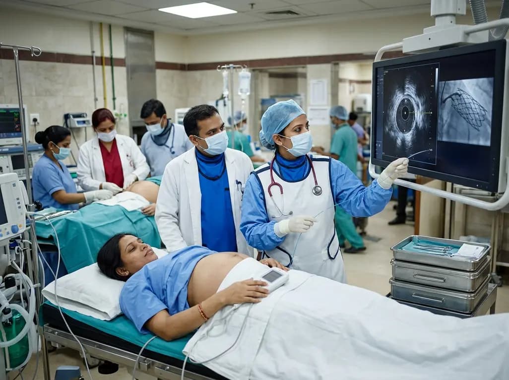
A 32-year-old gravida at 38 weeks with anterior STEMI underwent emergent IVUS-guided PCI and successful cesarean delivery under uninterrupted DAPT, highlighting cardio-obstetric care.
Today
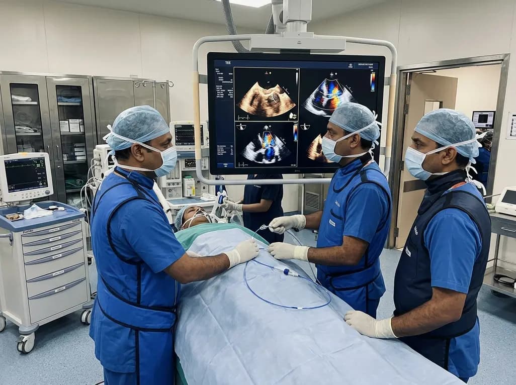
Transcatheter edge-to-edge repair (TEER) successfully treated severe mitral regurgitation from a posterior leaflet perforation. Guided by 3D multiplanar reconstruction, operators sandwiched the defect using two MitraClip devices, providing safe hemodynamic recovery without requiring open-heart surgery.
Today

A landmark biomechanical study demonstrates that sports bra support and running shoe cushioning interact significantly to influence tibial acceleration, breast motion, and comfort in female runners, providing crucial insights for clinical counseling in sports medicine and women's health.
Yesterday

A systematic review and meta-analysis of 5,406 participants reveals that tirzepatide significantly reduces hsCRP by 34.3%, improves cardiorenal biomarkers, and enhances functional capacity and exercise endurance in patients with heart failure.
Today
