.webp&w=384&q=75)

Loading, please wait...
Loading, please wait...
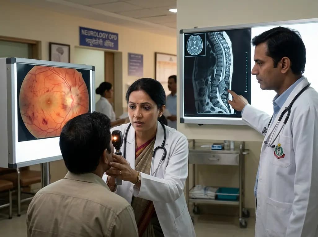
Clinicians frequently associate papilloedema with intracranial pathologies such as brain tumours, venous sinus thrombosis, or idiopathic intracranial hypertension. However, medical literature identifies a rare but critical association between spinal tumours and papilloedema. This presentation often occurs in the absence of ventriculomegaly or classical hydrocephalus, making it a significant diagnostic challenge for even the most experienced neurologists and ophthalmologists. When a patient presents with bilateral optic disc swelling but has a clear brain MRI, the spine is rarely the first place a physician looks. This oversight can lead to severe diagnostic delays, potentially resulting in irreversible visual impairment. Specifically, intradural spinal tumours can manifest with isolated vision-threatening papilloedema, where the only symptoms are visual obscurations and headaches. Consequently, understanding this unique clinical entity is paramount for preserving patient vision. Furthermore, the absence of limb neurology—such as weakness, sensory loss, or gait instability—can further mislead the initial diagnostic effort. Doctors must maintain a high index of suspicion and 'think outside the box' when intracranial causes are ruled out. By recognising spinal pathology as a potential differential for unexplained intracranial hypertension, healthcare providers can transform the patient journey from one of misdiagnosis to one of timely, life-changing intervention.
The exact pathophysiology of how a distal spinal lesion causes proximal intracranial hypertension remains a subject of intense clinical study. Most researchers agree that the mechanism involves complex cerebrospinal fluid (CSF) dynamics and protein concentration. Intradural spinal tumours, such as schwannomas or ependymomas, often secrete high levels of protein or breakdown products into the subarachnoid space. This secretion significantly increases the viscosity of the cerebrospinal fluid, creating a 'sludge' effect. Subsequently, this thickened fluid creates resistance at the arachnoid villi, which are the primary sites where CSF is absorbed into the venous system. When the absorption rate slows down, the intracranial pressure rises, even if there is no physical blockage within the brain itself. Additionally, some tumours may cause a 'communicating' hydrocephalus by interfering with the delicate balance of CSF production and resorption. Furthermore, venous stasis caused by tumour compression of the spinal venous plexuses may lead to an unfavourable hydrostatic pressure gradient. This pressure is then transmitted upward through the continuous subarachnoid space, leading to optic nerve head edema. Understanding these fluid dynamics is essential because it explains why a patient might have high opening pressures on a lumbar puncture despite having no visible brain lesions on neuroimaging.
One of the most treacherous aspects of spinal tumours and papilloedema is the potential lack of traditional spinal symptoms. Many clinicians expect a spinal tumour to present with limb weakness, paresthesia, or bladder dysfunction. However, in several documented case series, patients presented with isolated visual symptoms. This atypical presentation occurs because the tumour may be small or slow-growing, failing to compress the motor or sensory tracts significantly while still altering CSF composition. Consequently, the clinical focus remains stuck on the intracranial compartment. Patients may undergo multiple brain scans, lumbar punctures, and even treatments for idiopathic intracranial hypertension (IIH) without success. Moreover, if the CSF protein levels are high but the pressure is managed with medications like acetazolamide, the underlying tumour remains hidden. Therefore, it is vital to perform a full neuraxis MRI in any case of unexplained papilloedema where brain imaging is unremarkable. Specifically, contrast-enhanced imaging of the entire spine can reveal intradural extramedullary or intramedullary lesions that would otherwise be missed. Early detection through comprehensive imaging prevents the 'diagnostic odyssey' that many of these patients endure. Ultimately, recognizing that spinal pathology can masquerade as a primary brain disorder is a critical step in modern neuro-ophthalmic practice.
When vision-threatening papilloedema is identified, the immediate priority must be the preservation of sight. The acuity of visual deterioration often determines the clinical course. In cases where visual fields are rapidly constricting or visual acuity is plummeting, emergency cerebrospinal fluid (CSF) diversion is mandatory. This may involve the placement of an external ventricular drain (EVD) or a ventriculoperitoneal shunt, even before the spinal tumour is addressed surgically. While the tumour is the root cause, the immediate threat to the patient's quality of life is irreversible optic nerve damage. Furthermore, the management strategy must be tailored to the individual's degree of intracranial hypertension. For some patients, lumbar punctures provide temporary relief, but for those with severely elevated pressures, more permanent diversion is necessary. Physicians should also be aware that traditional treatments for IIH may be insufficient when high protein levels are the driving force behind the hypertension. Consequently, surgical intervention becomes the definitive treatment. However, the timing of these procedures requires careful coordination between the neurosurgical and ophthalmological teams. By prioritising the optic nerves, clinicians can buy the necessary time to plan and execute a safe spinal tumour resection without the risk of the patient waking up with permanent blindness.
The definitive treatment for intracranial hypertension caused by spinal tumours is the surgical resection of the lesion itself. Once the tumour is removed, the source of the excess protein or the mechanical obstruction is eliminated. In most cases, the CSF dynamics gradually normalise, leading to the resolution of papilloedema. However, the recovery of visual function depends heavily on the duration and severity of the pressure before intervention. If the optic nerves have already undergone significant atrophy, the visual gains may be limited. Therefore, post-operative monitoring by an ophthalmologist is essential to track the resolution of disc swelling and ensure that no residual hypertension remains. Additionally, long-term follow-up with spinal MRI is necessary to monitor for tumour recurrence, particularly with certain histologies like myxopapillary ependymomas. Furthermore, the psychological impact of such a diagnostic journey should not be underestimated. Patients who spent months seeking answers for their vision loss often require comprehensive support. Specifically, the success of the surgery is measured not just by the 'total resection' shown on a post-operative scan, but by the restoration or stabilisation of the patient's sight and quality of life. Modern neurosurgery, combined with early diagnostic awareness, offers these patients an excellent prognosis, provided the spinal origin is recognised early enough.
Navigating the complexities of unexplained disc swelling requires a structured and multidisciplinary approach. Clinicians must first rule out common intracranial causes using high-resolution MRI and MR venography. If these investigations are negative but papilloedema is confirmed, the next logical step is a lumbar puncture to measure opening pressure and CSF composition. Finding significantly elevated protein levels in the CSF should be a 'red flag' that points away from idiopathic causes and toward a potential spinal source. At this juncture, the healthcare team must order a contrast-enhanced MRI of the cervical, thoracic, and lumbar spine. Moreover, the importance of this 'thinking outside the box' approach cannot be overstated in a clinical setting where time is vision. Additionally, transition from conservative management to aggressive surgical planning is necessary if the vision continues to decline. Specifically, the integration of neuro-ophthalmology into the surgical planning phase ensures that visual field tests and optical coherence tomography (OCT) guide the urgency of the intervention. By adopting these clinical strategies, physicians can ensure that spinal tumours and papilloedema are diagnosed promptly. Ultimately, this proactive stance reduces the risk of permanent morbidity and ensures that the underlying pathology is addressed with the precision it requires. Consistent education regarding these rare presentations is the best defense against diagnostic delay in neurology.
Spinal tumours can significantly increase the protein content in the cerebrospinal fluid. This high protein concentration increases CSF viscosity and obstructs the arachnoid villi, which are responsible for CSF absorption. This results in raised intracranial pressure and subsequent papilloedema, even though the brain and ventricles appear normal on initial imaging.
Key indicators include unexplained papilloedema with a normal brain MRI, significantly elevated CSF protein levels on lumbar puncture, and persistent headaches. While limb weakness or sensory changes are traditional spinal signs, their absence does not exclude spinal pathology, making full neuraxis imaging essential in these cases.
The immediate priority is the preservation of vision. If visual deterioration is acute, emergency CSF diversion, such as shunting or drainage, may be required to lower intracranial pressure. Once the visual status is stabilised, the focus shifts to the surgical resection of the spinal tumour to resolve the underlying cause.
Disclaimer: This content is for informational and educational purposes only. It does not constitute medical advice or establish a doctor-patient relationship. Refer to the latest local and national guidelines for clinical practice.
References
Sun R et al. Intra-cranial hypertension and vision-threatening papilloedema caused by intradural spinal tumours: a case series of three. Br J Neurosurg. 2025 Aug. doi: 10.1080/02688697.2023.2263087. PMID: 37807636.
Petrovic D. Idiopathic Intracranial Hypertension and Vision: Understanding Papilledema and its Ocular Consequences. J Eye Dis Disord. 2025. 10:275. doi: 10.35248/2684-1622.25.10.275.
Xie JS et al. Papilledema: A review of etiology, pathophysiology, diagnosis, and management. Eye (Lond). 2022;36(1):1-10. doi: 10.1038/s41433-021-01746-z.

Read summarized clinical updates, watch expert medical content, and earn CME certifications right from your smartphone.
Open your smartphone camera to scan and install.





Discover how intradural spinal tumours can lead to vision-threatening papilloedema without brain lesions. This guide covers pathophysiology, diagnostic challenges, and the urgent need for vision preservation in unexplained intracranial hypertension.
2 weeks back
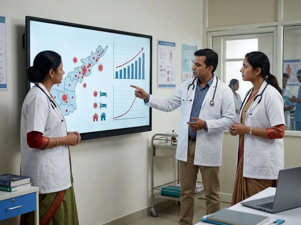
Andhra Pradesh reported 10 new Covid-19 cases, taking the state tally to 49 while deaths remain at four. With 24 patients hospitalized and 16 under home isolation, the Health Department has intensified monitoring. Medical professionals should review regional distribution, diagnostic protocols, and management plans.
Today

An 11-year Swedish registry study of 618 uterine sarcoma patients found that minimally invasive surgery yielded survival comparable to open surgery in early stages. However, adjuvant chemotherapy conferred no survival benefit in localized or advanced disease, highlighting stage and histology as key outcomes.
3 days back

A cross-sectional study evaluates post-intensive care syndrome in cardiac patients 2-4 weeks post-ICU discharge, highlighting cognitive, psychological, and functional impairments and the need for structured multidisciplinary rehabilitation.
3 days back

Anterior cruciate ligament reconstruction failure lacks uniform definition. A narrative review proposes an integrative framework incorporating objective and subjective instability, persistent pain, restricted motion, graft rupture, and secondary meniscal injury to standardize clinical reporting.
3 days back
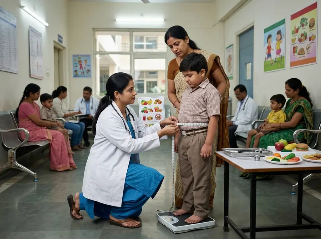
With World Obesity Atlas data warning that over 41 million Indian children are overweight or obese, ICMR and NIN have unveiled a 10-point policy roadmap. The initiative calls for mandatory front-of-pack labeling, HFSS taxes, strict marketing bans, and healthier school environments to curb non-communicable diseases.
Today
