.webp&w=384&q=75)

Loading, please wait...
Loading, please wait...
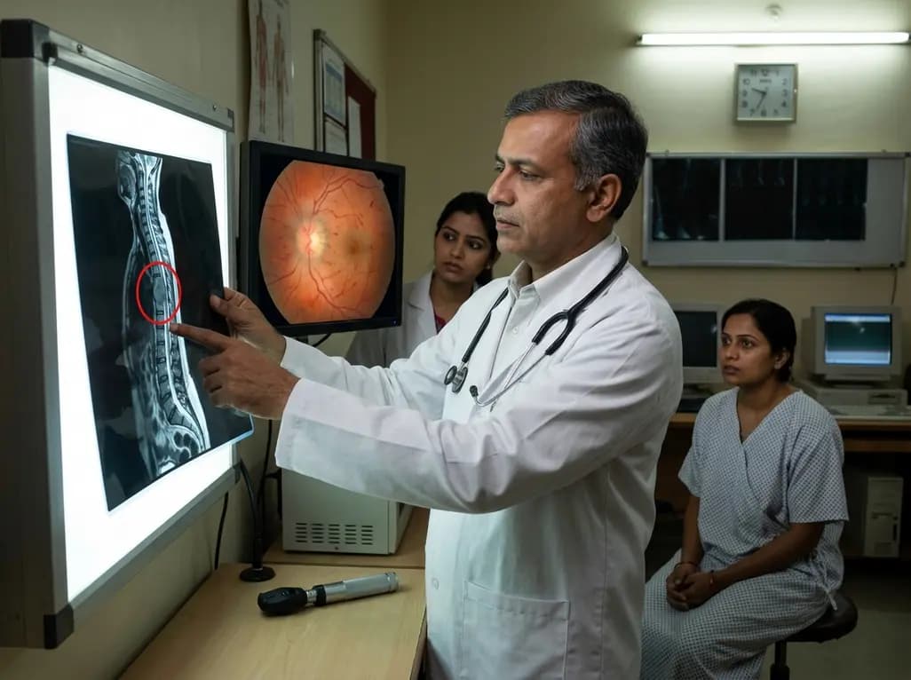
Physicians typically associate papilloedema with primary intracranial pathologies such as brain tumors, venous sinus thrombosis, or idiopathic intracranial hypertension. However, recent clinical evidence confirms that spinal tumors causing papilloedema represent a rare but significantly critical diagnostic challenge for modern practitioners. This phenomenon occurs when intradural spinal masses trigger raised intracranial pressure, often in the total absence of ventriculomegaly or hydrocephalus. Because these patients frequently lack typical limb neurology, such as weakness or sensory loss, initial diagnostic efforts often focus exclusively on the brain. Consequently, this narrow focus can lead to severe diagnostic delays, misdiagnosis, and potentially permanent vision loss. Recognizing that the cause of optic nerve swelling may lie far below the skull is essential for every neurologist and ophthalmologist. Furthermore, timely intervention can transform the clinical course, shifting the prognosis from certain blindness to full visual recovery. Therefore, clinicians must maintain a high index of suspicion when intracranial imaging fails to explain significant disc swelling. Specifically, understanding the subtle links between spinal pathology and cranial pressure is the first step toward effective management.
The exact mechanism behind how a spinal mass influences cranial pressure remains a subject of intense medical discussion. Notably, several complex dynamics involving cerebrospinal fluid (CSF) appear to be at play. Many researchers suggest that high CSF protein levels secreted by the spinal tumor significantly increase fluid viscosity. This thickened fluid subsequently obstructs the arachnoid villi, which are responsible for CSF absorption into the venous system. When absorption fails, intracranial pressure rises despite a lack of physical obstruction within the brain itself. Additionally, malignant cells can directly infiltrate the subarachnoid pathways, causing increased resistance to flow. Interestingly, some cases involve chronic micro-haemorrhages from the tumor, which further clog the drainage pathways with blood breakdown products. These physiological changes explain why papilloedema can manifest even when ventricles appear normal in size on a CT scan. Moreover, the venous congestion caused by a large spinal mass might retrograde affect the intracranial venous pressure. Understanding these diverse mechanisms helps physicians realize why the spinal column should be part of the investigative roadmap. Without addressing the underlying spinal pathology, intracranial interventions may only offer temporary relief.
One of the most confounding aspects of this condition is the frequent absence of classic spinal symptoms. Specifically, patients often present with isolated visual disturbances, such as blurred vision, transient visual obscurations, or persistent headaches. Because they do not report back pain or motor deficits, the clinician might not instinctively order spinal imaging. In many documented cases, the acuity of visual deterioration serves as the primary red flag. For instance, a patient might experience rapid vision loss over a matter of days, necessitating urgent evaluation. Similarly, the presence of nausea and vomiting alongside bilateral optic disc swelling strongly suggests raised intracranial pressure. However, the lack of ventriculomegaly on initial brain imaging often leads doctors toward a diagnosis of idiopathic intracranial hypertension. This misstep can result in inappropriate long-term medical management while the actual spinal tumor continues to grow. Therefore, every case of unexplained papilloedema deserves a thorough neurological examination that includes a search for subtle spinal signs. Clinicians should specifically check for dermatomal sensory changes or hyperreflexia, which might hint at a spinal origin despite the patient's primary ocular complaints.
To accurately diagnose spinal tumors causing papilloedema, a comprehensive imaging strategy is mandatory. While a brain MRI is the standard first step, practitioners must expand the search if the findings are negative for a cranial mass. Specifically, a total neural axis MRI is the gold standard for identifying intradural spinal lesions. This imaging should ideally include the cervical, thoracic, and lumbar regions to ensure no mass is overlooked. Furthermore, lumbar puncture remains a vital diagnostic tool to measure opening pressure and analyze CSF composition. Often, the CSF protein levels in these patients are exceptionally high, a finding known as Froin’s syndrome. This biochemical clue can distinguish the condition from idiopathic intracranial hypertension, where protein levels are typically normal. Additionally, neuro-ophthalmological assessments, including visual field testing and optical coherence tomography, provide a baseline for visual function. These tests help the surgical team determine the urgency of the intervention. Consequently, integrating results from diverse diagnostic modalities allows for a more precise treatment plan. By combining high-resolution imaging with detailed CSF analysis, the medical team can pinpoint the spinal source of the intracranial hypertension.
When papilloedema reaches a vision-threatening stage, clinicians must prioritize the preservation of sight above all other concerns. If visual deterioration is rapid, emergency CSF diversion becomes the most critical immediate step. This can involve the placement of an external ventricular drain or a shunt to bypass the obstruction and reduce pressure on the optic nerves. Consequently, these measures buy valuable time for the surgical team to plan the definitive removal of the spinal tumor. Moreover, medications such as acetazolamide or high-dose steroids might provide some temporary reduction in pressure, although they rarely address the root cause. Specifically, the medical team must coordinate closely between neurosurgery and ophthalmology to monitor visual fields daily. If the pressure remains uncontrolled, the optic nerve may suffer irreversible ischemic damage, leading to permanent blindness. Therefore, the acuity of the patient's visual loss often dictates the speed of the surgical response. Similarly, in cases where the spinal tumor is malignant, early diversion prevents the catastrophic complications of herniation. By acting decisively to manage the intracranial pressure, doctors significantly improve the chances of visual recovery post-resection.
The definitive treatment for intracranial hypertension caused by these lesions is the surgical resection of the spinal tumor. Once surgeons successfully remove the intradural mass, the CSF dynamics typically return to normal, and papilloedema begins to resolve. However, the speed of recovery depends on the duration of the pressure and the type of tumor involved. For example, benign schwannomas or ependymomas often show excellent post-operative results with total symptom resolution. Conversely, malignant lesions may require adjuvant therapies such as radiation or chemotherapy to prevent recurrence and maintain stable pressure. Following surgery, patients require long-term follow-up with serial imaging and eye exams to ensure the intracranial hypertension does not return. Additionally, many patients experience a significant improvement in their headaches and visual clarity within weeks of the procedure. Ultimately, the successful management of these complex cases depends on the clinician’s ability to 'think outside the box.' By recognizing the spine as a potential cause for cranial symptoms, the medical community can prevent the devastating consequences of misdiagnosis. This multidisciplinary approach ensures that patients receive the right treatment at the right time, effectively safeguarding their long-term health and vision.
Yes, intradural spinal tumors can occasionally present with isolated intracranial hypertension and papilloedema. Many patients do not exhibit motor or sensory deficits in their limbs initially. This absence of typical spinal neurology often leads to diagnostic delays if clinicians only focus on the brain during the initial medical workup.
The mechanism usually involves elevated CSF protein levels or tumoral cells that obstruct the arachnoid villi. This obstruction impairs the normal absorption of cerebrospinal fluid into the venous system, leading to raised intracranial pressure. Consequently, the high pressure is transmitted to the optic nerve head, causing bilateral papilloedema despite a normal brain.
The immediate priority is to preserve vision through emergency cerebrospinal fluid diversion if papilloedema is severe. Rapid visual deterioration requires urgent surgical intervention, such as shunting or tumor resection. Following stabilization, clinicians focus on removing the spinal mass to normalize CSF dynamics and prevent a recurrence of the hypertension.
Disclaimer: This content is for informational and educational purposes only. It does not constitute medical advice or establish a doctor-patient relationship. Refer to the latest local and national guidelines for clinical practice.
References
Sun R et al. Intra-cranial hypertension and vision-threatening papilloedema caused by intradural spinal tumours: a case series of three. Br J Neurosurg. 2025 Aug. doi: 10.1080/02688697.2023.2263087. PMID: 37807636.
Al-Houshimy A, et al. Hydrocephalus and Papilledema in Spinal Cord Tumors: A Report of Two Cases. PMC. 2023.
Petrovic D. Idiopathic Intracranial Hypertension and Vision: Understanding Papilledema and its Ocular Consequences. J Eye Dis. 2025; 10:275.

Read summarized clinical updates, watch expert medical content, and earn CME certifications right from your smartphone.
Open your smartphone camera to scan and install.





Intradural spinal tumors can rarely present with isolated intracranial hypertension and papilloedema. Clinicians must recognize this rare association, often occurring without limb neurology, to ensure timely surgical intervention and preserve patient vision through emergency CSF management.
2 weeks back
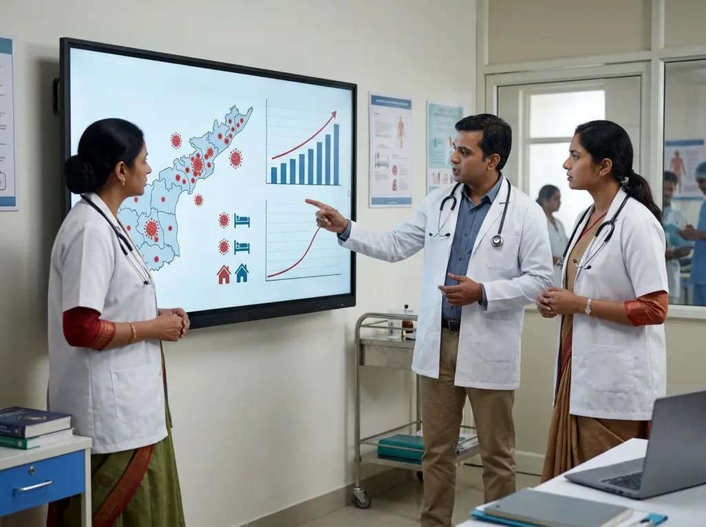
Andhra Pradesh reported 10 new Covid-19 cases, taking the state tally to 49 while deaths remain at four. With 24 patients hospitalized and 16 under home isolation, the Health Department has intensified monitoring. Medical professionals should review regional distribution, diagnostic protocols, and management plans.
Today

An 11-year Swedish registry study of 618 uterine sarcoma patients found that minimally invasive surgery yielded survival comparable to open surgery in early stages. However, adjuvant chemotherapy conferred no survival benefit in localized or advanced disease, highlighting stage and histology as key outcomes.
3 days back

A cross-sectional study evaluates post-intensive care syndrome in cardiac patients 2-4 weeks post-ICU discharge, highlighting cognitive, psychological, and functional impairments and the need for structured multidisciplinary rehabilitation.
3 days back

Anterior cruciate ligament reconstruction failure lacks uniform definition. A narrative review proposes an integrative framework incorporating objective and subjective instability, persistent pain, restricted motion, graft rupture, and secondary meniscal injury to standardize clinical reporting.
3 days back

With World Obesity Atlas data warning that over 41 million Indian children are overweight or obese, ICMR and NIN have unveiled a 10-point policy roadmap. The initiative calls for mandatory front-of-pack labeling, HFSS taxes, strict marketing bans, and healthier school environments to curb non-communicable diseases.
Today
