.webp&w=384&q=75)

Loading, please wait...
Loading, please wait...
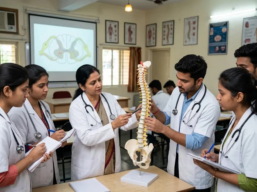
Spinal cord anatomy education represents a fundamental yet notoriously difficult component of the medical and dental curriculum. Students often struggle to visualize the complex spatial relationships between afferent and efferent pathways as they traverse the vertebral canal. Traditionally, educators have relied on two-dimensional slides and textbook diagrams to convey these concepts. However, these flat representations often fail to illustrate the three-dimensional reality of human anatomy. Consequently, many students memorize facts without truly grasping the physical orientation of neural structures. This gap in understanding can lead to difficulties in clinical practice, particularly when diagnosing neurological deficits or performing regional anesthesia. Therefore, researchers continue to seek innovative pedagogical tools that can bridge the divide between theoretical knowledge and spatial comprehension. Recent advancements in 3D printing technology have provided a promising solution to this long-standing challenge. By creating tangible, oversized models of anatomical structures, educators can offer a more tactile and immersive learning experience. This evolution in teaching methodology aims to improve student engagement and retention while simplifying the intricacies of neuroanatomy. Ultimately, the goal is to produce healthcare professionals who possess a deep, intuitive understanding of the human body.
The transition toward 3D models in spinal cord anatomy education marks a significant shift in medical pedagogy. Conventional slide-based instruction often utilizes high-quality images and diagrams, yet it limits the student to a single plane of view. In contrast, 3D-printed models allow students to interact with anatomical structures from multiple angles. For instance, a recent study utilized 3D-printed vertebrae at four times their natural size to enhance visibility and detail. By placing a clay spinal cord within the vertebral canal and using color-coded cables for neural pathways, instructors created a vivid representation of the nervous system. This hands-on approach encourages active learning, where students can physically trace the path of sensory and motor signals. Furthermore, the use of physical models helps demystify the relationship between the spinal cord and the surrounding skeletal structures. Specifically, students can see how nerve roots emerge and how pathways are organized within the white and gray matter. While digital 3D models also exist, physical models offer a tactile feedback that screens cannot replicate. This physical engagement often results in a more profound cognitive connection to the material. As a result, educational institutions are increasingly investing in 3D printing laboratories to supplement traditional lecture-based teaching.
To evaluate the efficacy of these new tools, researchers conducted a pre-test/post-test study comparing 3D models with traditional slides. The study involved sixty dental students divided into two instructional groups. Initially, both groups demonstrated similar baseline knowledge through a multiple-choice pre-test. Following the instructional sessions, both groups showed significant improvement in their scores, proving that both methods are effective to some degree. However, the group taught with the 3D model achieved significantly higher post-test scores compared to the slide-based group. Specifically, the mean score for the 3D model group reached 12.33, while the slide-based group averaged 8.47. This disparity highlights the superior instructional power of physical models in neuroanatomy. Moreover, the statistical analysis confirmed that the improvement in the 3D model group was not only significant but also substantially larger than that of their peers. These findings suggest that while slides are useful for providing a general overview, they cannot match the clarity provided by a physical model. Consequently, the study reinforces the argument for integrating more physical instructional aids into the curriculum. Such data-driven insights are crucial for curriculum planners who wish to optimize learning efficiency in high-stakes medical environments.
Understanding spatial relationships is a critical skill for any student pursuing spinal cord anatomy education. The human nervous system is not a series of isolated points but a complex, interconnected network. For example, the way the dorsal columns and spinothalamic tracts are situated relative to the ventral horns is vital for clinical localization. When students use 2D slides, they often struggle to translate those images into a three-dimensional patient. This difficulty is known as a high cognitive load, where the brain spends too much energy trying to rotate and visualize images. By providing a 3D model, instructors reduce this cognitive burden, allowing students to focus on the functional significance of the pathways. Additionally, the ability to see the spinal cord in the context of the C4 and T7 vertebrae provides essential landmarks. It allows students to visualize how the cord changes at different levels of the spine. Notably, the color-coded cables used in the study helped students differentiate between ascending sensory inputs and descending motor outputs. This visual clarity translates directly into better clinical reasoning. Therefore, the mastery of spatial anatomy is not just an academic exercise; it is a prerequisite for safe and effective patient care in fields like neurology and surgery.
Despite the clear benefits of 3D models in spinal cord anatomy education, implementation does face certain hurdles. One primary concern is the cost and technical expertise required for 3D printing. Designing accurate anatomical files and maintaining printing equipment can be resource-intensive for some departments. However, as the technology becomes more affordable and user-friendly, these barriers are steadily decreasing. Another challenge involves the time required to integrate these models into existing lesson plans. Faculty must be trained not only on how to use the models but also on how to facilitate interactive learning sessions. To address this, institutions can develop standardized teaching protocols that maximize the utility of each model. Furthermore, educators should consider a hybrid approach that combines the strengths of both slides and models. For instance, slides can provide the histopathological context while 3D models clarify the gross anatomy. Additionally, encouraging students to build their own simplified models using clay or other materials can further reinforce learning. By diversifying teaching methods, educators can cater to different learning styles, such as visual, auditory, and kinesthetic. Ultimately, the successful adoption of 3D technology requires a commitment to pedagogical innovation and continuous evaluation of student performance.
The future of spinal cord anatomy education is undoubtedly leaning toward more immersive and interactive technologies. Beyond physical 3D printing, we are seeing the rise of virtual reality (VR) and augmented reality (AR) in the classroom. These tools can allow students to virtually "walk through" the spinal cord or see neural pathways superimposed on a live patient. Nevertheless, the tangible nature of 3D-printed models remains uniquely valuable, especially in early medical training. They provide a sense of scale and presence that digital environments sometimes lack. As research continues to validate the effectiveness of these tools, we can expect a global shift in how anatomy is taught. This shift will likely lead to more standardized, high-quality educational resources that are accessible to students worldwide. Moreover, the integration of these models into clinical training for practicing doctors could improve surgical planning and patient communication. Patients, too, benefit when they can see a model of their condition rather than a confusing X-ray. In conclusion, the move toward 3D-based instruction represents a major leap forward in medical education. By focusing on spatial understanding and active engagement, we can ensure that the next generation of healthcare providers is better prepared for the complexities of modern medicine.
3D printing improves understanding by providing a tangible, spatial representation of complex neural structures. Unlike textbooks, which rely on two-dimensional images that require mental rotation, 3D models allow students to view pathways from multiple angles simultaneously. This reduces cognitive load and helps students visualize the precise location and relationship of afferent and efferent tracts within the vertebral canal, leading to significantly better retention and higher test scores.
Yes, slide-based teaching remains relevant as it provides a structured way to deliver large amounts of theoretical information and histopathological details quickly. Slides are excellent for showing cross-sections and microscopic views that are difficult to replicate in a physical model. Ideally, educators should use a blended approach where slides provide the conceptual foundation and 3D models provide the spatial and structural clarity needed for mastery.
Color-coded cables serve as a powerful visual aid to differentiate between distinct functional pathways. In neuroanatomy, it is crucial to distinguish between ascending sensory fibers and descending motor fibers. By using specific colors for afferent and efferent pathways, the model makes these abstract concepts concrete. This visual organization helps students quickly identify the direction of nerve impulses and understand the functional topography of the spinal cord more effectively.
Disclaimer: This content is for informational and educational purposes only. It does not constitute medical advice or a professional endorsement. Readers should consult with qualified healthcare professionals for diagnosis and treatment. Refer to the latest local and national guidelines for clinical practice.
References
Ömerli̇ A et al. Comparison of 3D model-based and slide-based teaching for afferent and efferent spinal cord pathways: a pre-test/post-test study. BMC Med Educ. 2026 Jul 04. doi: 10.1186/s12909-026-09398-y. PMID: 42401970.
Chen S, et al. The role of 3D printing in medical education: A systematic review. Journal of Anatomical Sciences. 2023;15(2):112-125.
Preece D, et al. Let's get physical: Advantages of a physical model over 3D computer models for learning anatomy. Anatomical Sciences Education. 2022;16(1):45-58.

Read summarized clinical updates, watch expert medical content, and earn CME certifications right from your smartphone.
Open your smartphone camera to scan and install.





A comparative study investigates 3D model-based versus slide-based teaching for spinal cord pathways. Results show that while both methods work, 3D models significantly outperform traditional slides in helping students master complex neuroanatomical spatial relationships.
3 months ago

Novel diimide and hydrazone scaffolds, particularly compound #C4, synergistically reactivate latent HIV-1 when combined with JQ1 or PKC activators. Demonstrating donor-matched proviral transcription ex vivo and non-canonical signaling, these findings advance shock and kill strategies toward reservoir eradication.
Today

A cross-sectional study reveals that maternal RSV vaccination coverage reached only 43.7% in Argentina, with maternal vaccine knowledge being the sole independent predictor of uptake. Clinicians play a crucial role in eliminating informational and structural barriers to optimize infant health.
Today

Review how tailored exercise programs benefit frail older patients with heart failure. Explore pathophysiological mechanisms, individualized training prescriptions, and strategies to overcome adherence barriers in clinical practice.
Yesterday

This comparative study evaluates posterior labral tears with and without glenohumeral instability, highlighting distinct demographic patterns, MRI underestimation in stable shoulders, and successful arthroscopic repair outcomes.
Yesterday

A clinical study demonstrates that early intervention with tocilizumab, either as monotherapy or bridged with corticosteroids, achieves significant proptosis reduction and resolves active inflammation in treatment-naive thyroid eye disease.
Yesterday
