

Loading, please wait...
Loading, please wait...

The spectrum of choroiditis encompasses a heterogeneous collection of posterior uveitic conditions that primarily damage the choroidal vasculature and stroma. Clinicians historically struggled with diagnostic precision because many chorioretinal lesions share overlapping clinical presentations. However, advanced retinal diagnostic tools have clarified these complex disease processes. Ophthalmologists now differentiate distinct entities through targeted pathophysiology and imaging biomarkers. Furthermore, recognizing specific presentations prevents irreversible visual disability. Consequently, clinicians must appreciate the anatomical differences, etiological triggers, and individualized treatment pathways across both infectious and non-infectious inflammatory disorders.
Choroidal inflammation involves distinct vascular layers within the posterior segment of the eye. Therefore, contemporary classification schemes subdivide these inflammatory processes into choriocapillaritis and stromal choroiditis. Indocyanine green angiography has driven this fundamental diagnostic distinction. In choriocapillaritis, inflammatory occlusion directly compromises the inner choroidal capillary bed. This primary vascular insult leads to secondary retinal pigment epithelium alterations and outer retinal atrophy. For instance, classic serpiginous choroiditis, multiple evanescent white dot syndrome, and punctate inner choroiditis represent primary choriocapillaropathies. In contrast, stromal choroiditis originates within the deeper choroidal vessel network and connective tissue. Inflammatory infiltrates and granulomas accumulate within the choroidal stroma, causing diffuse thickening and secondary choriocapillaris compression. As a result, diseases such as Vogt-Koyanagi-Harada syndrome, sympathetic ophthalmia, and sarcoidosis demonstrate primary stromal involvement. In addition, mixed variants frequently emerge during systemic dissemination. Clinicians must establish the exact anatomical layer of maximal inflammation during the baseline evaluation. Thus, identifying whether disease starts in the capillaries or the deep stroma guides appropriate therapy and prognosis.
Multimodal imaging remains crucial when evaluating active posterior uveitis. Specifically, indocyanine green angiography serves as the diagnostic cornerstone for evaluating choroidal microcirculation and vascular integrity. Clinicians observe that fluorescein angiography predominantly highlights retinal vascular leaks and superficial pigment changes. However, infrared wavelengths utilized during indocyanine green angiography penetrate the pigmented ocular layers with minimal attenuation. Consequently, the test reveals occult subclinical lesions that standard slit-lamp examinations frequently miss. In choriocapillaritis, early hypocyanescent patches reflect localized perfusion deficits within capillary networks. These hypocyanescent areas typically persist during the late angiographic phases without significant enlargement. Conversely, stromal choroiditis manifests as diffuse, regularly spaced hypocyanescent spots that represent active inflammatory granulomas. Furthermore, late-phase imaging demonstrates hypercyanescent vessel leakage and choroidal hyperfluorescence during severe active phases. Clinicians simultaneously integrate optical coherence tomography scans to assess neurosensory detachment and ellipsoid zone disruption. In addition, fundus autofluorescence identifies active border expansion through distinct hyperautofluorescent rims. Ultimately, serial angiographic evaluations track disease regression and prevent premature treatment discontinuation.
Distinguishing autoimmune serpiginous choroiditis from infectious tubercular choroiditis represents a critical diagnostic challenge in clinical practice. In endemic regions such as India, Mycobacterium tuberculosis drives a substantial proportion of posterior uveitis presentations. Infectious serpiginous-like choroiditis and multifocal serpiginoid choroiditis often mimic autoimmune disease phenotypes. However, key clinical and morphological distinctions guide accurate discrimination between these conditions. Autoimmune serpiginous choroiditis typically produces geographic, contiguous lesions that emerge around the optic disc and march centrifugally. In contrast, tubercular serpiginous-like choroiditis frequently presents as multifocal, non-contiguous lesions that involve the retinal periphery before reaching the peripapillary region. Furthermore, tubercular entities frequently exhibit significant vitreous cells and active retinal vasculitis, whereas autoimmune forms demonstrate minimal vitreous reaction. Clinicians must confirm tuberculin skin tests, interferon-gamma release assays, and chest computed tomography before prescribing heavy immunosuppression. Prescribing systemic corticosteroids alone in occult tubercular choroiditis can trigger disastrous infectious proliferation. Therefore, targeted phenotypic differentiation protects vision and prevents systemic morbidity.
Systemic inflammatory disorders frequently target the choroidal stroma and trigger bilateral visual symptoms. For example, Vogt-Koyanagi-Harada disease causes profound, diffuse stromal inflammation mediated by autoimmune T lymphocytes directed against melanocytes. Patients initially present with headache, meningismus, auditory disturbances, and bilateral visual blur. Ophthalmic examination demonstrates exudative retinal detachments, diffuse choroidal thickening, and optic disc hyperemia. Similarly, sympathetic ophthalmia shares nearly identical histopathological and angiographic characteristics following penetrating trauma or intraocular surgery. In both conditions, indocyanine green angiography reveals innumerable, evenly distributed hypocyanescent dark dots across the midperiphery. In addition, sarcoid choroiditis presents with distinct choroidal granulomas, candle-wax retinal periphlebitis, and elevated serum angiotensin-converting enzyme levels. Clinicians also identify birdshot chorioretinopathy through characteristic cream-colored fundus spots and positive human leukocyte antigen A29 testing. Because these conditions harbor progressive courses, clinicians must coordinate comprehensive systemic assessments. Early identification of bilateral stromal involvement prevents structural chorioretinal atrophy and preserves central visual acuity.
Successful therapeutic outcomes depend entirely upon etiological precision and timely therapeutic intervention. Therefore, clinicians must avoid empirical monotherapy before confirming or excluding underlying infectious pathogens. When ophthalmologists confirm tubercular choroiditis, they prescribe multi-drug anti-tubercular therapy combined with judicious systemic corticosteroids. The antimicrobial regimen controls the active bacillary load, while concomitant oral corticosteroids suppress excessive tissue-destructive inflammation. Conversely, clinicians manage non-infectious autoimmune choroiditis through rapid immunosuppressive protocols. High-dose oral corticosteroids provide initial disease suppression, but clinicians introduce conventional steroid-sparing immunosuppressants early in the disease course. For instance, methotrexate, azathioprine, and mycophenolate mofetil maintain stable remission and reduce steroid toxicity. In refractory or vision-threatening cases of autoimmune choroiditis, biologic agents such as adalimumab and infliximab deliver targeted cytokine inhibition. Furthermore, clinicians monitor treatment response objectively through serial optical coherence tomography and indocyanine green angiography rather than relying solely on visual acuity. Ultimately, individualized management protocols minimize disease recurrence, prevent choroidal neovascularization, and optimize long-term ocular health.
Indocyanine green angiography evaluates choroidal circulation through deep infrared light penetration. In choriocapillaritis, localized ischemic nonperfusion produces geographic hypocyanescent patches that remain nonfluorescent throughout early and late angiographic frames. Conversely, stromal choroiditis produces multiple, evenly distributed hypocyanescent dark dots representing inflammatory stromal granulomas. Furthermore, stromal disease frequently exhibits late choroidal vessel leakage and diffuse hyperfluorescence. Therefore, these distinct patterns accurately differentiate primary capillary closure from deep stromal infiltrative disorders.
Tubercular serpiginous-like choroiditis clinically mimics autoimmune serpiginous choroiditis but requires completely different medical management. If clinicians inadvertently administer high-dose corticosteroid monotherapy or systemic immunosuppressants to a patient with occult tuberculosis, the infection can rapidly proliferate, causing severe ocular necrosis and systemic dissemination. Consequently, physicians must systematically perform tuberculin skin testing, interferon-gamma release assays, and chest imaging to exclude active or latent tuberculosis before initiating immunosuppressive therapy in endemic populations.
Biologic agents provide targeted immunomodulatory action for refractory or vision-threatening non-infectious choroiditis. Tumor necrosis factor-alpha inhibitors, such as adalimumab and infliximab, suppress recalcitrant choroidal inflammation when conventional immunosuppressive drugs fail to achieve remission. Furthermore, these targeted agents allow clinicians to taper oral corticosteroids safely, preventing long-term steroid-induced ocular and systemic complications. Clinicians regularly employ biologics to preserve structural macular architecture and prevent recurrent choroidal neovascularization in sight-threatening autoimmune presentations.
Disclaimer: This content is for informational and educational purposes only... Refer to the latest local and national guidelines for clinical practice.
References

Read summarized clinical updates, watch expert medical content, and earn CME certifications right from your smartphone.
Open your smartphone camera to scan and install.





This review outlines the clinical spectrum of choroiditis, highlighting the role of indocyanine green angiography in distinguishing choriocapillaritis from stromal choroiditis. It emphasizes phenotype-driven workups for tuberculosis and tailored anti-infective versus immunosuppressive treatment strategies.
Today

New evidence demonstrates that ageing and female sex remodel lymphocyte compartments, weakening peripheral T-cell tolerance and lowering the threshold for red blood cell autoantibody production in warm autoimmune haemolytic anaemia.
Today

Large genomic databases like gnomAD and All of Us offer crucial insights into the population prevalence of partial RHD genotypes. Identifying these variants helps clinicians mitigate anti-D alloimmunization risks in transfusion medicine and obstetric care across diverse ancestral populations.
Today
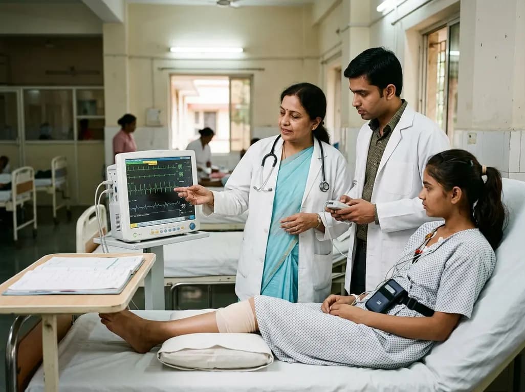
A 15-year-old patient presented with knee arthralgia and acute rheumatic fever, exhibiting complex arrhythmias including AV blocks and non-sustained ventricular tachycardia despite lacking severe valvulitis. This highlights the importance of extended Holter monitoring in pediatric rheumatic carditis.
Today
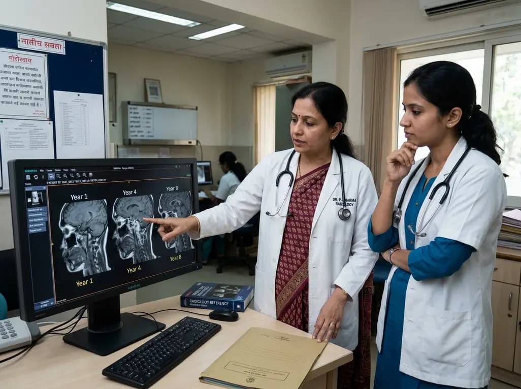
An eight-year longitudinal imaging follow-up reveals severe temporomandibular joint remodeling in nodulosis-arthropathy-osteolysis (NAO) syndrome. Despite extensive osseous destruction, the patient maintained functional jaw opening without TMJ interventions, highlighting the role of conservative management.
Today

Discover essential clinical insights into AGLP1-type amyloidosis, a rare localized complication of glucagon-like peptide-1 analog injections. Learn how histopathology and mass spectrometry differentiate benign iatrogenic amyloidomas from systemic disease, preventing misdiagnosis and unwarranted therapies.
Today
