.webp&w=384&q=75)

Loading, please wait...
Loading, please wait...
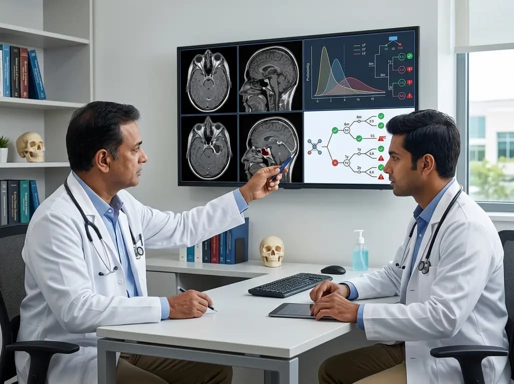
Managing resected intracranial neoplasms requires precision, particularly when establishing long-term radiological monitoring. Modern skull base meningioma surveillance relies on balancing early detection of tumor progression against the costs, inconvenience, and psychological impact of unnecessary neuroimaging. Skull base meningiomas present unique surgical challenges because of their close proximity to critical neurovascular structures, cranial nerves, and major vascular sinuses. Complete surgical resection is frequently impossible without risking significant neurological deficits. Consequently, clinicians need evidence-based post-operative monitoring schedules that reflect individual recurrence risks rather than arbitrary calendar intervals. A recent multi-center study published in the Journal of Neuro-Oncology utilized Bayesian conditional probability modeling to establish an optimized, data-driven follow-up protocol for post-surgical patients.
Skull base meningiomas account for a substantial proportion of intracranial tumors treated by neurosurgeons. Achieving gross total resection in this anatomical region is often hindered by tumor adherence to vital neural and vascular structures. Surgical decisions frequently favor subtotal resection or subtotal decompression to preserve patient functional status and quality of life. However, leaving residual microscopic or macroscopic tumor tissue inherently increases the risk of long-term disease progression.
Traditionally, post-operative surveillance protocols have relied heavily on institutional preference or generalized neuro-oncological consensus guidelines. These empirical schedules often recommend uniform, annual magnetic resonance imaging scans regardless of the actual extent of surgical resection or individual risk profile. Such standardized approaches can lead to over-imaging in low-risk individuals and under-imaging in high-risk patients. Establishing a tailored surveillance schedule is vital for identifying disease progression at a stage where re-intervention, such as stereotactic radiosurgery or revision microsurgery, remains safest and most effective.
Furthermore, long-term follow-up data show that skull base meningiomas can progress many years after initial surgical management. Therefore, clinicians require long-term risk assessment models. Data-driven surveillance algorithms offer a structured approach to clinical decision-making. By applying mathematical modeling to multi-center clinical cohorts, researchers can derive conditional probability schedules that maintain predictable safety margins throughout extended post-operative observation periods.
Understanding the specific risk factors that drive disease progression is fundamental to constructing accurate post-operative follow-up models. The multi-center Bayesian study evaluated 358 patients undergoing skull base meningioma resection over nearly two decades, with a median follow-up period exceeding eight years. The cohort demonstrated a median patient age of 57 years, with a classic female predominance of 76 percent. Histologically, WHO Grade 1 tumors accounted for 80.3 percent of cases, while WHO Grade 2 atypical meningiomas constituted 19.7 percent.
Overall, progression requiring re-intervention occurred in 14.1 percent of patients during the follow-up window. Extensive statistical analysis identified the extent of surgical resection as the strongest independent predictor of tumor progression. Subtotal resection significantly elevated the hazard of requiring secondary intervention compared to gross total resection, with a hazard ratio of 2.62. Conversely, an incidental tumor presentation was strongly associated with a lower risk of progression, demonstrating a hazard ratio of 0.11.
Interestingly, a higher baseline comorbidity burden was also associated with a reduced likelihood of re-intervention. This phenomenon likely reflects a higher threshold for undertaking invasive secondary treatments in medically fragile patients rather than a biological suppression of tumor growth. Overall, these predictive factors emphasize that residual tumor burden remains the primary determinant governing post-operative surveillance intensity.
To transition from simple predictive factors to actionable clinical guidelines, researchers applied conditional probability modeling. Bayesian statistical methods allow clinicians to calculate the updated risk of tumor progression at any given follow-up interval based on the absence of progression up to that time point. This conditional risk model formed the foundation for deriving a targeted schedule designed to keep the annual risk of progression requiring re-intervention at or below a strictly defined three percent threshold.
The resulting mathematical model clearly demonstrated that patients who underwent subtotal resection experience a persistently higher annual risk of tumor progression throughout their follow-up duration. Consequently, these patients require more frequent imaging evaluations to ensure timely detection of regrowth. In contrast, patients who achieved gross total resection demonstrate a much lower annual risk trajectory, allowing for safely extended interval scans.
By adopting this Bayesian conditional-risk framework, neuro-oncology teams can systematically replace standardized, rigid imaging intervals with dynamic, risk-stratified schedules. This methodology ensures that radiological surveillance remains proportional to actual clinical risk while optimizing healthcare resource utilization. Patients benefit from reduced anxiety and fewer diagnostic visits when complete tumor clearance is achieved, while those with residual disease receive vigilant, focused monitoring.
The empirical output of the Bayesian conditional probability model offers distinct, evidence-based imaging schedules based on the surgical outcome achieved. For patients undergoing gross total resection, the recommended magnetic resonance imaging schedule is remarkably streamlined. Scans are recommended at 3 months post-operatively, followed by surveillance at 2 years, 5 years, 8 years, and 10 years. This schedule provides essential early confirmation of surgical success while avoiding unnecessary annual imaging during middle follow-up years when risk remains exceptionally low.
For patients who undergo subtotal resection, the higher persistent progression risk necessitates a significantly more intensive surveillance schedule. The recommended protocol dictates magnetic resonance imaging scans at 3 months, 1 year, and 18 months post-surgery. Subsequently, annual scans are required from year 2 through year 8, followed by a final scheduled milestone scan at 10 years.
This stark contrast highlights the profound clinical impact of surgical extent on long-term management strategies. By implementing these stratified protocols, clinicians ensure that patients with subtotal resections receive close monitoring during the critical peak window of recurrence risk. Simultaneously, gross total resection patients avoid unnecessary radiological exposure, reducing personal burden and healthcare costs while maintaining complete clinical safety.
Integrating Bayesian surveillance protocols into routine clinical practice requires close coordination among neurosurgeons, neuro-oncologists, neuroradiologists, and radiation oncologists. Multidisciplinary tumor boards can utilize these evidence-based guidelines to standardize post-operative care plans across diverse healthcare settings. Establishing clear imaging timelines immediately following surgery enhances patient compliance and provides patients with predictable long-term care pathways.
Furthermore, these risk-stratified protocols help guide adjuvant treatment decisions. When residual tumor is known to exist after subtotal resection, clinicians can pair the intensive imaging schedule with early discussions regarding stereotactic radiosurgery or fractionated radiotherapy. Early identification of minimal progression on 18-month or annual scans enables non-invasive radio-surgical interventions before significant mass effect or neurological deterioration occurs.
In contrast, confirming stability in gross total resection cases at the 2-year and 5-year milestones reassures both clinicians and patients. This structured reassurance facilitates eventual discharge or transition to long-interval monitoring. Ultimately, adopting data-driven protocols improves quality of care, standardizes clinical documentation, and fosters transparent communication between specialists and patients regarding long-term brain tumor management strategies.
Implementing Bayesian risk-based surveillance protocols represents a major advancement in personalized neuro-oncology care. Transitioning from arbitrary annual scanning to quantitative, stratified algorithms enhances diagnostic precision while optimizing healthcare resources. Surgical extent remains the primary factor dictating post-operative surveillance intensity. By maintaining a maximum annual progression risk threshold of three percent, these evidence-based protocols offer clinicians high statistical confidence. Patients achieving gross total resection benefit from extended imaging intervals, whereas those undergoing subtotal resection receive appropriate vigilance during high-risk post-operative periods, ensuring optimal long-term outcomes.
The extent of surgical resection is the primary determinant of progression risk. Patients receiving subtotal resection have a significantly higher risk of progression requiring re-intervention compared to those achieving gross total resection, with studies showing a hazard ratio of 2.62.
According to the Bayesian risk-stratified protocol, patients achieving gross total resection require magnetic resonance imaging scans at 3 months post-operatively, followed by milestone surveillance scans at 2 years, 5 years, 8 years, and 10 years post-surgery to ensure long-term stability.
Subtotal resection leaves residual tumor tissue, resulting in a higher persistent annual progression risk. The recommended surveillance schedule includes scans at 3 months, 1 year, 18 months, annually from years 2 to 8, and at 10 years to catch growth early.
Disclaimer: This content is for informational and educational purposes only and does not constitute medical advice, diagnosis, or treatment. Always seek the advice of your physician or other qualified health provider with any questions you may have regarding a medical condition. Refer to the latest local and national guidelines for clinical practice.
References

Read summarized clinical updates, watch expert medical content, and earn CME certifications right from your smartphone.
Open your smartphone camera to scan and install.





A multi-center Bayesian study established a data-driven MRI surveillance protocol for resected skull base meningiomas, stratifying follow-up frequency by extent of resection to maintain a progression risk under 3%.
2 months ago

Discover the crucial role of glymphatic dysfunction in stroke, where perivascular clearance failure and neuroinflammation exacerbate cerebral edema and post-stroke brain injury.
Today

A prenatal case highlights severe glutaric acidemia type II presenting as bilateral hyperechoic cystic kidneys at 18 weeks. Whole-exome sequencing revealed a novel homozygous ETFDH frameshift variant, demonstrating the power of rapid genetic testing in fetal renal anomalies.
Today

Review how tailored exercise programs benefit frail older patients with heart failure. Explore pathophysiological mechanisms, individualized training prescriptions, and strategies to overcome adherence barriers in clinical practice.
Yesterday

This comparative study evaluates posterior labral tears with and without glenohumeral instability, highlighting distinct demographic patterns, MRI underestimation in stable shoulders, and successful arthroscopic repair outcomes.
Yesterday

A clinical study demonstrates that early intervention with tocilizumab, either as monotherapy or bridged with corticosteroids, achieves significant proptosis reduction and resolves active inflammation in treatment-naive thyroid eye disease.
Yesterday
