.webp&w=384&q=75)

Loading, please wait...
Loading, please wait...
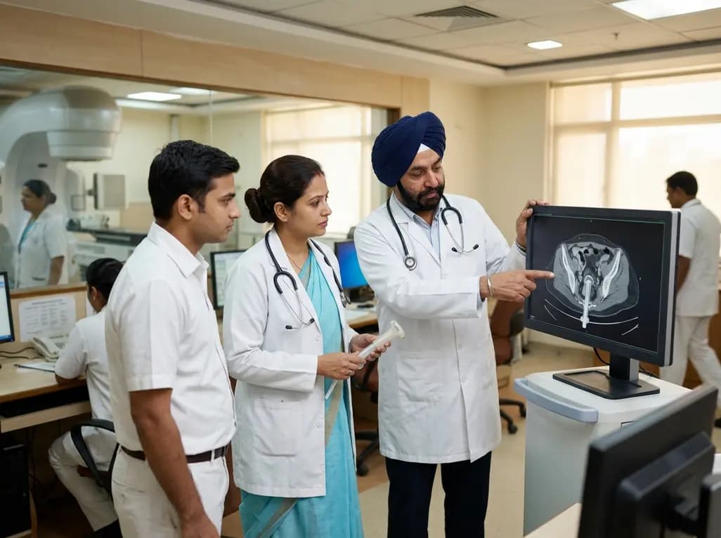
Adjuvant vaginal cuff brachytherapy serves as an established standard of care to prevent local vault recurrence in postoperative women diagnosed with high-intermediate risk endometrial cancer. Historically, radiation oncologists utilize radiographic simulation—via computed tomography or orthogonal radiographs—to verify cylinder applicator seating, assess vaginal mucosa contact, and evaluate organ-at-risk dosimetry. However, routine simulation demands additional equipment time, increases patient discomfort, and generates healthcare expenses. Recent clinical evidence now evaluates whether omitting routine imaging simulation impacts therapeutic efficacy.
In a large single-institution retrospective cohort study, investigators examined long-term oncologic outcomes in 255 women with high-intermediate risk endometrial cancer treated with vaginal cuff brachytherapy without radiographic simulation. Over a median follow-up period of 60.1 months, the researchers documented 11 vaginal recurrence events. Consequently, the 5-year cumulative incidence of vaginal recurrence was calculated at 3.6% (95% CI = 1.2–6.5%). These results provide reassuring real-world evidence supporting clinical cylinder placement techniques when managed by experienced multidisciplinary teams.
To contextualize these findings, researchers benchmarked their outcomes against the standard brachytherapy arm of the landmark PORTEC-4a trial, which mandated strict imaging simulation and quality assurance for applicator positioning. In PORTEC-4a, the 5-year cumulative incidence of vaginal recurrence was 1.6% (95% CI = 0.00–3.32%). Statistical analysis indicated that the recurrence risk observed in the simulation-free cohort remained clinically comparable to the protocol-driven simulation cohort. Furthermore, subgroup analyses demonstrated that recurrence risks did not differ significantly across various clinical risk categories or specific applicator designs.
Vaginal cylinder brachytherapy typically delivers highly localized surface or depth doses to the upper vaginal vault. When clinicians insert an appropriately sized cylinder that matches vaginal apical anatomy, adequate mucosal apposition is achieved reliably. While volumetric 3D image-guided brachytherapy provides detailed dosimetric verification for complex multi-channel or interstitial geometries, simple single-channel cylinder treatments follow standardized dose distributions. Therefore, experienced clinical insertion, careful cylinder diameter selection, and secure immobilization can achieve robust local coverage without mandatory per-fraction or baseline simulation scans.
Streamlining adjuvant radiation therapy pathways offers significant advantages for high-volume cancer centers, especially in resource-constrained environments across low- and middle-income countries. Eliminating routine simulation scans reduces scanner bottlenecks, minimizes radiation exposure from repeated planning imaging, and decreases overall treatment expenditure. Moreover, shorter procedure times substantially improve patient comfort and compliance during multi-fraction courses. Thus, this pragmatic treatment strategy expands treatment capacity without apparent compromises in local disease control.
While the elimination of simulation provides workflow efficiency, patient selection remains critical. Modern gynecologic oncology increasingly incorporates molecular classification, including POLE mutations, mismatch repair deficiency, and p53 status, to guide adjuvant decisions. Patients harboring adverse biology or complex vaginal anatomy may still warrant customized volumetric imaging. Moving forward, prospective trials evaluating verification strategies will clarify the exact patient subsets that benefit from advanced image guidance versus those who can safely undergo streamlined clinical treatment.
Vaginal cuff brachytherapy is an internal radiation technique commonly delivered after hysterectomy for endometrial cancer. It utilizes an intra-vaginal applicator cylinder connected to a high-dose-rate radioactive source. This approach targets the upper vaginal apex, which represents the most frequent site of local disease relapse, while sparing adjacent pelvic normal tissues.
Simulation imaging with pelvic computed tomography or orthogonal radiographs was historically performed to verify cylinder contact with the vaginal vault apex. This process ensures proper applicator seating, detects apical air pockets, confirms geometric alignment, and calculates exact radiation dose constraints to adjacent sensitive organs, including the rectum and bladder.
Recent long-term clinical data demonstrate that omission of simulation does not compromise local cancer control in appropriately selected patients. With careful clinical sizing and proper physical insertion technique, 5-year vaginal recurrence rates remain below 4%, which is clinically comparable to recurrence rates observed in trial cohorts utilizing mandatory simulation verification.
Disclaimer: This content is for informational and educational purposes only. It does not constitute medical advice, diagnosis, or treatment recommendations. Always consult a qualified healthcare professional regarding individual clinical decisions. Refer to the latest local and national guidelines for clinical practice.
References
1. Eckelmann B et al. Simulation-free vaginal cuff brachytherapy for high-intermediate risk endometrial cancer. Brachytherapy. 2026 Aug 14. doi: undefined. PMID: 42601313.
2. van den Heerik ASVM, Horeweg N, Nout RA, et al. Molecular-integrated risk profile to determine adjuvant radiotherapy in endometrial cancer: PORTEC-4a trial. Lancet Oncol. 2025;26(1):45-56.
3. Harkenrider MM, Block AM, Alektiar KM, et al. American Brachytherapy Task Group Report: Adjuvant vaginal brachytherapy for early-stage endometrial cancer: A comprehensive review. Brachytherapy. 2017;16(1):95-108.

Read summarized clinical updates, watch expert medical content, and earn CME certifications right from your smartphone.
Open your smartphone camera to scan and install.





A retrospective cohort study reveals that simulation-free vaginal cuff brachytherapy achieves low 5-year vaginal recurrence rates comparable to simulation-verified protocols in high-intermediate risk endometrial cancer, supporting streamlined radiation oncology workflows.
last month

Evaluating concordance between patient-reported and clinician-assessed modified Rankin Scale scores reveals how nonmotor symptoms, caregiver reliance, and cultural factors affect functional disability ratings in chronic neurologic disorders.
Today

A scoping review evaluates 116 studies on AI in women's reproductive health, finding that 83.6% carry high risk of bias. While ensemble models show promise, disparities and lack of validation hinder LMIC deployment.
Today

Differential oxygenation during peripheral TA-NRP can cause undetected coronary hypoxemia due to arch vessel clamping. A novel circuit with continuous aortic root monitoring detects desaturation early, facilitating left ventricular unloading and preventing allograft dysfunction and unnecessary heart discard.
Today

A longitudinal study reveals that patients undergoing staged bilateral hip arthroscopy experience progressive contralateral labral deterioration during surgical intervals. However, structural degradation does not compromise 2-year postoperative clinical outcomes or patient-reported satisfaction scores.
Today

A post hoc analysis of the PARM-T2D study shows pemafibrate exerts baseline-dependent effects on LDL-C and particle size in type 2 diabetes and hypertriglyceridemia. It lowers triglycerides and remnants while favorably remodeling atherogenic LDL particles based on initial lipid control status.
Today
