

Loading, please wait...
Loading, please wait...

Traumatic brain injury remains a primary cause of worldwide morbidity and mortality, especially in resource-constrained emergency environments. Clinicians traditionally depend on the Glasgow Coma Scale and computed tomography to assess acute cerebral trauma. However, these conventional modalities present considerable limitations during early resuscitation. Intubation, pharmacologic sedation, and systemic trauma frequently confound physical examinations. Furthermore, repeat neuroimaging remains logistically challenging and resource-intensive in underserved healthcare centers. Consequently, measuring serum biomarkers in TBI has emerged as an essential diagnostic strategy. Circulating proteins provide objective pathophysiological insight into acute intracranial structural pathology. Recently, investigators conducted a prospective clinical trial at the National Hospital Abuja to validate specific blood tests. They evaluated glial fibrillary acidic protein, neuron-specific enolase, and S100B protein among patients suffering moderate and severe injuries. These biomarkers demonstrated immediate elevation upon hospital admission, clearly distinguishing injury severities. Therefore, objective blood-based diagnostics offer invaluable adjunctive data when clinical examination remains compromised. In addition, rapid biochemical triage optimizes urgent allocation of critical intensive care resources. Thus, integrating biochemical profiles with bedside assessments bridges vital diagnostic gaps during acute neurotrauma management.
Different circulating markers reflect distinct mechanical damage patterns within neural tissue compartments. Glial fibrillary acidic protein serves as an intermediate filament protein highly specific to astrocytic cytoskeletal architecture. Following acute mechanical shear stress, injured astrocytes rapidly shed these structural proteins through compromised blood-brain barrier membranes into peripheral circulation. Consequently, elevated levels strongly indicate substantial parenchymal brain damage and severe structural disruption. In contrast, neuron-specific enolase originates within neuronal cytoplasm, acting as an intracytoplasmic glycolytic enzyme. Marked elevations of this marker directly signify extensive neuronal membrane damage and axonal destruction. Meanwhile, S100B represents a low-molecular-weight calcium-binding peptide concentrated primarily within astroglial and Schwann cells. Astrocytes release this protein during acute metabolic stress and cellular injury. Moreover, circulating S100B concentrations correlate closely with blood-brain barrier permeability alterations. Accordingly, measuring all three molecules simultaneously yields a comprehensive biochemical picture of both glial and neuronal degradation. Clinicians can therefore track distinct cellular injury pathways dynamically rather than relying on static anatomical snapshots. Furthermore, this multi-marker panel provides superior pathophysiological granularity compared to single-protein assessments.
Clinical investigations demonstrate strong associations between biomarker release and initial clinical severity scores. In the prospective cohort, baseline concentrations of GFAP, NSE, and S100B were profoundly elevated across all admitted patients. Furthermore, patients presenting with severe injuries exhibited significantly higher baseline values than individuals presenting with moderate injuries. Specifically, mean admission levels reached 1.85 ng/ml for GFAP, 14.25 ng/ml for NSE, and 0.60 ng/ml for S100B. Serum concentrations demonstrated robust inverse correlations with Glasgow Coma Scale scores documented at emergency presentation. In addition, initial biomarker elevations corresponded reliably with radiological damage severity on baseline computed tomography scans. Patients presenting with midline shift, basal cistern effacement, or diffuse axonal lesions displayed substantially higher circulating protein concentrations. Consequently, these blood markers provide objective biological confirmation of radiographic brain injury extent. This correlation proves particularly beneficial when immediate advanced imaging is unavailable or delayed. Therefore, acute biomarker quantification directly enhances clinical triage accuracy during the critical initial resuscitation window.
Understanding the post-injury temporal kinetics of brain biomarkers provides essential prognostic clarity for critical care teams. In the clinical trial, investigators systematically tracked biomarker concentrations at hospital admission, day three, and day seven post-injury. Astrocytic markers and neuronal enzymes demonstrated distinct clearance kinetics and persistence profiles over the first week. S100B concentrations typically peaked early and cleared rapidly, reflecting acute blood-brain barrier reconstitution. In contrast, GFAP and NSE exhibited sustained elevations in patients experiencing progressive secondary cerebral insults or uncontrolled cerebral edema. Consequently, persistent biomarker elevations at day three and day seven indicated continuous ongoing neurodestruction rather than isolated initial trauma. Patients who achieved neurological stability showed steady biomarker clearance toward normal physiological levels over the evaluation period. Conversely, secondary biomarker surges reliably flagged secondary complications, including intracranial hypertension, expanding contusions, and delayed cerebral ischemia. Therefore, serial biomarker sampling offers continuous biochemical surveillance that bedside examinations cannot match. Clinicians can accordingly adjust neurointensive monitoring strategies and medical interventions before irreversible catastrophic deterioration occurs.
Predicting functional recovery represents a major goal in modern neurotrauma critical care management. In this prospective investigation, researchers evaluated functional recovery at three months post-injury using the Glasgow Outcome Scale. Admission biomarker concentrations strongly predicted three-month functional performance and overall patient survival. Specifically, patients exhibiting markedly elevated initial levels of GFAP and NSE experienced significantly worse functional outcomes and higher mortality rates. Furthermore, sustained elevations at day seven served as robust independent predictors of persistent vegetative state or severe disability. In contrast, low initial biomarker concentrations combined with rapid clearance curves correlated with favorable neurological recovery. Thus, early biochemical testing assists neurotrauma teams in formulating realistic prognoses and guiding family discussions. Moreover, these objective prognostic profiles guide timely decisions regarding advanced surgical decompression or aggressive medical neuroprotection. Healthcare teams can tailor long-term neurorehabilitation pathways based on expected functional trajectories. Ultimately, incorporating serum biomarker stratification into standardized protocols refines prognostic precision and optimizes resource utilization across trauma networks.
Trauma centers in low- and middle-income regions face unique obstacles in providing timely neurotrauma care. Many facilities experience significant delays in obtaining emergency computed tomography scans due to financial constraints or scanner downtime. Consequently, point-of-care or laboratory-based ELISA biomarker assays represent an accessible, objective triage solution. By utilizing rapid serum testing, emergency physicians can swiftly identify patients at highest risk for surgical intracranial mass lesions. In addition, objective biomarker readings assist clinicians in prioritizing transfer logistics to tertiary neurosurgical facilities. Standardizing biomarker testing requires established laboratory infrastructure, quality control measures, and clear diagnostic cutoffs. Nevertheless, incorporating these blood assays alongside existing guidelines will significantly reduce preventable morbidity. As portable testing platforms become increasingly available, decentralized trauma centers can integrate molecular assessments directly into emergency algorithms. Furthermore, international neurotrauma registries should validate these protocols across diverse demographic cohorts. Therefore, blood-based diagnostic tools promise to democratize modern neurocritical care delivery worldwide.
Serum biomarkers provide rapid, objective measurements of neuronal and glial tissue disruption. They bypass common examination barriers like pharmacological sedation or endotracheal intubation. Consequently, these tests aid early severity stratification, prioritize urgent neuroimaging, and guide critical resource allocation in resource-limited emergency settings.
Glial fibrillary acidic protein specifically indicates astrocytic cytoskeletal injury and parenchymal disruption. Neuron-specific enolase serves as an intracellular neuronal glycolytic enzyme that reflects direct neuronal and axonal destruction. Meanwhile, S100B is an astroglial calcium-binding protein that indicates acute glial cellular activation and blood-brain barrier permeability breakdown.
Yes, serial biomarker sampling effectively identifies evolving secondary brain damage. While initial levels reflect primary impact severity, persistent or secondary surges at day three or seven signal progressive cerebral edema, expanding contusions, or elevated intracranial pressure. Therefore, serial trends alert neurointensive clinicians to intervene before irreversible deficits develop.
Disclaimer: This content is for informational and educational purposes only and does not constitute medical advice, diagnosis, or treatment. Healthcare professionals should exercise their independent clinical judgment when interpreting clinical evidence and adapting management strategies. Refer to the latest local and national guidelines for clinical practice.
References

Read summarized clinical updates, watch expert medical content, and earn CME certifications right from your smartphone.
Open your smartphone camera to scan and install.





A prospective study evaluates serum GFAP, NSE, and S100B in moderate and severe traumatic brain injury. Elevated levels significantly correlate with injury severity, CT findings, and 3-month functional outcomes, offering critical diagnostic and prognostic value in resource-constrained trauma centers.
Today

A Bayesian component network meta-analysis of 130 RCTs reveals that self-monitoring and gamification provide the clearest positive effects in mHealth physical activity interventions, while social comparison may have adverse psychological trade-offs.
Today

The GESS-AEEM consensus establishes a clear clinical implementation framework for testosterone therapy in postmenopausal women with hypoactive sexual desire disorder, focusing on transdermal dosing, precise laboratory monitoring, and safety communication.
Today
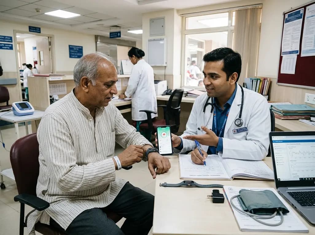
A multicenter observational study evaluated an 8-week digital health monitoring platform using wearable sensors in ambulatory heart failure. While participants achieved high daily wear-time of 11.7 hours and 80% completion, initial recruitment revealed significant technology-related onboarding barriers.
Today

A paired cadaveric study compared triple and quintuple percutaneous Achilles tendon lengthening. While quintuple hemisection showed numerically greater elongation, differences were not statistically significant.
Yesterday

A clinical decision-support framework using health checkup data from 231,683 adults predicts incident obesity with high discrimination (AUROC 0.918). The system integrates temporal survival analysis and risk-factor explanations to prioritize individualized preventive interventions.
Today
