.webp&w=384&q=75)

Loading, please wait...
Loading, please wait...

Acute cholangitis typically presents with fever, right upper quadrant tenderness, and jaundice. However, atypical biliary infections can manifest insidiously as pyrexia of unknown origin. In these complex scenarios, unrecognized bacterial migration into the portomesenteric circulation can induce septic pylephlebitis, an infectious suppurative thrombosis of the portal vein. Clinicians frequently encounter severe diagnostic dilemmas when standard signs of ascending cholangitis remain absent. Consequently, delayed intervention may allow vascular thrombosis to progress toward chronic portal hypertension and cavernous transformation.
Suppurative infection within the abdominal cavity can seed bacteria directly into the draining mesenteric and portal vasculature. While diverticulitis and appendicitis historically represent the most common triggers, occult ascending cholangitis remains an underrecognized culprit. Furthermore, inflamed biliary ductal walls share intimate microvascular channels with the adjacent portal venous network. When bacterial proliferation overwhelms local immune defenses, thrombogenic cytokines and endotoxins promote endothelial injury. This pathological cascade rapidly induces platelet aggregation and localized fibrin deposition. Therefore, microvascular thrombosis extends into the main portal vein trunk or intrahepatic branches. In addition, polymicrobial pathogens, predominantly Gram-negative enteric bacilli and anaerobes, sustain persistent endophlebitis. If clinicians do not halt this progressive inflammatory process, total portal occlusion impairs hepatic perfusion. Consequently, patients develop portal biliopathy, non-cirrhotic portal hypertension, and secondary gastrointestinal varices despite normal hepatic parenchymal stiffness.
Classic biliary sepsis presents with Charcot triad or Reynolds pentad, but subacute presentations often diverge substantially. Patients with occult biliary sepsis may present solely with prolonged intermittent fever, profound malaise, and progressive weight loss. Moreover, laboratory investigations frequently demonstrate marked systemic inflammation, such as extreme elevations in C-reactive protein, serum ferritin, and leukocytosis with neutrophilia. Hepatic panels typically reveal a mixed cholestatic-hepatitic pattern with elevated alkaline phosphatase and transaminases, whereas synthetic function remains entirely preserved. Blood and urine cultures often yield negative results due to previous empirical antibiotic exposure or intermittent bacteremic showering. Consequently, physicians may misdiagnose the condition as an occult malignancy, an autoimmune disease, or non-cirrhotic portal hypertension. Furthermore, the development of esophageal or gastric varices on routine endoscopic evaluation may mislead clinicians into suspecting chronic cryptogenic liver disease instead of active vascular infection.
Transabdominal ultrasonography functions as the conventional first-line modality for evaluating elevated liver enzymes and biliary symptoms. However, transabdominal ultrasound exhibits notable limitations in detecting early septic pylephlebitis and occult intraductal pathology. Acoustic shadowing from overlying bowel gas frequently obscures the confluence of the portal vein and distal common bile duct. In addition, early non-occlusive thrombi or subtle wall thickening along mesenteric vessels can evade sonographic detection. Although ultrasound may reveal mild intrahepatic or extrahepatic ductal dilatation, it often fails to identify the underlying obstructing lesion or microscopic biliary stricture. Therefore, normal or equivocal Doppler signals can create false reassurance, delaying appropriate therapeutic decisions. Clinicians must recognize that a negative sonogram cannot definitively exclude suppurative portal thrombophlebitis when patients present with persistent inflammatory markers and unexplained fevers.
Prompt cross-sectional imaging provides definitive diagnostic clarity in suspected portomesenteric infections. Specifically, contrast-enhanced computed tomography (CECT) and magnetic resonance cholangiopancreatography (MRCP) accurately delineate both the biliary tree and the vascular architecture. CECT scans reliably demonstrate luminal filling defects, portal vein wall enhancement, and surrounding mesenteric fat stranding. Furthermore, MRCP precisely visualizes subtle biliary strictures, intraductal sludge, and compression from periportal vascular networks. When portal thrombosis becomes chronic or complete, periportal venous collaterals rapidly develop, resulting in cavernous transformation. These extensive serpentine collateral vessels can encase and compress the extrahepatic bile ducts, producing secondary portal biliopathy. Consequently, early imaging prevents progressive vascular remodeling, minimizes ischemic hepatic injury, and guides targeted therapeutic drainage procedures when biliary obstruction is present.
Aggressive and targeted antimicrobial therapy constitutes the absolute cornerstone of managing portal vein bacteremia. Because portal infections typically arise from gastrointestinal flora, initial empirical regimens must provide comprehensive coverage against enteric Gram-negative bacilli, streptococci, and anaerobes. Clinicians commonly initiate intravenous broad-spectrum beta-lactams with beta-lactamase inhibitors or third-generation cephalosporins combined with metronidazole. In critically ill patients, carbapenems provide dependable monotherapy. Furthermore, physicians should adjust antimicrobial regimens promptly according to blood culture isolates or aspirated biliary fluid. Intravenous therapy should generally continue for at least two to three weeks until systemic inflammatory markers and fevers resolve completely. Subsequently, clinicians transition patients to oral agents to complete a total antimicrobial course of four to six weeks, thereby preventing septic recurrence and metastatic hepatic abscess formation.
The clinical decision to initiate anticoagulation requires careful individualized risk assessment, balancing recanalization benefits against hemorrhagic risks. Emerging evidence strongly supports early therapeutic anticoagulation to arrest thrombus extension and prevent complete mesenteric venous occlusion. Low-molecular-weight heparin or unfractionated heparin represents the initial therapy of choice, which physicians later transition to direct oral anticoagulants or vitamin K antagonists. Moreover, timely anticoagulation significantly enhances the rates of complete or partial portal vein recanalization. Follow-up Doppler ultrasonography or contrast imaging at three months helps evaluate venous patency and guide the overall duration of anticoagulation. However, patients with prominent esophageal varices require careful endoscopic surveillance and prophylaxis, such as endoscopic band ligation, to prevent life-threatening variceal hemorrhage during anticoagulation therapy.
Occult cholangitis triggers severe localized inflammation within intrahepatic and extrahepatic biliary ducts. Bacterial endotoxins and pro-inflammatory cytokines penetrate adjacent portomesenteric venules, causing endothelial damage and activating the coagulation cascade. Consequently, suppurative thrombosis develops within the portal venous system. This infectious thrombosis sustains bacteremia and causes persistent pyrexia, even when classic signs of acute biliary obstruction remain absent.
Contrast-enhanced computed tomography (CECT) and magnetic resonance cholangiopancreatography (MRCP) represent the most reliable imaging modalities. While transabdominal ultrasound often misses non-occlusive thrombi due to bowel gas, CECT clearly depicts intraluminal filling defects and perivascular inflammation. Additionally, MRCP accurately delineates subtle biliary abnormalities, portal biliopathy, and cavernous transformation, ensuring prompt and accurate clinical diagnosis.
Although antibiotic therapy is the primary treatment, clinicians strongly recommend therapeutic anticoagulation for acute septic pylephlebitis unless severe contraindications exist. Early anticoagulation prevents thrombus propagation into the mesenteric veins, lowers bowel infarction risk, and promotes venous recanalization. However, physicians must carefully screen for gastrointestinal varices and implement bleeding prophylaxis before initiating sustained anticoagulant regimens.
Disclaimer: This content is for informational and educational purposes only and does not constitute medical advice, diagnosis, or treatment. Healthcare professionals should rely on their clinical judgment and verify information independently. Refer to the latest local and national guidelines for clinical practice.
References

Read summarized clinical updates, watch expert medical content, and earn CME certifications right from your smartphone.
Open your smartphone camera to scan and install.





Septic pylephlebitis can present atypically as pyrexia of unknown origin without classic biliary symptoms. Early cross-sectional imaging, targeted broad-spectrum antimicrobial therapy, and prompt anticoagulation are crucial to reverse thrombosis, prevent portal hypertension, and avert cavernous transformation.
last month

Systemic amyloidosis management is undergoing a therapeutic revolution. This clinical review synthesizes updates from the International Society of Amyloidosis, detailing novel plasma cell therapies, TTR stabilizers, silencers, translational models, and artificial intelligence in drug development.
Today

A retrospective study of 824 patients undergoing over 2,200 intradetrusor onabotulinumtoxinA injections identifies sex- and etiology-specific predictors of 2-week post-void residual, highlighting the influence of baseline residual volumes, toxin dosage, and distinct urodynamic parameters on urinary retention risk.
Today
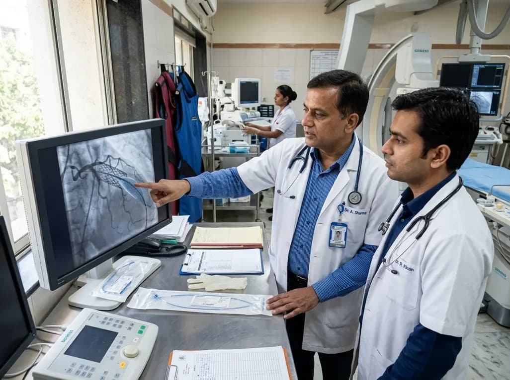
The US FDA has granted approval to the SELUTION SLR drug-eluting balloon for treating coronary in-stent restenosis. This milestone introduces a sustained-release sirolimus platform that eliminates the need for additional permanent metallic stents while maintaining robust vascular healing and luminal patency.
Today

The Thumbs-Out Classification (TOC) is a reliable screening tool for detecting thumb-in-palm deformity in children with cerebral palsy. Utilizing traffic-light tiers for radial abduction, TOC enables clinicians to monitor upper limb function and initiate timely therapeutic or surgical interventions.
Today

A comprehensive review analyzes periconceptional GLP-1 receptor agonist exposure, revealing reassuring data on congenital anomalies and adverse perinatal outcomes while emphasizing the need for cautious patient counseling.
Today
