

Loading, please wait...
Loading, please wait...

Myocardial contractility reflects the intrinsic capacity of cardiac muscle fibres to generate force and shorten against a load. Clinicians traditionally evaluate this function under resting conditions. However, evaluating dynamic contractile reserve offers far deeper diagnostic insight. The force-frequency relation describes how myocardial contractile performance changes as heart rate escalates. In healthy human hearts, contractility increases progressively with higher pacing rates, a phenomenon known as the Bowditch staircase. Consequently, intracellular calcium handling accelerates, which promotes stronger cross-bridge cycling and efficient systolic contraction. Conversely, pathological states disrupt this vital physiological mechanism. In failing myocardium, sarcoplasmic reticulum calcium ATPase activity declines significantly. As a result, the force-frequency relation flattens or reverses into a negative slope during tachycardia. This blunted inotropic response severely compromises cardiac reserve during physical stress. Therefore, accurate measurement of the force-frequency relation provides clinicians with an early, sensitive marker of subclinical myocardial dysfunction. Detecting these subtle contractile abnormalities allows timely risk stratification before overt ventricular dilation occurs. Furthermore, understanding this physiological relationship helps cardiologists tailor therapeutic regimens, particularly when optimising beta-blocker dosages or device-based pacing strategies in patients with cardiomyopathy.
Diagnostic imaging laboratories historically served as the cornerstone for investigating contractile reserve. In stress echocardiography laboratories, clinicians determine the force-frequency relation by calculating the ratio of systolic blood pressure to end-systolic volume index across progressive exercise or pacing stages. This non-invasive assessment effectively identifies impaired myocardial reserve in coronary artery disease and dilated cardiomyopathy. Similarly, nuclear laboratories utilise gated radionuclide angiography or positron emission tomography to evaluate ventricular volume changes alongside perfusion deficits. These nuclear modalities provide high diagnostic reproducibility. Nevertheless, conventional laboratory evaluations possess distinct operational constraints. Stress echocardiography remains highly operator-dependent, requiring exceptional sonographer expertise and clear acoustic windows. Moreover, pharmacological stress agents such as dobutamine or dipyridamole may not accurately mirror everyday physiological exertion. Physical stress protocols often introduce motion artefacts that hinder precise endocardial border delineation during peak exercise. In addition, routine lab-based tests capture only isolated snapshots of cardiac performance within artificial clinical settings. Patients rarely experience real-world emotional stress or environmental variations during laboratory assessments. Therefore, while stress imaging provides valuable baseline diagnostic data, cardiologists increasingly require longitudinal tools that extend dynamic contractile evaluation beyond traditional laboratory walls.
Recent bioengineering innovations have introduced transcutaneous force sensors that capture cardiac mechanics directly from the chest surface. Forcecardiography and seismocardiography record the subtle mechanical vibrations generated during isovolumic myocardial contraction and ejection. Specially calibrated piezoelectric elements and force-sensing resistors measure chest wall displacement with millisecond precision. Consequently, these cutaneous sensors generate operator-independent signals that closely correlate with invasively measured contractility indices. Clinicians can continuously track mechanical force alongside heart rate changes during spontaneous ambulatory movement. Furthermore, miniaturised wearable patches integrate multi-axial accelerometers and single-lead electrocardiography into cohesive, lightweight systems. These modern devices eliminate the need for bulky equipment or continuous ultrasound probes. As a result, patients can perform daily household routines without physical encumbrance or discomfort. Machine learning algorithms filter movement artefacts and extract clean cardiac contraction signatures in real time. Thus, ambulatory sensors transform contractile reserve monitoring from an episodic laboratory procedure into an unobtrusive, continuous physiological evaluation. In addition, these wearable systems capture hemodynamic fluctuations during natural circadian cycles. This continuous monitoring reveals transient episodes of contractile blunting that standard resting clinics would otherwise overlook entirely.
Implantable electronic devices represent another powerful platform for evaluating myocardial force under physiological conditions. Contemporary pacemakers and cardiac resynchronisation therapy systems continuously record intracardiac electrograms and hemodynamic metrics. For instance, integrated intracardiac accelerometers measure peak endocardial acceleration during early systole. This measurement reflects the maximal rate of ventricular pressure rise, serving as a reliable surrogate for contractility. Consequently, smart pacemakers can construct dynamic force-frequency curves across fluctuating heart rates during day-to-day physical activity. Clinicians can remotely monitor whether pacing-induced rate acceleration enhances or diminishes inotropic output. Moreover, pulmonary artery pressure sensors deliver continuous hemodynamic data that complement contractile tracking. When myocardial contractility declines, filling pressures rise days before symptoms emerge. Remote monitoring networks transmit these synchronized diagnostic metrics directly to cloud-based clinical platforms. Therefore, care teams receive timely notifications regarding deteriorating cardiac performance long before overt decompensation occurs. In addition, implantable monitors eliminate common adherence issues associated with patient-worn wearables. Patients receive continuous, passive physiological surveillance without altering their daily behaviour. Accordingly, implantable tracking bridges the gap between acute intervention and chronic disease surveillance.
Tracking contractile dynamics provides profound therapeutic utility for patients suffering from chronic heart failure and coronary artery disease. In chronic heart failure with reduced ejection fraction, a downward force-frequency curve heralds impending clinical worsening. Because wearable sensors detect blunted contractility during modest exertion, cardiologists can intervene preemptively. Specifically, clinicians can adjust neurohormonal blockade, up-titrate mineralocorticoid receptor antagonists, or modify diuretic dosages well before hospitalisation becomes necessary. Furthermore, continuous inotropic assessment guides optimal beta-blocker titration. Physicians can determine the precise resting heart rate that maximizes cardiac output without precipitating contractile exhaustion. Similarly, in patients with obstructive coronary artery disease, regional ischemia triggers immediate contractile deceleration during tachycardia. Sensor-derived force monitoring identifies ischemia-induced contractility failure during routine daily exertion. Consequently, this technology uncovers silent myocardial ischemia that routine resting electrocardiograms frequently miss. In addition, post-infarction risk stratification benefits from objective contractile reserve tracking. Patients displaying preserved force-frequency slopes achieve superior long-term survival compared to those exhibiting flat or inverted curves. Ultimately, incorporating sensor-based functional metrics transforms cardiology practice from reactive crisis management into proactive, personalised chronic disease care.
Deploying sensor-based contractile monitoring across diverse clinical environments presents distinct operational challenges and immense opportunities. In developing healthcare ecosystems like India, heart failure manifests at a significantly younger age due to high rates of premature coronary disease and uncontrolled hypertension. Remote hemodynamic monitoring could dramatically reduce the overwhelming burden of hospital readmissions across tertiary and rural medical centers. However, broad translation requires overcoming several technical hurdles. First, motion artefacts caused by rigorous physical activity can degrade cutaneous signal quality. Device engineers must refine adaptive filtering algorithms to preserve precision. Second, continuous data generation creates substantial transmission bandwidth and processing requirements. Clinics need robust digital infrastructure and secure cloud interfaces to prevent physician cognitive overload. Furthermore, commercial device cost and health insurance coverage remain critical barriers for widespread adoption across socioeconomic tiers. Frugal engineering approaches, utilizing cost-effective wearable patches integrated with ubiquitous smartphone networks, offer a viable solution for the Indian subcontinent. In addition, professional societies must establish standardized clinical validation protocols and clear diagnostic thresholds. As sensor technology matures, remote contractile monitoring will empower Indian physicians to deliver predictive, home-based cardiovascular care to millions of vulnerable patients.
In a healthy heart, progressive increases in heart rate induce stronger myocardial contractions due to accelerated calcium cycling and enhanced inotropic reserve. Consequently, the force-frequency curve exhibits a positive upward slope. In contrast, failing myocardium suffers from impaired calcium handling and down-regulated beta-adrenergic receptors. Therefore, the curve flattens or reverses into a negative slope during exertion. This blunted inotropic response leads to rapid hemodynamic exhaustion and symptom exacerbation during daily activities.
Standard stress echocardiography requires expensive ultrasound equipment, expert operators, and dedicated laboratory space, providing only brief snapshots under artificial stress. In contrast, wearable cutaneous force sensors record continuous, operator-independent measurements of cardiac mechanical vibrations during natural daily routines. Furthermore, these lightweight patches eliminate acoustic window limitations and enable long-term remote hemodynamic surveillance. Consequently, clinicians gain longitudinal physiological data, facilitating early detection of subclinical cardiac decompensation without requiring frequent hospital visits.
Yes, continuous force-frequency monitoring provides an early warning mechanism for impending heart failure decompensation. Pathological contractile blunting and subtle hemodynamic derangements typically appear several days before overt fluid overload or physical symptoms develop. By detecting these early inotropic shifts, wearable and implantable sensor platforms trigger automated alerts for healthcare providers. Consequently, physicians can proactively adjust oral diuretic doses and neurohormonal medications at home, successfully preventing acute disease destabilization and reducing costly emergency admissions.
Disclaimer: This content is for informational and educational purposes only... Refer to the latest local and national guidelines for clinical practice.
References

Read summarized clinical updates, watch expert medical content, and earn CME certifications right from your smartphone.
Open your smartphone camera to scan and install.





Discover sensor-based force-frequency relation monitoring, bridging stress labs and wearables to advance personalised care in heart failure and coronary disease.
Today
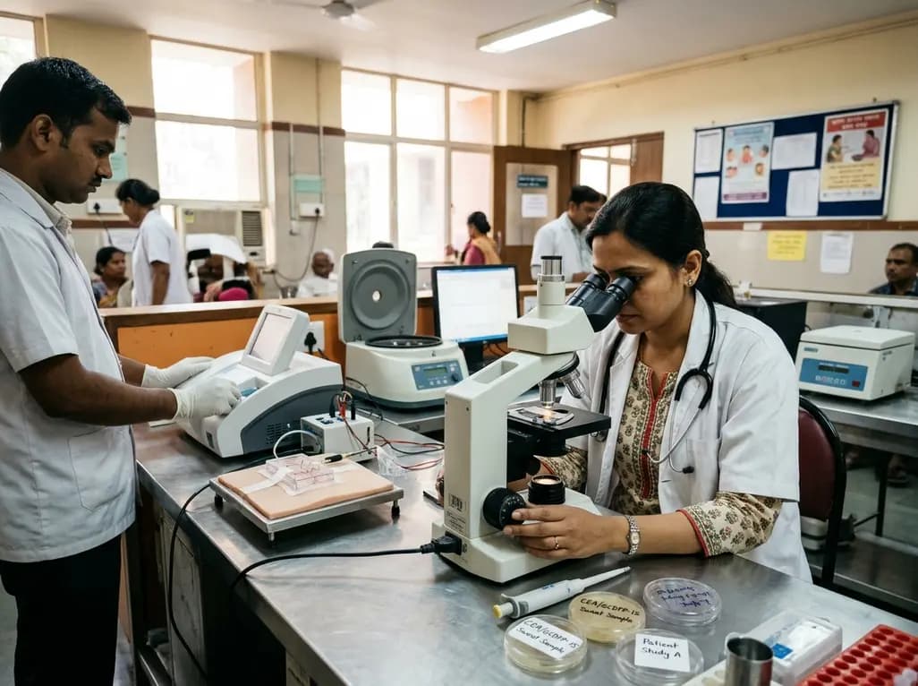
A pilot study evaluated WearDOXXN, a capillary microfluidic sweat patch, for noninvasive breast cancer monitoring by comparing sweat and serum CEA and PIP/GCDFP-15 levels.
Yesterday
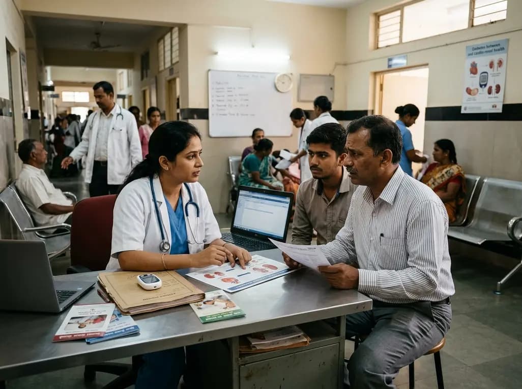
India faces a massive diabetes burden with over 89 million adults affected. Managing blood sugar in isolation fails to prevent severe cardiorenal complications. Clinicians must adopt an integrated metabolic ecosystem that prioritizes early detection, multifactorial risk assessment, and personalized therapies.
Yesterday
A qualitative study highlights the clinical role of MRI in axial spondyloarthritis, exposing disparities in access, reporting gaps in private healthcare, and the urgent need for standardized imaging protocols and multidisciplinary collaboration between rheumatologists and radiologists.
Today
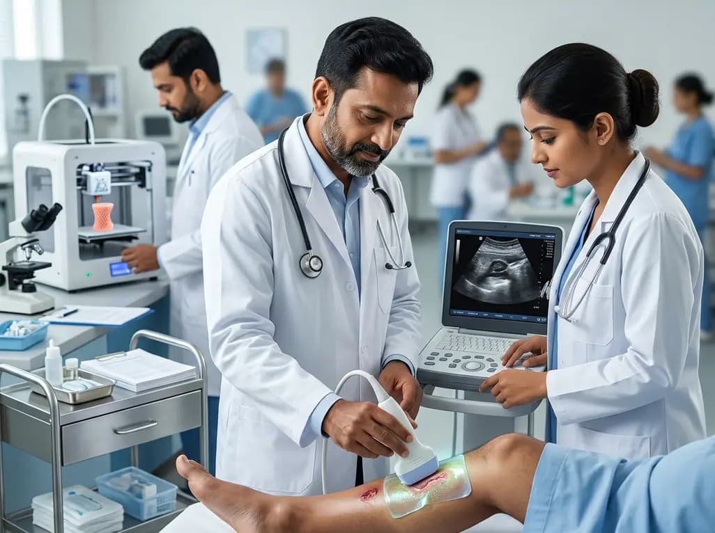
A breakthrough 3D-printed piezoelectric collagen hydrogel activated by ultrasound accelerates diabetic wound repair, demonstrating 98.5% wound closure, robust antibacterial action, and enhanced vascularization without toxic chemical crosslinkers.
Yesterday
