

Loading, please wait...
Loading, please wait...

Axial spondyloarthritis represents a painful chronic inflammatory condition that predominantly targets the sacroiliac joints and spine. Clinicians increasingly rely on advanced imaging to identify active inflammatory sacroiliitis long before structural deformities appear on plain radiographs. Consequently, the optimal utilization of MRI in axial spondyloarthritis has emerged as a cornerstone for timely diagnosis and therapeutic decision-making. However, daily clinical practice reveals striking variability in how physicians request, acquire, and interpret these scans across different healthcare systems.
Early identification of active sacroiliitis remains paramount for preventing irreversible structural fusion and persistent disability. Therefore, rheumatologists utilize magnetic resonance imaging to capture subchondral bone marrow edema alongside capsulitis and enthesitis. These acute inflammatory lesions appear prominently on fat-suppressed T2-weighted or short tau inversion recovery sequences. In contrast, conventional radiography only displays chronic, late-stage alterations like erosions, sclerosis, and ankylosis. Thus, cross-sectional imaging closes a critical diagnostic window in patients presenting with non-radiographic disease.
Nevertheless, clinicians must interpret active bone marrow edema with caution. Similar inflammatory signals frequently appear in healthy mechanical stress, postpartum osteitis condensans ilii, degenerative changes, and intense athletic training. For this reason, practitioners should carefully correlate imaging findings with HLA-B27 status, inflammatory back pain symptoms, and elevated acute-phase reactants. When radiologists identify structural damage such as fatty marrow metaplasia and erosions alongside active inflammation, diagnostic specificity increases significantly. Ultimately, a holistic clinical evaluation prevents the pitfalls of overreliance on isolated signal hyperintensities.
Beyond initial diagnosis, physicians also employ magnetic resonance imaging to monitor therapeutic responses to biologic and targeted synthetic disease-modifying antirheumatic drugs. For example, tumor necrosis factor inhibitors and interleukin-17 inhibitors routinely reduce active osteitis within months of initiation. Accordingly, imaging can confirm whether persistent symptoms reflect active inflammatory flare or concomitant fibromyalgia and mechanical wear. Hence, objective scan data often guides crucial decisions regarding biologic switching or dose escalation.
However, routine protocol scanning in asymptomatic patients remains contentious among international experts. Many clinical guidelines argue against repetitive imaging when patients demonstrate sustained clinical and biochemical remission. Furthermore, residual bone marrow changes do not always correlate perfectly with patient-reported pain scores or validated disease activity instruments. Consequently, rheumatologists reserve surveillance scans primarily for ambiguous clinical presentations, suspected paradoxical flare-ups, or prior to medication tapering. By exercising judicious clinical restraint, physicians avoid unnecessary interventions and unnecessary medical expenses.
Recent qualitative investigations uncover notable discrepancies between public academic centres and ambulatory private radiology facilities. Specifically, clinicians report that private healthcare facilities often supply descriptive but non-standardized narrative reports. These ambiguous summaries frequently fail to address the validated Assessment of SpondyloArthritis international Society criteria. As a result, treating rheumatologists must repeatedly re-evaluate original digital imaging files to determine whether true sacroiliitis exists.
Moreover, significant inter-centre differences in scanner hardware access create prolonged diagnostic delays for referred patients. Rural and peripheral secondary health centres often struggle with lengthy waiting times for specialized scans. In response, some patients seek rapid evaluations in independent private clinics, which can introduce conflicting interpretations. Consequently, fragmented documentation undermines continuity of care and delays the initiation of essential biological therapy. Addressing these institutional gaps requires unified regional imaging standards that transcend individual hospital sectors.
Heterogeneity in imaging protocols represents another substantial barrier to reliable disease management across centers. Inappropriate slice orientation or omission of semi-coronal views along the sacroiliac axis frequently causes inconclusive studies. Therefore, expert consensus mandates precise acquisition planes angled parallel and perpendicular to the long axis of the sacral bone. Omitting dedicated T1-weighted sequences also hampers the comprehensive detection of structural fatty lesions, backfill, and erosive defects.
Furthermore, physicians occasionally order extensive whole-spine imaging when focused pelvic views would provide definitive diagnostic confirmation. Although thoracic and cervical evaluations prove indispensable for advanced ankylosing spondylitis, isolated sacroiliac studies typically suffice for early non-radiographic presentations. Standardized prescription templates can guide general practitioners, orthopedists, and internists toward proper initial imaging orders. Consequently, adopting harmonized electronic requisition pathways enhances diagnostic yield while optimizing valuable radiologic machine time.
Collaborative communication between rheumatologists and musculoskeletal radiologists plays an indispensable role in diagnostic accuracy. Qualitative studies consistently show that both specialties crave structured platforms for interdisciplinary case discussions. Unfortunately, tight clinical schedules and compartmentalized department workflows often limit routine multidisciplinary interactions. When radiologists lack adequate clinical history regarding inflammatory back pain features, they naturally produce overly defensive or broad differential lists.
Similarly, rheumatologists who do not communicate specific clinical hypotheses miss the opportunity for targeted radiological consultation. Implementing standardized structured reporting templates substantially resolves these communication barriers. Structured reports systematically grade the location, depth, and extent of bone marrow edema while explicitly evaluating structural hallmarks. Consequently, joint clinical-radiological grand rounds and shared digital reading interfaces create mutual learning environments that enhance clinical decision confidence.
From the patient perspective, undergoing repeated magnetic resonance scans can trigger considerable psychological anxiety and physical discomfort. Many individuals experience claustrophobia within enclosed scanner bores or endure severe back pain while lying completely motionless on rigid tables. Moreover, patients frequently encounter deep distress when confronting conflicting terminology between medical consultations and technical imaging reports. For instance, benign mechanical degenerative findings are easily misinterpreted by patients as signs of aggressive, irreversible spinal damage.
Therefore, proactive patient education represents a fundamental component of compassionate chronic disease care. Clinicians must clearly explain the rationale behind ordering each scan and outline realistic expectations regarding scan duration and noise. Additionally, physicians should take time to translate technical imaging terminology into reassuring, actionable language during follow-up visits. When healthcare teams address patient concerns openly, they foster stronger therapeutic alliances and encourage sustained long-term treatment adherence.
Clinicians should order an MRI when a patient under 45 presents with chronic inflammatory back pain lasting over three months, especially if plain radiographs appear normal. Because early non-radiographic axial spondyloarthritis lacks radiographic structural damage, semi-coronal MRI of the sacroiliac joints detects active subchondral bone marrow edema. This allows early diagnosis and timely therapeutic intervention before irreversible spinal fusion develops.
Standard evaluation requires dedicated semi-coronal views angled parallel to the sacral slope. Clinicians must request both fluid-sensitive sequences, such as STIR or fat-suppressed T2-weighted scans, and anatomical T1-weighted turbo spin-echo sequences. Fluid-sensitive sequences highlight active inflammatory bone marrow edema, while T1-weighted images reveal chronic structural changes, including subchondral sclerosis, erosions, fatty marrow metaplasia, and ankylosis.
Yes, MRI effectively distinguishes inflammatory sacroiliitis from mechanical conditions when experts evaluate the distribution and type of lesions. Axial spondyloarthritis typically exhibits intense, multi-quadrant subchondral bone marrow edema associated with erosions and structural fatty transformation. Conversely, mechanical stress, osteitis condensans ilii, or degenerative disc disease produce localized, triangular, or superficial anterior changes without true inflammatory erosions.
Disclaimer: This content is for informational and educational purposes only. It is not intended to be a substitute for professional medical advice, diagnosis, or treatment. Always seek the advice of your physician or other qualified health provider with any questions you may have regarding a medical condition. Refer to the latest local and national guidelines for clinical practice.
References

Read summarized clinical updates, watch expert medical content, and earn CME certifications right from your smartphone.
Open your smartphone camera to scan and install.





A qualitative study highlights the clinical role of MRI in axial spondyloarthritis, exposing disparities in access, reporting gaps in private healthcare, and the urgent need for standardized imaging protocols and multidisciplinary collaboration between rheumatologists and radiologists.
Today
In cognitive impairment and dementia, residents often lose the ability to articulate discomfort. Consequently, clinicians frequently misinterpret pain indicators as behavioral disturbances. Discover how structured observational pain assessment protocols improve clinical outcomes and curb inappropriate psychotropic use.
Today
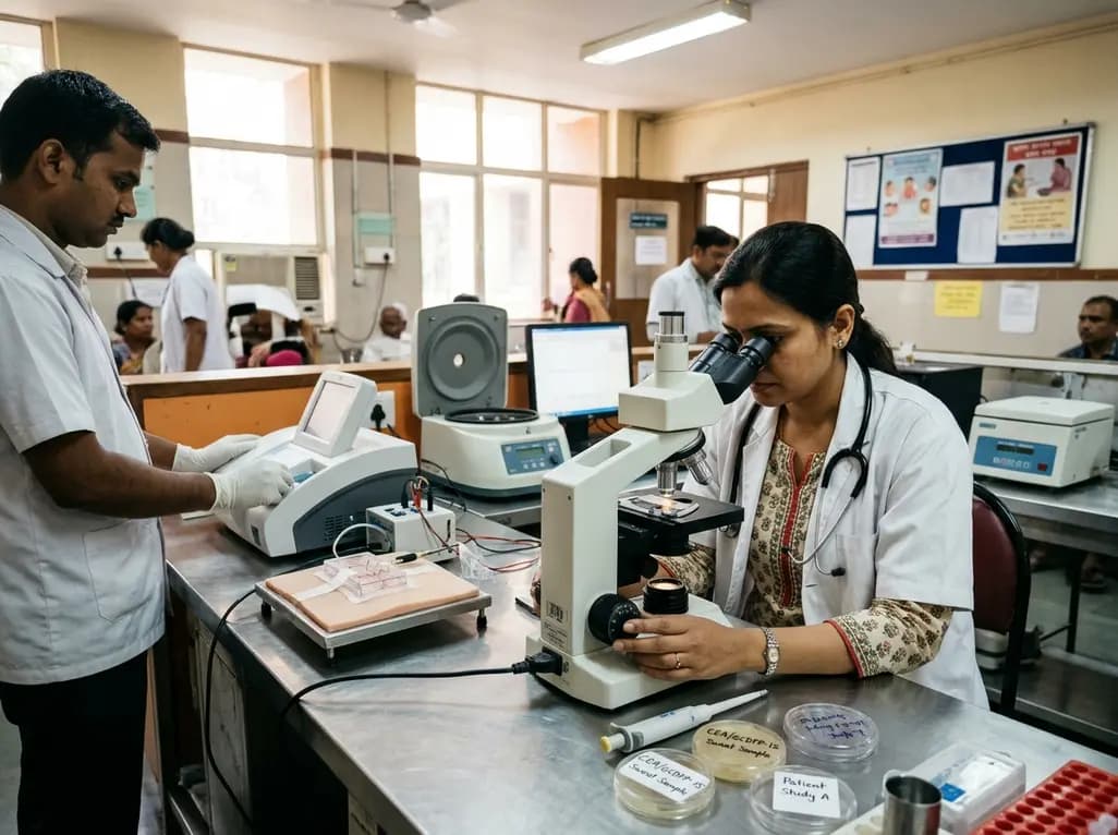
A pilot study evaluated WearDOXXN, a capillary microfluidic sweat patch, for noninvasive breast cancer monitoring by comparing sweat and serum CEA and PIP/GCDFP-15 levels.
Yesterday

India faces a massive diabetes burden with over 89 million adults affected. Managing blood sugar in isolation fails to prevent severe cardiorenal complications. Clinicians must adopt an integrated metabolic ecosystem that prioritizes early detection, multifactorial risk assessment, and personalized therapies.
Yesterday

A breakthrough 3D-printed piezoelectric collagen hydrogel activated by ultrasound accelerates diabetic wound repair, demonstrating 98.5% wound closure, robust antibacterial action, and enhanced vascularization without toxic chemical crosslinkers.
Yesterday
