

Loading, please wait...
Loading, please wait...

Precise asterion localization remains an essential component of posterior cranial fossa surgery. Neurosurgeons frequently perform retrosigmoid craniotomies to access lesions in the cerebellopontine angle, such as acoustic neuromas, meningiomas, and neurovascular compression syndromes. During these operative procedures, surgeons must expose the pathology while meticulously protecting the transverse-sigmoid sinus complex. The venous sinus junction represents an anatomical boundary that guides bone flap design. Consequently, inadvertent sinus puncture can induce catastrophic venous hemorrhage, sinus thrombosis, or air embolism. Anatomists and surgeons historically identified the asterion—the junction where the lambdoid, parietomastoid, and occipitomastoid sutures meet—as a reliable surface proxy for the underlying venous sinus confluence. Therefore, craniotomy planning often begins by identifying this junction on the skull surface. However, locating the asterion through an intact scalp poses substantial clinical challenges. Modern surgical teams increasingly depend on advanced neuronavigation systems to map these structures. Nevertheless, navigation equipment is not universally accessible in resource-constrained environments. Additionally, registration errors or brain shift can compromise navigation reliability. As a result, robust surface-based cranial topography remains a vital clinical tool for safe operative planning.
Although the bony landmarks provide clear anatomical markers on dry skulls, translating these landmarks to the intact scalp presents clear difficulties. Soft tissue thickness varies significantly across patient demographics. Moreover, the thick musculature of the suboccipital and retroauricular regions obscures the underlying cranial sutures during the initial surgical incision. Historically, surgeons relied on palpable external markers, including the mastoid tip, the inion, and the zygomatic arch. However, standard anthropometric planes often fail to provide straightforward intraoperative utility. For instance, the classical Frankfort horizontal plane uses the superior margin of the external auditory meatus and the inferior orbital rim. Unfortunately, locating the osseous porion during surgery without extensive imaging is challenging. Furthermore, placing measuring instruments directly into the ear canal causes patient discomfort and increases contamination risks. Consequently, clinicians need a reliable, superficial surrogate that preserves anatomical rigor. Recent investigative work demonstrates that modern three-dimensional computed tomography volume rendering bridges this critical gap. Fused imaging techniques allow researchers to coregister skin surfaces with the deep osseous and vascular anatomy. Thus, morphologists can define external reference lines that clinicians can effortlessly replicate on the surgical drape.
To overcome traditional cephalometric hurdles, investigators developed the modified Frankfort horizontal plane (mFHP). This scalp-based surrogate connects the palpable infraorbital rim directly to the tragion on the lateral cranial view. Because both landmarks sit superficially, operative teams can palpate and mark them quickly without invasive instruments. Additionally, researchers constructed the asterion line, known as the A-line, which passes through the asterion perpendicular to the mFHP. In a comprehensive morphometric analysis, researchers evaluated 278 asterions across 140 adult patients using fused three-dimensional computed tomography reconstructions. The investigative team applied age-adjusted linear mixed-effects models with patient-specific random intercepts. Furthermore, the statistical framework incorporated Holm corrections for multiple comparisons to ensure rigorous data validation. The measurements demonstrated consistent geometric patterns across the entire cohort. Specifically, the perpendicular A-line successfully established the horizontal distance from the tragion to the asterion axis. Consequently, this coordinate system provides a practical grid for estimating posterior cranial coordinates. By aligning scalp landmarks with intracranial anatomy, the method establishes an intuitive roadmap for skull base approaches.
The morphometric evaluation revealed statistically significant differences between sexes regarding cranial dimensions. Specifically, the horizontal tragion-asterion distance measured 47.0 ± 4.2 mm in male patients. In contrast, female patients demonstrated a significantly shorter horizontal distance of 43.3 ± 4.1 mm. This sex-based divergence maintained strong statistical significance after Holm adjustment. However, researchers observed no statistically significant variance between the left and right sides of the skull. Therefore, unilateral cranial asymmetry does not meaningfully impact the horizontal coordinates of the asterion. Furthermore, the asterion consistently resided superior to the horizontal reference plane across both cohorts. The vertical distance from the modified Frankfort horizontal plane to the asterion also demonstrated reproducible trajectories. Importantly, these findings underline the danger of adopting uniform cranial measurements across all patients. Because male skulls typically exhibit larger cranial vault diameters, surgeons must tailor incision sites accordingly. Tailoring preoperative incision designs to patient-specific morphology prevents inadequate bony exposure. Consequently, understanding these morphometric differences equips clinicians with the precision necessary to avoid unexpected vascular encounters during bone flap elevation.
Accurate scalp projection directly translates to superior operative safety during retrosigmoid craniotomy. When surgeons place the initial burr hole, they must uncover the margin of the sinus junction without entering the lumen. Positioning the burr hole too far anteriorly or superiorly risks catastrophic laceration of the transverse or sigmoid sinuses. Conversely, positioning the craniotomy too far inferiorly or medially restricts visualization of the internal acoustic meatus and cerebellopontine angle. As a result, the surgeon might require excessive cerebellar retraction, which elevates the risk of postoperative contusion and edema. By utilizing the modified Frankfort horizontal plane and the corresponding A-line, surgical teams can outline the expected asterion trajectory onto the scalp prior to sterile draping. In addition, this technique guides the initial skin incision, whether selecting a straight vertical incision, a lazy-S pattern, or a curved retroauricular flap. Thus, the surgeon can center the exposure directly over the burr hole target. Furthermore, confirming the asterion coordinates helps operating room teams verify digital neuronavigation trajectories before opening the calvarium.
Although high-resolution image guidance systems represent the gold standard in modern skull base centers, widespread global access remains limited. In many public healthcare facilities across developing nations, including district hospitals in India, advanced neuronavigation consoles are not routinely available in elective or emergency theaters. Therefore, simple, anatomy-based topographical systems provide life-saving utility. A sterile ruler and surgical marker allow teams to trace the modified Frankfort horizontal plane and locate the asterion line within minutes. Furthermore, this landmark-based strategy reduces operative setup duration and lowers overall procedural costs. Even in tertiary facilities equipped with intraoperative navigation, mechanical failure, optical line-of-sight obstruction, or soft-tissue shift can disrupt system tracking. Consequently, neurosurgeons should never rely exclusively on automated devices. Mastering superficial cranial morphometry provides a vital mental safety net. In summary, validating scalp proxies through modern three-dimensional imaging unites traditional anatomical rigor with contemporary clinical safety. This hybrid perspective ensures that skull base surgeons maintain spatial confidence and achieve excellent clinical outcomes across diverse surgical environments.
The standard Frankfort horizontal plane connects the skeletal porion and the orbitale, requiring bony visualization. In contrast, the modified Frankfort horizontal plane connects the palpable scalp tragion to the infraorbital rim. This modification allows clinicians to identify the reference plane directly on the patient's skin without invasive skeletal exposure.
The asterion classically corresponds to the underlying junction between the transverse and sigmoid venous sinuses. However, anatomical variations occur frequently. The asterion typically lies superficial to the sinus complex or just superior to the transverse sinus, making precise scalp localization vital to prevent inadvertent vascular laceration during initial burr hole placement.
Significant sex differences arise primarily from variations in overall craniofacial dimensions and calvarial vault size. Males exhibit significantly greater horizontal distances from the tragion to the asterion line compared to females. Recognizing these morphometric variations ensures that neurosurgeons appropriately tailor scalp incisions and burr hole trajectories to each patient.
Disclaimer: This content is for informational and educational purposes only. It is not intended to be a substitute for professional medical advice, diagnosis, or treatment. Always seek the advice of your physician or other qualified health provider with any questions you may have regarding a medical condition or procedure. Never disregard professional medical advice or delay in seeking it because of something you have read here. Refer to the latest local and national guidelines for clinical practice.
References

Read summarized clinical updates, watch expert medical content, and earn CME certifications right from your smartphone.
Open your smartphone camera to scan and install.





A 3D CT study defines asterion localization via a modified Frankfort horizontal plane to guide safe retrosigmoid craniotomy, demonstrating significant sex-based morphometric differences.
Today

A Bayesian component network meta-analysis of 130 RCTs reveals that self-monitoring and gamification provide the clearest positive effects in mHealth physical activity interventions, while social comparison may have adverse psychological trade-offs.
Today

The GESS-AEEM consensus establishes a clear clinical implementation framework for testosterone therapy in postmenopausal women with hypoactive sexual desire disorder, focusing on transdermal dosing, precise laboratory monitoring, and safety communication.
Today

A multicenter observational study evaluated an 8-week digital health monitoring platform using wearable sensors in ambulatory heart failure. While participants achieved high daily wear-time of 11.7 hours and 80% completion, initial recruitment revealed significant technology-related onboarding barriers.
Today
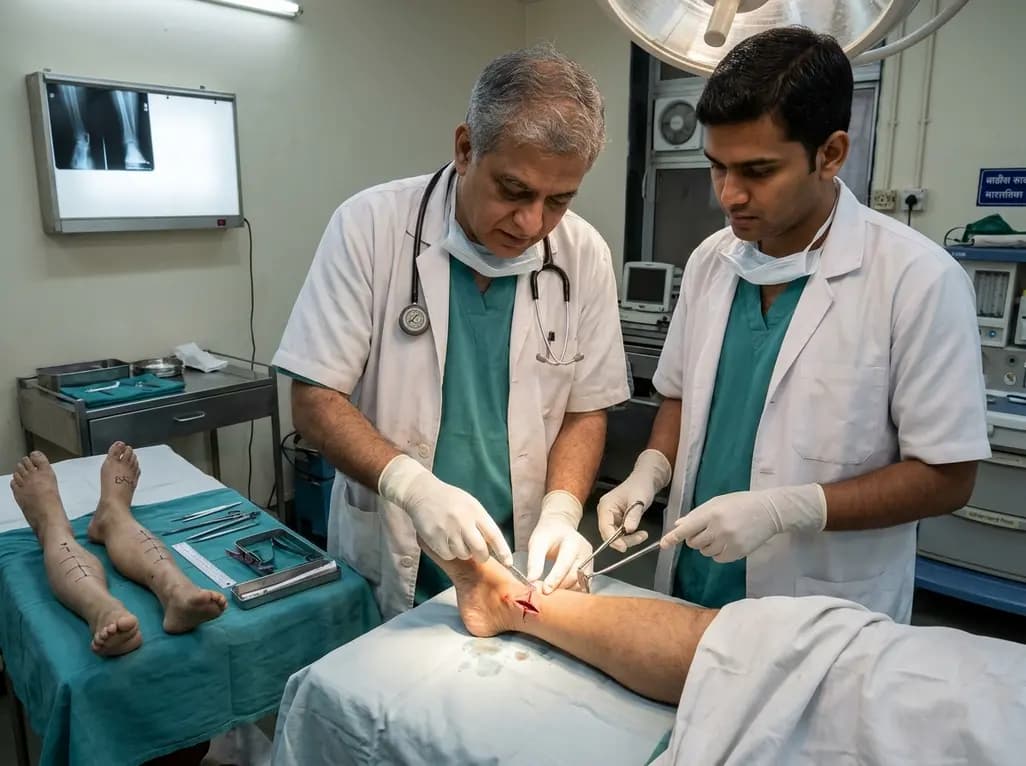
A paired cadaveric study compared triple and quintuple percutaneous Achilles tendon lengthening. While quintuple hemisection showed numerically greater elongation, differences were not statistically significant.
Yesterday

A clinical decision-support framework using health checkup data from 231,683 adults predicts incident obesity with high discrimination (AUROC 0.918). The system integrates temporal survival analysis and risk-factor explanations to prioritize individualized preventive interventions.
Today
