.webp&w=384&q=75)

Loading, please wait...
Loading, please wait...
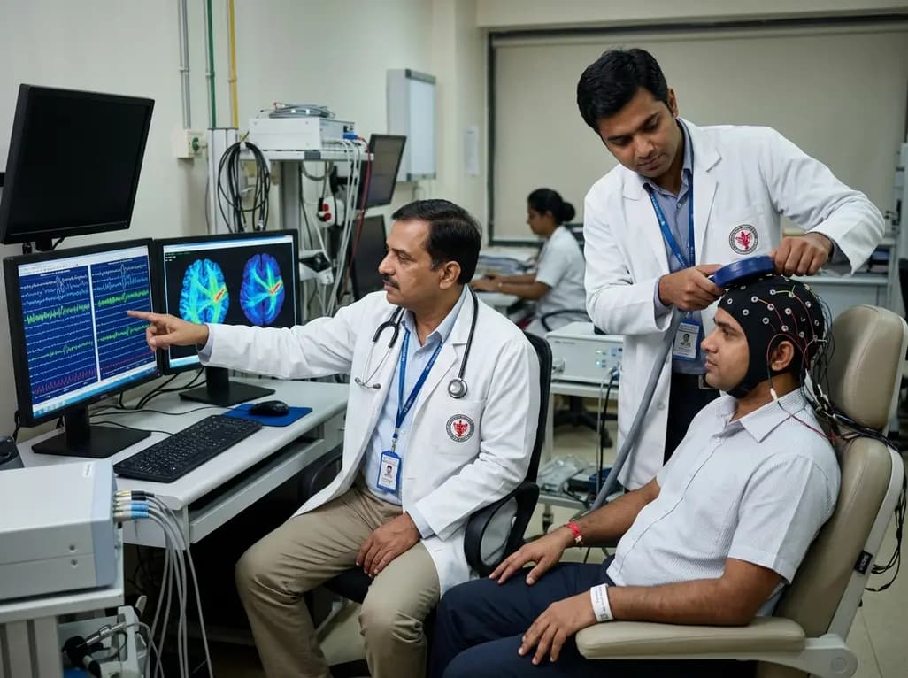
Clinicians and neurophysiologists increasingly recognise that dynamic brain states govern human neuroplasticity and therapeutic responses. Specifically, the sensorimotor mu rhythm represents an endogenous oscillatory rhythm between 8 and 13 Hertz. Transcranial magnetic stimulation delivers targeted pulses to quantify corticospinal excitability across these distinct oscillatory phases. When investigators stimulate the primary motor cortex during the trough of the mu rhythm, they typically record larger motor-evoked potentials than during the peak phase. However, considerable inter-individual variability has historically confounded the reproducibility of these neurophysiological measurements. While some individuals display robust phase-dependent facilitation, other subjects demonstrate blunted or atypical responses under identical stimulation protocols. Consequently, translational researchers have sought to identify structural neural markers that explain these disparate functional profiles. Intracortical microstructure and underlying white matter architecture offer plausible anatomical substrates for such physiological differences. By clarifying how structural connections modulate phase-dependent excitability, clinicians can transition from static stimulation regimens to responsive closed-loop paradigms. Therefore, understanding these fundamental sensorimotor relationships represents a major advancement for modern clinical neurophysiology. Ultimately, this knowledge empowers clinicians to refine diagnostic precision and therapeutic protocols in routine neurological practice.
The primary somatosensory cortex and primary motor cortex form highly integrated functional units that regulate voluntary motor execution. In particular, dense short-range U-fibers and intracortical horizontal axons support reciprocal communication across the central sulcus. Somatosensory afferent signals continuously shape motor output, adjusting corticospinal activation to meet ongoing environmental demands. Furthermore, advanced diffusion magnetic resonance imaging allows clinicians to examine the microstructural integrity of these connections in vivo. Quantitative indices such as fractional anisotropy and tract density provide robust estimates of axonal diameter, packing density, and myelination. When structural tracts exhibit high microstructural order, physiological signals traverse sensorimotor networks with exceptional temporal fidelity. In contrast, disorganized or degraded white matter pathways produce temporal dispersion and disrupt synaptic synchronization across regions. Neurologists frequently encounter this structural disruption in patients with stroke, traumatic brain injury, and neurodegenerative disorders. Consequently, evaluating white matter integrity between sensory and motor cortices clarifies why identical physiological stimuli provoke divergent functional responses. Thus, detailed structural characterization provides the anatomical foundation necessary for understanding individual oscillatory dynamics.
A recent investigation by Hougland and colleagues examined how somatosensory-to-motor structural connectivity predicts phase-dependent corticospinal excitability. The researchers combined real-time electroencephalography with single-pulse transcranial magnetic stimulation to measure motor-evoked potentials across distinct mu-rhythm phases. Simultaneously, they utilized high-resolution diffusion-weighted imaging to reconstruct microstructural pathways connecting sensory and motor regions. Interestingly, the experimental data revealed a strong positive correlation between structural tract connectivity and the magnitude of the mu-phase effect. Participants with higher microstructural organization between sensory and motor cortices exhibited significantly greater modulation of corticospinal excitability. In addition, the phase yielding peak excitability aligned consistently with the trough of the mu oscillation in these high-connectivity individuals. Conversely, participants with weaker microstructural connectivity demonstrated minimal phase sensitivity, displaying negligible amplitude differences across stimulation phases. Moreover, these anatomical relationships persisted independently of individual baseline motor thresholds or total oscillatory power. These pivotal findings confirm that structural tract properties directly govern real-time cortical excitability dynamics. Consequently, baseline structural connectivity serves as a reliable biomarker for predicting phase-dependent responsiveness.
Several distinct neurobiological mechanisms explain how somatosensory-to-motor connectivity modulates phase-dependent corticospinal excitability. First, somatosensory projections provide potent feedforward inhibition and facilitation directly onto motor corticospinal output neurons. Highly organized, myelinated axons ensure that rhythmic oscillatory signals propagate rapidly with minimal phase distortion. As a result, the sensorimotor network synchronizes local gamma-aminobutyric acid interneurons, establishing pronounced rhythmic variations in membrane potentials. During the mu-rhythm trough, diminished intracortical inhibition permits enhanced depolarization of corticospinal neurons, amplifying motor responses. In contrast, poorly organized axonal bundles introduce latency jitter and desynchronize incoming phase inputs. Consequently, the motor cortex fails to generate distinct intervals of heightened excitability, dampening observed phase effects. Furthermore, robust microstructural connections sustain recurrent functional loops between sensory and motor cortices, maintaining rhythmic stability across trials. When these structural pathways function optimally, external magnetic pulses elicit uniform, predictable motor-evoked potentials. Accordingly, microstructural organization operates as a vital physiological filter that dictates how sensorimotor oscillations govern corticospinal output.
These structural-functional discoveries offer major translational implications for modern neurorehabilitation and neurological practice. Historically, conventional repetitive transcranial magnetic stimulation protocols have applied rigid stimulation parameters without accounting for fluctuating endogenous brain states. Consequently, clinical trials in stroke rehabilitation and movement disorders have yielded inconsistent therapeutic benefits. Real-time closed-loop neuromodulation addresses this limitation by delivering stimulation precisely during peak-receptive oscillatory phases. However, the current findings demonstrate that clinicians cannot expect uniform responsiveness across all patients. Instead, clinical teams should assess individual structural connectivity using tractography prior to prescribing closed-loop neuromodulation. In Indian tertiary neurology centers, where stroke and neurotrauma burdens remain substantial, this biomarker-driven approach ensures rational patient selection. Patients with preserved sensory-motor connectivity can receive phase-locked stimulation protocols to accelerate motor recovery. Meanwhile, patients with severe microstructural disruption may require alternative therapeutic modalities or dual-site cortical priming. Therefore, integrating structural neuroimaging with real-time electrophysiology bridges the gap toward truly personalized neurorehabilitation. Ultimately, this approach optimizes clinical resource allocation while improving long-term neurological recovery.
The sensorimotor mu rhythm reflects rhythmic cycles of cortical excitation and inhibition. When magnetic pulses hit the primary motor cortex during the trough phase, local intracortical inhibition reaches its minimum. Consequently, pyramidal neurons depolarize more readily, generating significantly larger motor-evoked potentials compared to pulses delivered during the inhibitory peak phase.
Dense, highly organized axonal pathways between the somatosensory and motor cortices transmit oscillatory signals with superior temporal precision. In contrast, degraded structural tracts introduce conduction delays and signal desynchronization. Consequently, individuals with well-myelinated, organized sensory-motor connections maintain sharp oscillatory phase boundaries, enabling strong, predictable modulation of corticospinal output.
Clinicians can use diffusion tractography to evaluate sensory-motor structural integrity before prescribing phase-targeted brain stimulation. Patients with preserved microstructural connectivity will likely respond favorably to closed-loop EEG-TMS protocols. Conversely, patients with severe tract disruption may benefit more from priming strategies or combined multimodal therapies to optimize functional recovery.
Disclaimer: This content is for informational and educational purposes only and is intended for registered medical practitioners. It should not be used as a substitute for professional medical advice, diagnosis, or treatment. Healthcare providers must exercise independent clinical judgment when interpreting neurophysiological data or instituting neuromodulatory therapies. Refer to the latest local and national guidelines for clinical practice.
References

Read summarized clinical updates, watch expert medical content, and earn CME certifications right from your smartphone.
Open your smartphone camera to scan and install.





New research reveals that microstructural connectivity between primary somatosensory and motor cortices predicts the sensorimotor mu-rhythm phase effect on corticospinal excitability. These findings offer vital biomarkers for personalizing closed-loop EEG-TMS therapies in modern neurorehabilitation.
4 weeks back

Recent research defines ferro-aging as a conserved iron-lipid axis driving organ decline across primates. Age-related iron dyshomeostasis stimulates ACSL4-mediated lipid peroxidation, promoting cellular senescence. Notably, vitamin C directly inhibits ACSL4, suppressing phospholipid oxidation and multi-organ decay.
Today
A retrospective cohort study evaluated 1,000 low-risk singleton births to assess maternal and neonatal outcomes during office-hours versus off-hours. Off-hours deliveries were significantly associated with higher odds of intrapartum cesarean sections and episiotomies, highlighting critical obstetric staffing considerations.
Today

New clinical evidence demonstrates a shift away from strict exercise restrictions in genetic heart disease. While patients with channelopathies engage in more vigorous activity than those with cardiomyopathies, shared decision-making and individualized risk assessments ensure safe, beneficial exercise participation.
Today
Sarcopenia significantly impairs functional independence and amplifies fracture risks in patients with osteoarthritis and osteoporosis. Learn about modern diagnostic strategies, GLP-1 therapy considerations, and evidence-based non-pharmacological management across multidisciplinary clinical settings.
Today

Discover how to identify superimposed entrapment neuropathy in diabetic patients. This review details clinical evaluation, surgical decompression evidence, and a structured algorithm for primary care referrals to prevent ulcers and preserve limb function.
Today
