.webp&w=384&q=75)

Loading, please wait...
Loading, please wait...
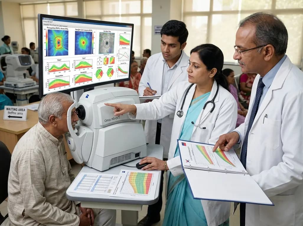
In the evolving landscape of ophthalmic diagnostics, optical coherence tomography (OCT) has emerged as the cornerstone for managing glaucoma and neurodegenerative conditions. The accurate assessment of RNFL and GCIPL thickness is vital because these layers represent the structural integrity of retinal ganglion cells and their axons. However, clinical interpretation often hinges on comparing a patient's results against a normative database. If the database is not representative of the patient's demographic, the risk of diagnostic error increases significantly. For clinicians in diverse settings, understanding the nuances of how ethnicity and age influence these measurements is not just academic; it is a practical necessity for preventing both overdiagnosis and underdiagnosis.
Recent research underscores that retinal structures are not static and are heavily influenced by the aging process. As we age, a natural attrition of neuronal tissue occurs, which can often mimic the early stages of glaucomatous damage. This is why establishing population-specific norms is critical. A study focusing on a large Korean cohort has provided much-needed clarity on these parameters. By defining decade-specific percentiles, medical professionals can now better distinguish between physiological aging and pathological thinning. This precision is especially important in the early detection of glaucoma, where structural changes often precede functional visual field loss. Using tailored data ensures that the "red disease" phenomenon—where healthy eyes are labeled as diseased—is minimized in Asian populations.
The strength of any normative database lies in the breadth and health of its study population. The investigation utilized data from the Korea National Health and Nutrition Examination Survey (KNHANES) spanning from 2017 to 2021. This cross-sectional, population-based study included a robust sample of 8,558 healthy Korean adults. Such a large sample size provides high statistical power to identify subtle trends that might be missed in smaller clinical trials. The researchers employed the Cirrus HD-OCT system, using specific protocols: the Optic Disc Cube 200×200 for retinal nerve fiber layer (RNFL) measurements and the Macular Cube 512×128 for ganglion cell-inner plexiform layer (GCIPL) thickness measurements.
The cohort was well-balanced, featuring 4,654 women and 3,904 men with a mean age of approximately 47.4 years. To ensure the findings reflected truly healthy individuals, the study excluded participants with intraocular pressure exceeding 21 mmHg, history of ocular surgery, or any signs of retinal or optic nerve disease. This rigorous selection process ensures that the resulting RNFL and GCIPL thickness percentiles are reliable benchmarks. By categorizing the data into decades, the study offers a granular view of how the retina transitions through various life stages. This detailed mapping is essential for practitioners who need to evaluate patients across a wide age spectrum, from young adults to the elderly.
One of the most significant contributions of this research is the quantification of age-related decline in retinal layers. Initial linear regression analysis revealed a steady, significant thinning of the retina over time. Specifically, the mean RNFL thickness was found to decrease at a rate of -0.116 μm per year, while the GCIPL thickness decreased by -0.106 μm per year. These figures provide a baseline for what can be considered "normal" aging in a healthy adult. However, the study went further by utilizing Simple Linear Regression Change-Point (SLRCP) analysis to identify specific ages where the rate of decline accelerates.
The discovery of these change points is a breakthrough for clinical monitoring. The analysis identified that the decline in RNFL and GCIPL thickness is not perfectly linear throughout life. For the GCIPL, a significant change point was found at 59.9 years. After this age, the rate of thinning nearly doubled, reaching -0.222 μm per year. For the RNFL, the change point occurred slightly later, at 65.6 years, after which the rate of decline accelerated sharply to -0.322 μm per year. Understanding that the retina begins to thin more rapidly after the age of 60 is vital for geriatric ophthalmology. It alerts the clinician that a certain degree of accelerated loss in older patients may still fall within the normative percentile, rather than necessarily indicating the onset of a new pathology.
For many years, the built-in normative databases of commercial OCT machines were largely derived from Caucasian populations. This led to significant challenges when applying these standards to Asian patients, who often exhibit different optic disc morphologies and retinal profiles. This study addresses this gap by providing normative percentiles specifically for healthy Korean adults. By establishing the 5th, 25th, 50th, 75th, and 95th percentiles, the research provides a comprehensive map of the "normal" range within this specific ethnic group. This ensures that the color-coded probability maps on OCT printouts are interpreted with cultural and biological context.
The differences in RNFL and GCIPL thickness across ethnicities can be attributed to variations in axial length, disc size, and even systemic factors. For instance, Korean eyes often have different patterns of RNFL distribution compared to Western counterparts. If a clinician uses a Caucasian-based database for an Asian patient, the machine might flag a healthy eye as "borderline" (yellow) or "outside normal limits" (red). This study's normative data allows for a more accurate application of these color codes. It provides a safer and more reliable diagnostic framework for doctors in India and other Asian regions, where population characteristics may align more closely with the Korean cohort than with Western databases. This move toward ethnicity-specific data is a major step toward personalized medicine in ophthalmology.
The primary goal of monitoring RNFL and GCIPL thickness is the early detection and management of glaucoma. Glaucoma is characterized by the progressive loss of retinal ganglion cells, and structural loss usually occurs before the patient notices any change in their vision. The percentile data generated by this study allows clinicians to pinpoint exactly where a patient stands relative to their healthy peers. If a patient's measurements fall below the 5th percentile, it serves as a strong indicator that further investigation for glaucoma is warranted. Conversely, if a patient is in their 70s and shows thin layers, the clinician can check if this fits the accelerated aging trend identified in the SLRCP analysis.
Furthermore, the study highlights that GCIPL and RNFL thinning do not always happen simultaneously or at the same rate. Since the change point for GCIPL occurs about five years earlier than for the RNFL, macula-centered scans might provide an earlier warning system for age-related changes. Clinicians should integrate both peripapillary and macular scans to get a holistic view of the retina. By comparing a patient's longitudinal data against these normative percentiles, doctors can distinguish between a stable, albeit thin, retina and one that is actively progressing due to disease. This evidence-based approach reduces the psychological burden on patients and the economic burden of unnecessary treatments.
As we move further into the era of big data and artificial intelligence in healthcare, studies like this serve as the foundation for advanced diagnostic algorithms. The large-scale nature of the KNHANES data provides a blueprint for how other nations, including India, can develop their own normative databases. While the current study focuses on Korean adults, the methodology—combining population-based surveys with high-definition imaging—is universally applicable. Future research may look at how systemic comorbidities, such as diabetes or hypertension, further alter these normative percentiles, even in the absence of overt retinopathy.
Integrating RNFL and GCIPL thickness data into AI models could eventually allow for automated risk stratification. Imagine a system where an OCT scan is automatically adjusted for the patient's age and ethnicity, providing a "risk score" based on the deviation from the precise change points identified here. For now, the takeaway for the medical educator and practitioner is clear: age and ethnicity are non-negotiable variables in the interpretation of retinal structure. By utilizing the decade-specific norms and being aware of the accelerated thinning that occurs after age 60, clinicians can provide more accurate, compassionate, and effective care. This research is a testament to the power of epidemiological data in refining our clinical tools and improving patient outcomes in ophthalmology.
The study identified specific ages—59.9 years for GCIPL and 65.6 years for RNFL—where the rate of retinal thinning significantly accelerates. For clinicians, this means that a faster rate of structural loss in patients over 60 might be part of the natural aging process rather than a sign of disease progression. Recognizing these change points helps in avoiding overdiagnosis of glaucoma in the elderly population and ensures that treatment is only initiated when pathological loss is confirmed.
Retinal structures like RNFL and GCIPL thickness vary naturally across different ethnicities due to factors like optic disc size and axial length. Most OCT machines use databases primarily derived from Caucasian populations, which can lead to misinterpretation of results in Asian patients. Using population-specific norms, such as the Korean data provided in this study, increases diagnostic accuracy by ensuring that a patient's results are compared to a biologically and anatomically relevant peer group.
Both layers are critical for glaucoma diagnosis, but they show different aging patterns. This study found that GCIPL thickness begins to decline more rapidly earlier (around age 60) compared to the RNFL (around age 65). In clinical practice, this suggests that macular GCIPL scans might show age-related changes sooner than peripapillary RNFL scans. Combining both measurements allows for a more comprehensive assessment of a patient's retinal health, helping to differentiate between global aging and localized glaucomatous damage.
Disclaimer: This content is for informational and educational purposes only. It is not intended as a substitute for professional medical advice, diagnosis, or treatment. Always seek the advice of your physician or other qualified health provider with any questions you may have regarding a medical condition. Refer to the latest local and national guidelines for clinical practice.
References
Song JR et al. Normative Percentiles and Age-Related Change Points for RNFL and GCIPL Thickness in Healthy Korean Adults Based on Cirrus HD‑OCT. Ophthalmic Epidemiol. 2026 Jul 05. doi: 10.1080/09286586.2026.2699400. PMID: 42402708.
Lee JY, Hwang YH, Lee SM, Kim YY. Age and Retinal Nerve Fiber Layer Thickness Measured by Spectral Domain Optical Coherence Tomography. KoreaMed Synapse. 2012;26(3):163-168.
Mehta N, Waheed NK. Diversity in optical coherence tomography normative databases: moving beyond race. Int J Retin Vitr. 2020;6:5. doi: 10.1186/s40942-020-0208-5.

Read summarized clinical updates, watch expert medical content, and earn CME certifications right from your smartphone.
Open your smartphone camera to scan and install.





This study provides decade-specific normative percentiles for RNFL and GCIPL thickness in 8,558 healthy Korean adults. It identifies significant age-related decline rates and change points, offering crucial reference data for accurate glaucoma diagnosis and monitoring using Cirrus HD-OCT.
3 months ago

As monsoon seasons elevate arboviral disease threats, Tamil Nadu has initiated an ICMR-monitored evaluation of the tetravalent dengue vaccine in Tenkasi district. Clinicians emphasize clinical monitoring, public prevention, and high-quality early pediatric care to reduce disease morbidity across endemic regions.
Today

Leucine Rich Bio has introduced South Asia's first comprehensive vaginal microbiome test, BugSpeaks. By profiling microbial dysbiosis, pathogens, and antimicrobial resistance, this next-generation diagnostic platform provides clinicians with deeper insights into recurrent infections, fertility, and reproductive health.
Today

Review how tailored exercise programs benefit frail older patients with heart failure. Explore pathophysiological mechanisms, individualized training prescriptions, and strategies to overcome adherence barriers in clinical practice.
Today

This comparative study evaluates posterior labral tears with and without glenohumeral instability, highlighting distinct demographic patterns, MRI underestimation in stable shoulders, and successful arthroscopic repair outcomes.
Today

A clinical study demonstrates that early intervention with tocilizumab, either as monotherapy or bridged with corticosteroids, achieves significant proptosis reduction and resolves active inflammation in treatment-naive thyroid eye disease.
Today
