.webp&w=384&q=75)

Loading, please wait...
Loading, please wait...
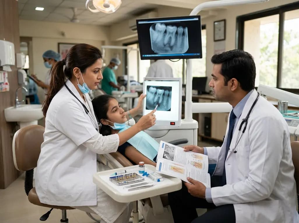
Over the past decade, revitalization treatment in endodontics has evolved into an essential biological protocol for immature teeth. Historically, dental practitioners relied on conventional apexification techniques with calcium hydroxide or apical mineral trioxide aggregate plugs. Although traditional modalities established an apical mechanical barrier, they consistently failed to stimulate continued physiological root development. Consequently, treated teeth remained structurally fragile and susceptible to cervical root fractures under masticatory loads. In contrast, modern revitalization harnesses host mesenchymal stem cells, biochemical signaling, and biological scaffolds. Clinicians primarily recruit stem cells from the apical papilla by deliberately inducing bleeding from periapical tissues into the lumen. Furthermore, dentin matrix proteins released during chemical conditioning provide essential bioactive growth factors that direct cellular differentiation. Therefore, this regenerative strategy establishes an active biological framework that supports periapical resolution and prospective dentinal wall thickening. In addition, preserving apical tissue vitality allows cellular cementum and bone-like structures to fortify root canals. Today, dentists approach infected open apices with sound biological rationale rather than purely mechanical obstruction. This modern biological paradigm offers young patients a conservative pathway that preserves natural dentition for decades.
Achieving predictable clinical success demands rigorous diagnostic assessment and strict patient selection criteria. Primarily, clinicians recommend revitalization for immature permanent teeth with necrosis caused by trauma, caries, or dens evaginatus. Clinicians must evaluate root maturation stages carefully before planning regenerative procedures. Wide open apices exceeding one millimeter facilitate stem cell recruitment and vascular ingrowth from periapical areas. In contrast, mature teeth present anatomical hurdles, although researchers actively explore extended applications in adult dentition. In addition, three-dimensional cone-beam computed tomography provides indispensable diagnostic insight regarding remaining dentinal thickness and apical anatomy. Clinicians must also appraise patient compliance, systemic health, and parental cooperation before commencing multi-visit regimens. Moreover, thermal and electric testing frequently yields unreliable responses in traumatized immature teeth. Therefore, clinicians should utilize laser Doppler flowmetry or pulse oximetry whenever possible. If extensive coronal destruction requires an intracanal post, practitioners should avoid revitalization because post preparation destroys regenerated tissues. Furthermore, thorough baseline documentation establishes an objective benchmark for monitoring subsequent healing. Thus, comprehensive preoperative evaluation prevents procedural failure and ensures appropriate therapy.
Eradicating intracanal infection while protecting delicate stem cells constitutes the central challenge in regenerative procedures. Because mechanical instrumentation thins fragile root walls and destroys apical cells, practitioners avoid mechanical filing. Instead, clinicians rely entirely on chemo-mechanical flushing strategies to clear bacterial contaminants. Operators deliver sodium hypochlorite at low concentrations, strictly between 1.5% and 2.5%, using side-vented safety needles. High concentrations destroy apical papilla stem cells and denature dentin signaling proteins. Therefore, gentle irrigation with seventeen percent ethylenediaminetetraacetic acid must follow sodium hypochlorite disinfection. This vital chelating agent neutralizes chlorine toxicity and demineralizes superficial dentin to release endogenous growth factors. Between visits, operators place inter-appointment medicaments like pure calcium hydroxide or low-dose antibiotic pastes. Specifically, practitioners avoid antibiotic concentrations exceeding one milligram per milliliter because concentrated pastes kill regenerative cells. In addition, dentists omit minocycline from antibiotic formulations to prevent unsightly crown staining. Copious sterile saline rinsing clears debris effectively before medicament placement. Furthermore, ultrasonic activation at low power enhances irrigant distribution without generating excessive heat. Consequently, this conservative disinfection protocol suppresses endodontic pathogens while fostering a hospitable microenvironment for stem cell survival.
Once clinicians establish complete clinical disinfection, the procedural focus shifts toward biological scaffold creation. Practitioners induce intracanal bleeding by gently inserting a sterile hand file beyond the apical foramen. Consequently, blood flows coronally to the cementoenamel junction, creating a natural fibrin mesh rich in platelets. Alternatively, clinicians may introduce autologous platelet-rich fibrin scaffolds when local bleeding proves difficult to elicit. Once the scaffold stabilizes, clinicians place a resorbable collagen matrix directly over the clot. Subsequently, practitioners condense a three-to-four-millimeter layer of hydraulic calcium silicate cement against this collagen barrier. Modern bioceramics like mineral trioxide aggregate or biodentine exhibit outstanding biocompatibility and stimulate hard tissue deposition. Furthermore, clinicians increasingly choose non-staining formulations containing zirconium oxide to preserve natural tooth shade. Finally, the practitioner places an adhesive resin restoration over the bioceramic barrier immediately. This definitive coronal seal prevents microleakage, which remains the leading trigger of post-treatment failure. In addition, high-strength restorative composites protect vulnerable cervical dentin against future functional fracture. Proper material handling ensures optimal curing and physical stability under masticatory stress. Therefore, meticulous adhesive techniques safeguard underlying regenerative outcomes.
Clinicians evaluate revitalization success through hierarchical clinical, radiographic, and patient-centered metrics. The primary clinical goal mandates resolving acute symptoms, eliminating sinus tracts, and achieving periapical bony healing. Fortunately, clinical trials show that revitalization achieves periapical healing rates exceeding ninety percent. The secondary goal targets continued root maturation, characterized by increased wall thickness and root elongation. Although root lengthening remains variable, histology demonstrates that reparative cementum-like tissues fortify thin canal walls. Meanwhile, the tertiary goal involves the recovery of neurosensory responses, which occurs in approximately half of treated cases. However, clinicians must also manage potential complications, including canal calcification and recurrent infection. Revitalized teeth safely tolerate physiological orthodontic forces once periapical tissues heal completely. If recurrent infection develops, clinicians can re-disinfect the canal space or pivot to an apical bioceramic plug. In addition, transparent communication with families builds trust during extended healing periods. Moreover, routine follow-up examinations should occur at six, twelve, and twenty-four months postoperatively. Prompt recognition of adverse changes ensures timely therapeutic adjustments. Thus, active recall monitoring and adaptable management protocols safeguard long-term tooth survival and patient satisfaction.
The primary candidates comprise immature permanent teeth with non-vital pulps and open apices, typically caused by physical trauma or dental anomalies like dens evaginatus. An apical opening wider than one millimeter permits abundant cellular migration from the apical papilla into the canal lumen. Furthermore, the tooth must have sufficient coronal structure remaining so that it does not require an intracanal post for long-term restoration.
Clinicians intentionally avoid mechanical filing because immature teeth feature exceedingly thin dentinal walls that mechanical files can easily perforate or further weaken. Furthermore, aggressive instrumentation destroys residual stem cells located in the apical papilla and apical canal third. Instead, practitioners utilize gentle chemical irrigation with sodium hypochlorite and EDTA to eliminate microbial biofilms, thereby preserving valuable dentin structure and host cellular vitality.
Evidence demonstrates that successfully revitalized teeth tolerate conventional orthodontic forces without experiencing accelerated root resorption or biological failure. However, clinicians must allow complete periapical bone healing before initiating orthodontic tooth movement. In addition, orthodontists should apply light, continuous physiological forces rather than heavy discontinuous loads. Regular clinical monitoring and periodic vitality assessments ensure the regenerating tissues remain healthy throughout active orthodontic realignment.
Disclaimer: This content is for informational and educational purposes only, and does not constitute medical advice or substitute for clinical judgement. Always validate findings independently. The author(s) and publisher are not liable for any consequences resulting from this material. Refer to the latest local and national guidelines for clinical practice.
References

Read summarized clinical updates, watch expert medical content, and earn CME certifications right from your smartphone.
Open your smartphone camera to scan and install.





Revitalization treatment in endodontics has evolved into an essential biologic protocol for immature necrotic teeth. This comprehensive review examines case selection criteria, modern disinfection techniques, biomaterial barriers, patient-centred prognostic factors, and complex interdisciplinary considerations.
Today
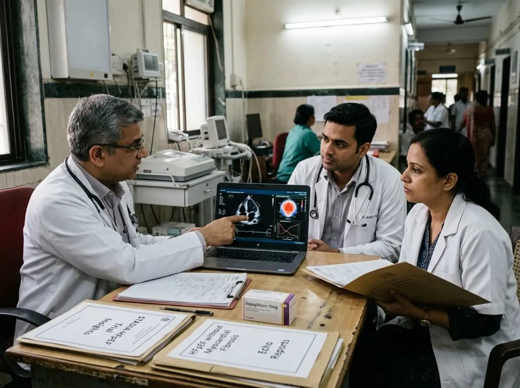
The STADIA-HFpEF trial evaluated whether dapagliflozin improves left ventricular distensibility in phenotypically stratified HFpEF patients without significant myocardial fibrosis. While primary diastolic endpoints were neutral, exploratory improvements in left atrial conduit strain and biomarkers emerged.
Today
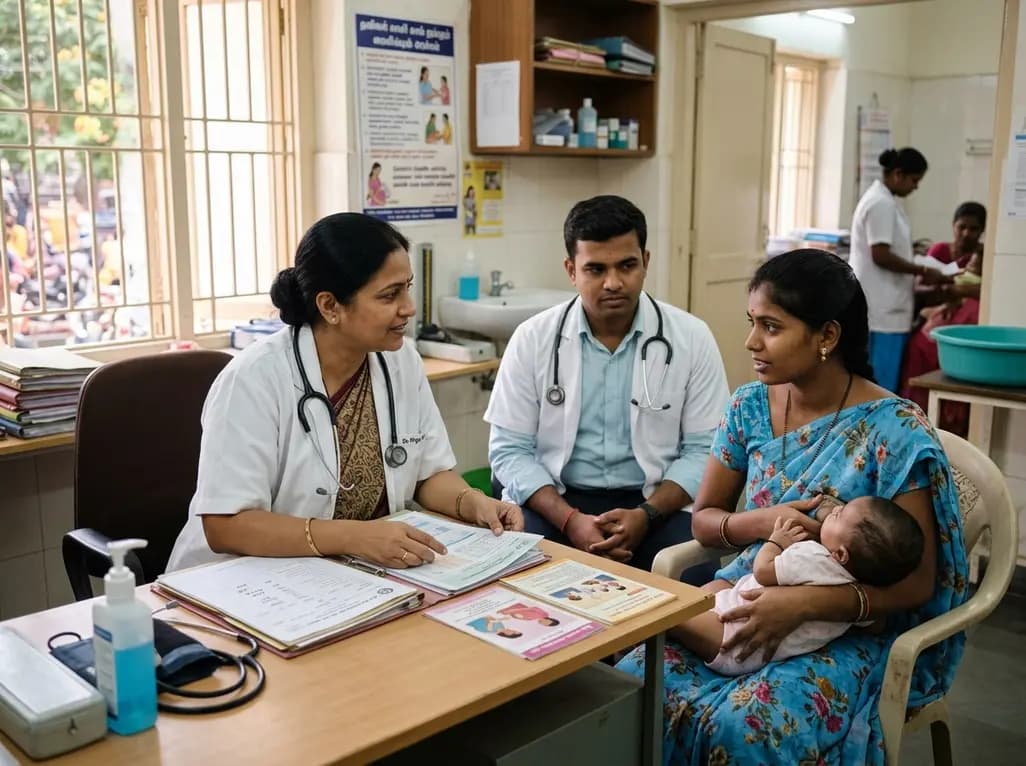
A Belgian study highlights maternal desires and healthcare provider perspectives on breastfeeding in women living with HIV. While viral suppression reduces transmission risk below 1%, significant clinical training and policy gaps remain, highlighting the urgent need for multidisciplinary counseling protocols.
Today

A landmark multi-ethnic study of 40,132 participants demonstrates that risk-weighted apoB (RW-apoB) captures coronary risk beyond measured apoB. By integrating the atherogenic potency of triglyceride-rich lipoproteins and Lp(a), RW-apoB improves cardiovascular risk discrimination across diverse populations.
Today
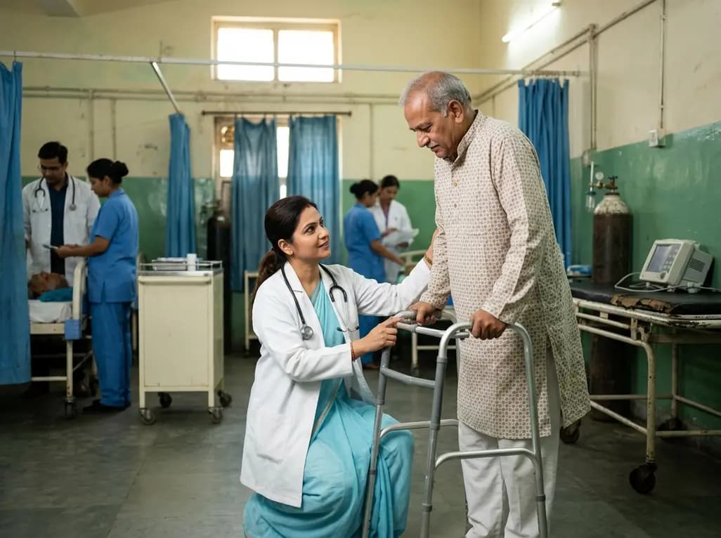
A systematic review demonstrates that emergency department physiotherapy safely matches physician care for 30-day re-presentations while reducing geriatric fall readmissions by 34%.
Today
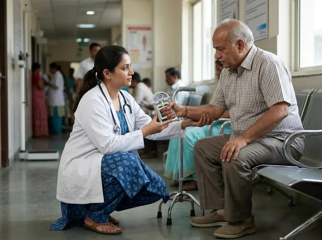
A cross-sectional study reveals that elevated BMI in type 2 diabetes correlates with reduced skeletal muscle mass, handgrip weakness, impaired walking capacity, balance deficits, and poor quality of life, highlighting the need for targeted rehabilitation.
Today
