.webp&w=384&q=75)

Loading, please wait...
Loading, please wait...

Retinal vasculature segmentation serves as a fundamental tool for evaluating clinical biomarkers in systemic diseases. Clinicians often rely on retinal artery and vein (A/V) analysis to assess cardiovascular risks. However, traditional automated methods frequently struggle with vessel caliber variations and low-contrast structures. Consequently, fragmented semantic representations often emerge in standard segmentation outputs, complicating diagnosis.
To solve these challenges, researchers proposed Ultra, a multi-granularity topological reasoning network. This system employs a two-stage architecture to ensure structural integrity and accuracy. First, PriorNet generates coarse, multi-scale priors to provide essential structural guidance. Subsequently, RefineNet performs refinement focused on topology-aware details. Furthermore, the network integrates a neighboring pixel connectivity regularization (NICER) layer. This layer utilizes a connectivity prediction union (CPU) module to selectively incorporate local connectivity information. Therefore, the system enforces anatomical consistency through a pixel-wise local connectivity loss, ensuring the network learns proper vessel shapes.
Accurate segmentation is particularly vital in ultra-wide field (UWF) imaging. These images often suffer from peripheral distortions and significant anatomical variability. However, the Ultra network demonstrates state-of-the-art performance and robust generalization capabilities. It performs exceptionally well on both UWF datasets and conventional color fundus photography (CFP). This versatility ensures that the tool remains useful across diverse clinical settings in India, from tertiary eye hospitals to primary screening centers.
The Ultra network introduces a multi-granularity topological reasoning approach. It specifically uses the NICER layer and CPU module to ensure that segmented vessels remain anatomically connected and consistent, avoiding the fragmentation seen in older AI models.
Retinal vessels are the only part of the human circulatory system that can be visualized non-invasively. Therefore, segmenting these vessels allows doctors to monitor biomarkers for conditions like hypertension, diabetes, and stroke risk more effectively.
Disclaimer: This content is for informational and educational purposes only. It does not constitute medical advice or a professional endorsement. Refer to the latest local and national guidelines for clinical practice.
References
Mou L et al. Multi-Granularity Topological Reasoning for Anatomically Consistent Vasculature Parsing. IEEE Trans Image Process. 2026 Apr 23. doi: 10.1109/TIP.2026.3683249. PMID: 42024936.
Sharma Research Group. Deep Retinal Vessel Segmentation For Ultra-Widefield Fundus Photography. University of Rochester Lab Sites. 2026.

Read summarized clinical updates, watch expert medical content, and earn CME certifications right from your smartphone.
Open your smartphone camera to scan and install.





Researchers introduce Ultra, a multi-granularity topological reasoning network for precise retinal artery and vein segmentation in fundus imaging....
6 months ago

Explore vital recurrent stroke predictors in lacunar stroke patients managed with intensive blood pressure lowering. This analysis examines trial findings from SPS3 to uncover residual vascular risks, metabolic factors, and targeted secondary prevention strategies for high-risk neurological patients.
Today
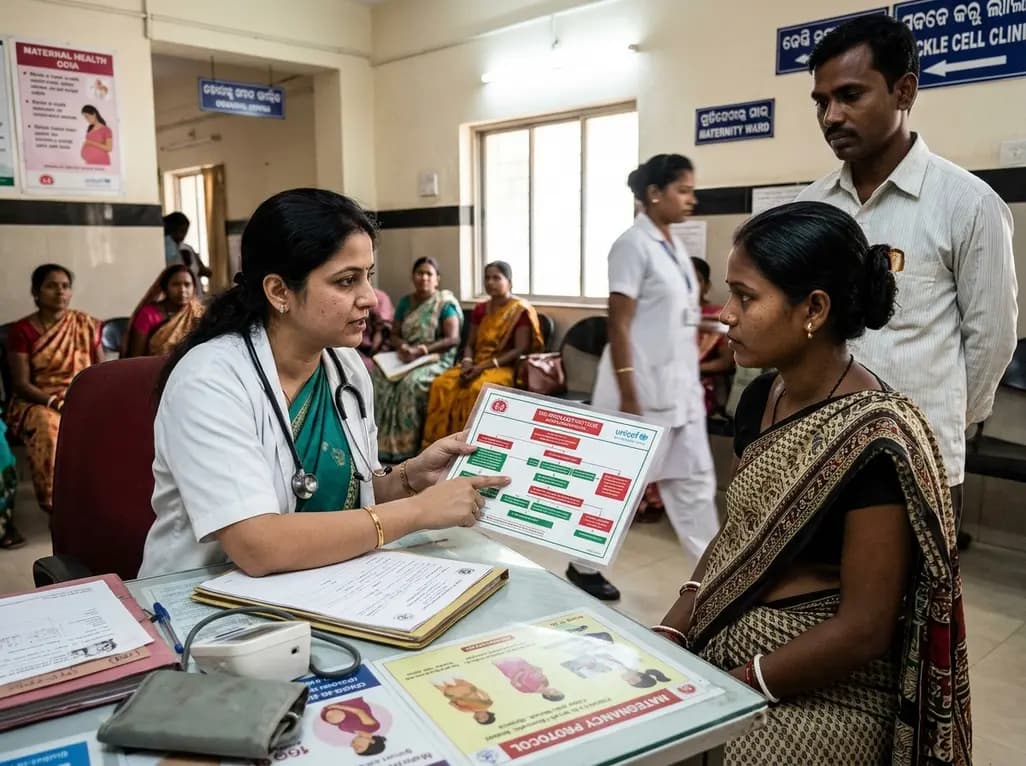
Odisha has established India's first state-level clinical protocol specifically tailored for pregnant women with sickle cell disease. Developed with NHM and UNICEF, the framework provides structured antenatal screening, crisis mitigation, standardized referral pathways, and specialized interdisciplinary care.
Today

India is advancing preventive healthcare by integrating genomics, artificial intelligence, and quantum technologies. With the SANGAM programme sequencing one million Indian genomes, clinicians can unlock personalized therapies, detect cardiometabolic risks early, and curb the burden of diabetes and heart disease.
Today
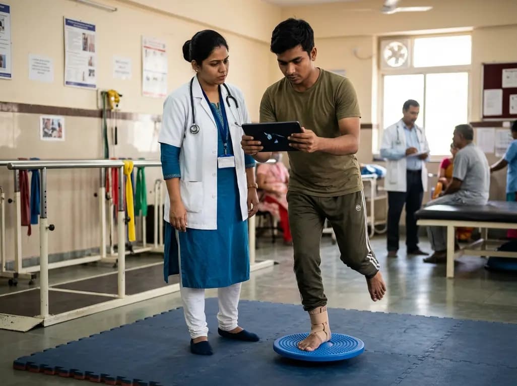
A randomized controlled trial demonstrates that an eight-week dual-task training intervention significantly improves static and dynamic postural control over standard balance exercises in individuals with chronic ankle instability, addressing critical neurocognitive deficits during athletic and daily activities.
Today
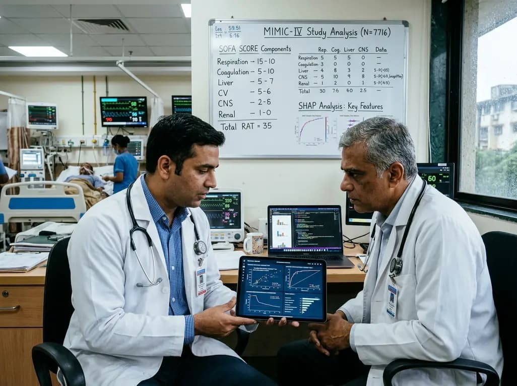
A retrospective cohort study of 7,716 ICU patients from the MIMIC-IV database demonstrates that Gradient Boosting Decision Trees significantly outperform traditional SOFA scores in predicting in-hospital mortality among patients with sepsis and diabetes, with SHAP analysis providing clinical interpretability.
Today
