

Loading, please wait...
Loading, please wait...

Traumatic spinal cord trauma inflicts profound physical devastation, disrupting communication channels between the brain and peripheral nervous system. Clinicians frequently observe that these injuries induce extensive supraspinal neuroplasticity alongside severe motor impairment. Recent clinical research indicates that resting-state functional connectivity experiences significant remodeling across multiple cerebral areas following myelopathy. Historically, neuroscientists believed that central cortical reorganization remained largely restricted to primary motor and sensory neocortices. However, modern functional magnetic resonance imaging shows that distributed neural assemblies undergo profound spontaneous synchronization adjustments. Because afferent ascending signals diminish abruptly, downstream cerebral hubs alter their metabolic coordination patterns. Furthermore, maladaptive reorganization frequently drives debilitating secondary sequelae, including intractable neuropathic pain, autonomic failure, and subtle cognitive decline. Therefore, clinicians must reevaluate spinal cord trauma through a comprehensive whole-brain systems lens. In addition, recognizing early network disruptions enables multidisciplinary care teams to anticipate complex neurological symptoms. Consequently, resting-state functional connectivity serves as an objective window into systemic cerebral restructuring after direct cord injury.
A landmark systematic review recently synthesized whole-brain neuroimaging datasets to map functional reorganizational architecture in spinal cord injury. Researchers comprehensively evaluated eleven distinct cohorts comprising 151 injured individuals and 208 non-injured matched controls. Crucially, the investigators observed remarkable alterations that extended far beyond the primary somatomotor cortex. For example, substantial dysconnectivity emerged between the somatomotor network and the ventral attentional network. Moreover, pronounced connectivity shifts occurred between the ventral attention network and the frontal pole. In contrast to isolated motor cortex dampening, these widespread alterations highlight intricate cortico-cortical disturbances. Additionally, the systematic synthesis revealed marked decoupling between cerebellar hubs and the default mode network. Because previous studies produced conflicting conclusions regarding isolated regional activation, this network-based approach provides vital clarity. Consequently, the findings confirm that traumatic myelopathy alters global brain topography rather than isolated motor projection zones. In fact, these extensive circuit disruptions mirror the multidimensional challenges patients encounter during rehabilitation. Therefore, comprehensive neuroimaging provides essential anatomical validation for secondary cognitive and sensory complications.
Secondary neuropathic pain represents one of the most debilitating chronic sequelae following serious spinal trauma. Notably, up to seventy percent of injured patients suffer from persistent central or neuropathic dysesthesias. Aberrant resting-state communication between attentional networks and somatosensory territories offers a compelling mechanistic explanation for this persistent distress. When ascending sensory inputs terminate abruptly, thalamocortical rhythms lose their physiological inhibitory calibration. Consequently, the ventral attention network demonstrates hyper-responsiveness toward aberrant internal sensory stimuli. Furthermore, disrupted frontal pole connectivity impairs descending antinociceptive modulation systems. As a result, innocuous stimuli or spontaneous central discharges are perceived as excruciating discomfort. Similarly, altered cerebellar crosstalk with attentional systems exacerbates affective pain processing and emotional exhaustion. Pharmacological analgesics frequently provide inadequate relief precisely because they fail to recalibrate disrupted large-scale networks. Therefore, mapping resting-state functional connectivity gives neurologists an objective biological marker for refractory pain phenotypes. Ultimately, tracking these neural circuits can facilitate more targeted neuromodulatory therapies, such as transcranial magnetic stimulation.
Although clinical rehabilitation traditionally emphasizes motor restitution, cognitive deficits frequently burden individuals surviving serious spinal trauma. Specifically, patients often experience noticeable declines in sustained attention, executive functioning, and mental processing speed. In the systematic review, researchers documented prominent dysconnectivity between the cerebellum and regions of the default mode network. Historically, clinicians regarded the cerebellum merely as a coordinator of voluntary muscular activity and balance. However, contemporary cognitive neuroscience demonstrates that cerebellar loops modulate prefrontal networks governing working memory and executive control. When spinal afferents disconnect, cerebellar input diminishes, which destabilizes synchronized default mode oscillations. In addition, altered coupling between the ventral attention network and frontal poles degrades top-down cognitive governance. Consequently, patients find multitasking, emotional regulation, and rapid decision-making extraordinarily fatiguing during daily routines. Moreover, chronic systemic inflammation and nocturnal sleep fragmentation exacerbate this progressive supraspinal network degradation. Therefore, rehabilitation specialists must incorporate systematic neuropsychological testing alongside motor evaluations during routine follow-up consultations.
Integrating functional neuroimaging findings into daily clinical workflows creates promising opportunities for personalized neurorehabilitation. Currently, clinicians evaluate spinal recovery using conventional motor scores and sensory testing batteries. However, these peripheral scales cannot quantify the extent of supraspinal functional network reorganization. In contrast, resting-state functional connectivity provides quantifiable objective biomarkers reflecting central nervous system plasticity. For instance, advanced neuroimaging can distinguish patients exhibiting adaptive cortical reorganization from those developing maladaptive connectivity patterns. Furthermore, early identification of abnormal frontoparietal connectivity can guide customized cognitive rehabilitation protocols before deficits become entrenched. Clinicians can also combine connectivity maps with non-invasive brain stimulation to modulate dysfunctional cortical targets. Specifically, applying repetitive transcranial stimulation over hypoactive frontal hubs might normalize disrupted network synchronization. Additionally, virtual reality training and targeted sensorimotor biofeedback actively encourage healthy supraspinal circuit reintegration. Thus, functional imaging transitions from a specialized research curiosity into a practical therapeutic navigation system.
Overcoming current limitations in neurotrauma management requires comprehensive longitudinal investigations across diverse clinical populations. Most published resting-state fMRI studies remain limited by small sample cohorts and cross-sectional study designs. Therefore, prospective multi-center studies must track patients from acute trauma through long-term chronic rehabilitation phases. In particular, correlating temporal connectivity fluctuations with specific neurological milestones will illuminate critical therapeutic windows for intervention. Moreover, integrating resting-state functional imaging with structural tractography and fluid biomarkers will refine prognostic models. Clinical teams must also investigate how pharmacological agents and spinal cord neuromodulation devices alter supraspinal synchronization. In routine practice, interdisciplinary collaboration between neurologists, spine surgeons, physiatrists, and radiologists remains indispensable. As clinicians better comprehend the widespread brain remodeling that accompanies spinal injuries, treatment paradigms can expand meaningfully. Consequently, addressing both spinal cord pathology and widespread cerebral network alterations will dramatically elevate patient outcomes and functional autonomy.
Yes, resting-state functional connectivity demonstrates significant utility in characterizing chronic neuropathic pain mechanisms. Altered spontaneous communication between the somatomotor cortex, thalamocortical loops, and the ventral attentional network correlates with central pain amplification. By identifying these maladaptive supraspinal signatures, functional neuroimaging enables clinicians to tailor targeted neurostimulation protocols for refractory post-injury neuropathic syndromes.
Cognitive deficits emerge primarily because severed spinal pathways disrupt distributed supraspinal networks governing executive control and attention. Reduced afferent signaling destabilizes coordinated interactions between the cerebellum, default mode network, and frontal pole. Furthermore, concurrent secondary factors such as chronic neuroinflammation, persistent neuropathic distress, and sleep disruption accelerate cognitive fatigue and impair working memory capacity.
Clinicians can employ multimodal therapeutic interventions to foster positive supraspinal neuroplasticity and rebalance connectivity. Non-invasive brain stimulation techniques, such as repetitive transcranial magnetic stimulation, directly modulate dysregulated frontoparietal circuits. Additionally, combining immersive virtual reality training, robotic locomotor therapy, and targeted cognitive rehabilitation encourages active sensory reintegration and enhances functional cortical reorganization over time.
Disclaimer: This content is for informational and educational purposes only and does not constitute medical advice, diagnosis, or treatment. Clinicians must exercise independent judgment and verify clinical protocols. Refer to the latest local and national guidelines for clinical practice.
References

Read summarized clinical updates, watch expert medical content, and earn CME certifications right from your smartphone.
Open your smartphone camera to scan and install.





A systematic review reveals that spinal cord injury alters resting-state functional connectivity far beyond the sensorimotor cortex, implicating attentional, cerebellar, and default mode networks in secondary complications like chronic pain and cognitive dysfunction.
Today

A retrospective CAHAL study comparing Amplatzer and Occlutech devices in 413 patients found no significant difference in moderate-to-large residual shunt rates at 6 months follow-up after percutaneous PFO closure, supporting clinical equivalence between both platforms.
Today

AIIMS researchers suggest yoga may alleviate gut dysbiosis in Polyendocrine Metabolic Ovarian Syndrome (PMOS) by modulating the gut-brain axis, lowering chronic inflammation, and enhancing parasympathetic tone to support metabolic and hormonal equilibrium.
Today
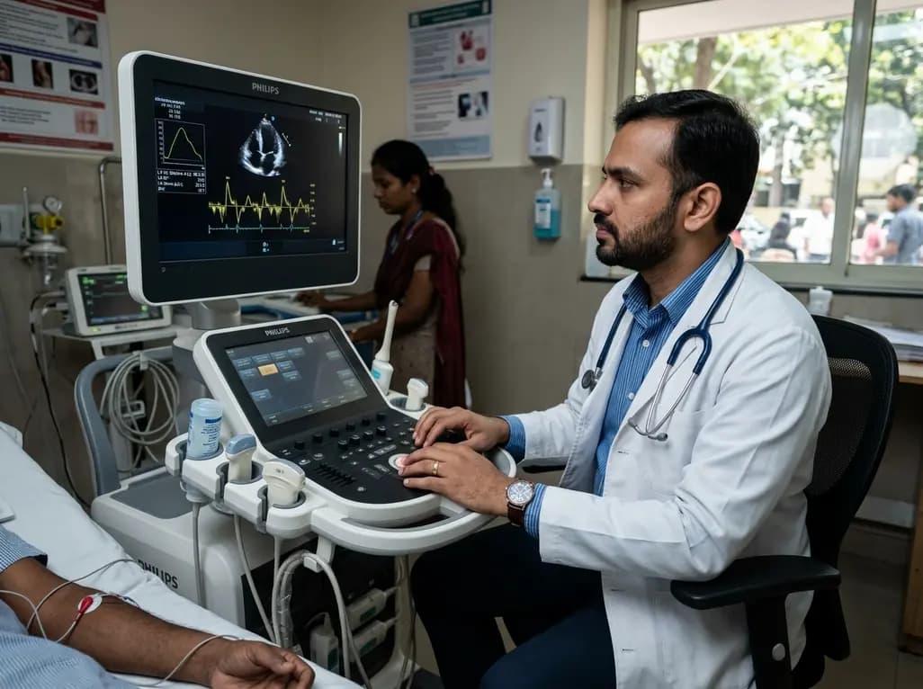
A structured practical guide to left atrial echocardiographic assessment covering biplane volumetry, phasic physiology, speckle-tracking strain, and hemodynamic filling pressure estimation for routine cardiovascular practice.
Today

Age-related spinal kyphosis is driven by vertebral fragility, disc degeneration, and paraspinal muscle dysfunction. Coexisting with locomotive syndrome and frailty, its management requires combined anti-osteoporotic therapy, spinal extensor rehabilitation, and comprehensive nutritional optimization.
Today

A longitudinal feasibility pilot study highlights that combining polygenic risk score disclosure with in-person lifestyle counseling significantly enhances genomic literacy in individuals at risk for type 2 diabetes, overcoming educational disparities and reinforcing patient motivation.
Today
