

Loading, please wait...
Loading, please wait...

Dental practitioners routinely rely on direct resin restorations for anterior and posterior teeth. However, successful clinical outcomes depend directly on effective resin composite light curing. When composite materials fail to polymerize uniformly, restorations suffer from premature wear, secondary caries, marginal breakdown, and increased monomer leaching. Recent laboratory evidence reveals that optical beam heterogeneity and chemical initiator design significantly dictate conversion quality across the entire restoration. Therefore, understanding the intricate optical and thermal interactions during light activation is essential for clinical excellence.
Modern light-emitting diode (LED) curing devices emit radiation across multiple spectral wavelengths to activate diverse photoinitiators. However, many commercial light-curing units generate localized hot spots and non-uniform spectral distributions across their output tips. When an LED tip lacks optical homogenization optics, blue light and violet light exit from isolated, separate micro-diodes. Consequently, the radiant exposure delivered to the resin surface varies significantly depending on the exact spatial position beneath the tip. Researchers examined how non-homogenized light delivery units project irregular light profiles through filled resin specimens. The experimental findings demonstrated that non-homogenizing tips create dramatic localized disparities in transmitted energy. In contrast, light guides equipped with homogenizing optical elements blend distinct wavelengths into a uniform beam profile. Therefore, homogenizing tips ensure that every square millimeter of the resin restoration receives an equivalent spectral balance. Clinicians often assume that high overall radiant output guarantees uniform polymerization across the restoration. In reality, an unblended light beam can leave adjacent microscopic areas severely under-cured. This optical phenomenon explains why restorations sometimes display localized marginal degradation despite adequate overall exposure time. Clinicians must recognize that beam uniformity represents an essential determinant of predictable curing.
Dental composite formulations historically relied almost exclusively on camphorquinone (CQ) paired with tertiary amine co-initiators. Camphorquinone absorbs light energy primarily within the blue spectrum near 468 nanometers. Because camphorquinone possesses an intense yellow tint, manufacturers increasingly incorporate alternative photoinitiators like diphenyl(2,4,6-trimethylbenzoyl)phosphine oxide (TPO) for lighter aesthetic shades. TPO absorbs shorter violet wavelengths, with peak absorbance occurring near 380 to 410 nanometers. Furthermore, TPO functions as a Type I cleavage photoinitiator that reacts rapidly without requiring an amine co-initiator. Nevertheless, matching the photoinitiator absorption spectrum to curing unit emission profiles remains crucial for complete conversion. When researchers activated CQ-only composites with a non-homogenized multi-peak LED, polymerization proceeded well under blue diodes but stalled completely beneath violet diodes. Specifically, degree of conversion reached sixteen percent near 456 nanometer emitters after three seconds, but registered zero percent beneath 408 nanometer emitters. Conversely, when researchers tested TPO-containing composites, curing occurred efficiently under violet diodes but decreased under blue diodes. Hybrid initiator systems containing both CQ and TPO balanced these spectral discrepancies across broader exposure fields. Therefore, selecting composite materials with complementary initiator systems provides greater tolerance against spectral inhomogeneities in curing devices.
Real-time Fourier-transform infrared spectroscopy and spatial micro-mapping offer profound insights into the kinetics of monomer conversion. Studies demonstrate that polymerization occurs heterogeneously across both time and space. During brief three-second curing protocols, non-homogenized multi-peak lights created stark disparities across resin samples. Even when clinicians extended curing exposure to ten seconds, significant spatial conversion deficits persisted in non-homogenized groups. For instance, adjacent regions separated by merely a few millimeters exhibited degree of conversion differences greater than twenty-five percent. In addition, peripheral zones consistently exhibited lower monomer conversion compared to central areas across all test combinations. This edge deficit occurs because light scattering disperses irradiance toward specimen boundaries while ambient atmospheric oxygen inhibits surface cross-linking. As a consequence, margin areas experience inferior physical properties, including reduced microhardness and accelerated enzymatic degradation. Incomplete cross-linking also leaves residual unreacted dimethacrylate monomers, which can elute into pulpal tissues or saliva. Homogenizing tips effectively eliminated these localized micro-domains of incomplete polymerization by spreading spectral irradiance symmetrically. Consequently, clinicians must appreciate that curing time alone cannot overcome poor beam architecture or severe optical mismatching. Achieving uniform conversion across deep and peripheral margins requires stable, homogeneous radiant distribution.
Polymerization of dimethacrylate resin matrices releases substantial exothermic energy while absorbing heat from intense curing lights. Infrared thermal cameras reveal that local temperature rises directly mirror the spatial irradiance distribution of the curing unit. Specifically, non-homogenized curing tips generate intense thermal hot spots corresponding precisely with high-output diode positions. When composite formulations cure rapidly under peak irradiance, local exothermic reactions produce steep temperature spikes exceeding safe biological thresholds. Excessive thermal transfer across thin remaining dentin barriers poses a clear risk of irreversible pulpitis or pulpal necrosis. Furthermore, resin chemistry dictates the total exothermic heat release during network formation. Rapid cleavage photoinitiators such as TPO initiate rapid chain propagation, which concentrates exothermic heat generation into brief time windows. In contrast, camphorquinone-amine systems demonstrate slightly slower polymerization kinetics, spreading heat release over longer intervals. Homogenizing tips mitigate dangerous thermal peaks by diffusing high-energy photon clusters across a broader surface area. Therefore, uniform beam distribution protects pulpal vitality while still delivering adequate overall energy for polymer network cross-linking. Dentists must remain vigilant regarding pulpal temperatures, particularly when restoring deep cavity preparations with high-power curing devices.
Clinicians can implement several evidence-based protocols to minimize cure heterogeneity and enhance restoration longevity. First, dentists should inspect their light-curing units to verify beam homogeneity and optical tip integrity. Devices incorporating optical diffuser lenses or optimized light guides ensure even energy transmission across the entire restoration footprint. Second, practitioners must carefully evaluate composite photoinitiator chemistry before choosing light curing parameters. Restorations containing alternative initiators like TPO necessitate multi-wavelength curing units that deliver sufficient violet irradiance. Third, clinicians should avoid relying on ultra-short three-second curing protocols for critical bonding layers or deep increments. Although high-power short exposures seem efficient, they frequently produce steep conversion gradients and excessive polymerization shrinkage stress. Instead, delivering radiant energy over ten to twenty seconds promotes gradual network formation and minimizes unreacted monomer residues. Additionally, dentists must position the curing tip perpendicular and as close as possible to the composite surface. Because light intensity attenuates rapidly with increasing distance, careful positioning ensures adequate peripheral and bottom-layer conversion. Finally, regular radiometer calibration verifies that light units maintain consistent output over repeated clinical sterilization cycles. Implementing these practical steps ensures predictable polymer conversion and superior clinical performance.
Beam homogeneity ensures that light-curing units deliver uniform spectral irradiance and energy density across the entire light tip. Non-homogenized light guides produce localized optical hot spots and cold zones, creating severe degree of conversion disparities within a single composite increment. Consequently, under-cured marginal regions experience accelerated wear, microleakage, and premature restoration failure. Homogenizing optics eliminate these localized defects, ensuring reliable, uniform polymer cross-linking across all cavity margins.
Traditional dental resin composites utilize camphorquinone, which absorbs blue light between 450 and 470 nanometers. However, modern aesthetic and bleach-shade composites often incorporate alternative initiators like TPO, which absorb shorter violet wavelengths around 390 to 410 nanometers. Single-peak blue LED lights cannot activate TPO effectively. Therefore, practitioners must utilize polywave or multi-peak LED units with appropriate spectral outputs to ensure complete monomer conversion in composites formulated with alternative photoinitiator systems.
Extending curing duration from three to ten seconds improves overall conversion but cannot completely resolve localized cure deficiencies caused by non-homogenized multi-peak beams. Spatial discrepancies in radiant exposure still leave significant degree of conversion variations across the resin surface. Furthermore, excessive exposure times with high-intensity curing units dramatically increase pulp chamber temperatures without guaranteeing peripheral margin cure. Using a curing light equipped with homogenizing optics remains the most reliable solution for uniform cross-linking.
Disclaimer: This content is for informational and educational purposes only and does not constitute medical advice. Consult qualified healthcare professionals for diagnosis and treatment. Refer to the latest local and national guidelines for clinical practice.
References

Read summarized clinical updates, watch expert medical content, and earn CME certifications right from your smartphone.
Open your smartphone camera to scan and install.





A recent dental study reveals that light curing beam profiles and photoinitiator chemistry create spatial cure heterogeneity and thermal spikes in resin composites, highlighting the need for homogenized light guides.
Today
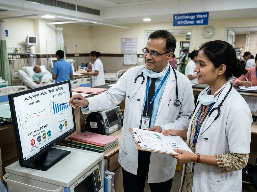
The VOCAL-AHF study reveals that most acute heart failure patients across reduced and non-reduced ejection fraction categories are eligible for in-hospital GDMT initiation. Notably, nearly two-thirds of HFrEF patients qualify for quadruple therapy, while non-reduced EF patients show high eligibility for dual therapy.
Today

Ascertainment bias in cancer case-control studies inflates odds ratios and penetrance estimates when cases are drawn from high-risk clinics. A landmark modeling study demonstrates substantial bias ranging up to 23.72, highlighting the critical need for ascertainment adjustment in genetic risk counseling.
Today
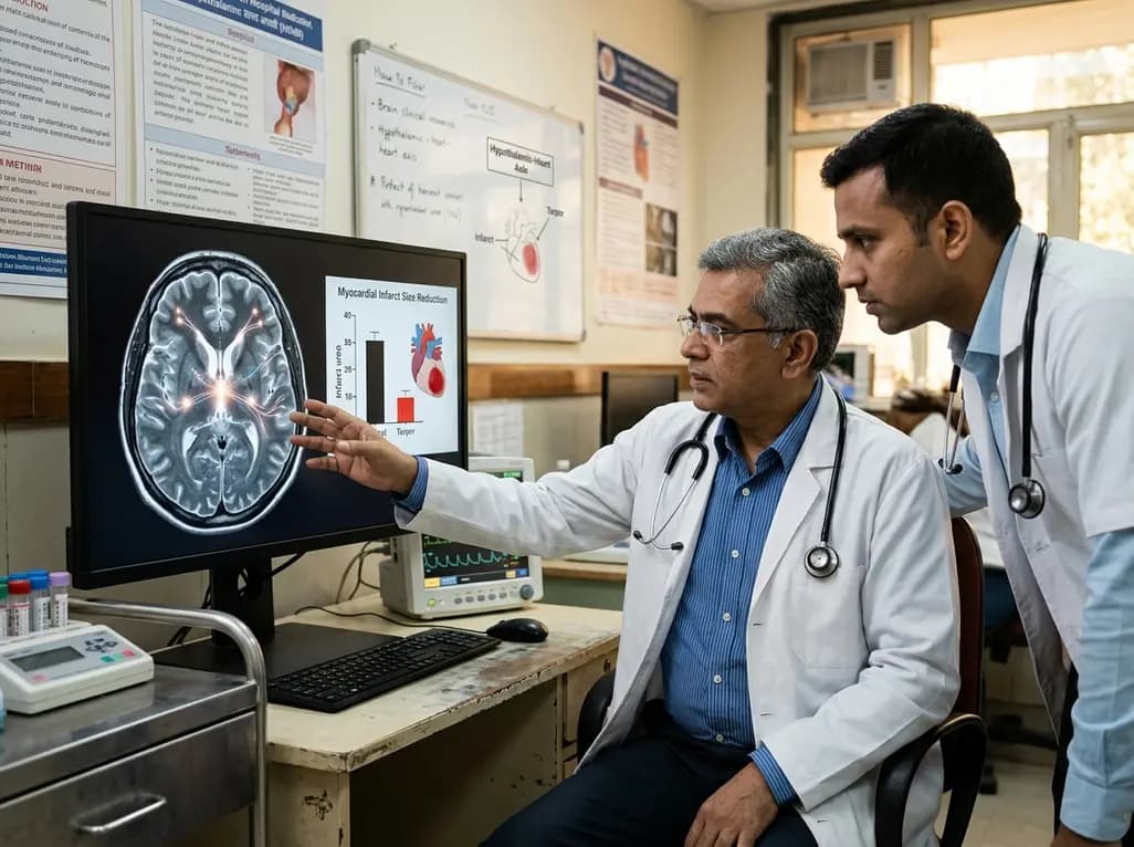
Researchers demonstrate that central induction of synthetic torpor in non-hibernating rats provides striking cardioprotection, reducing myocardial infarct size by ~40% during ischaemia-reperfusion injury through endogenous survival signaling, independent of hypothermia.
Today
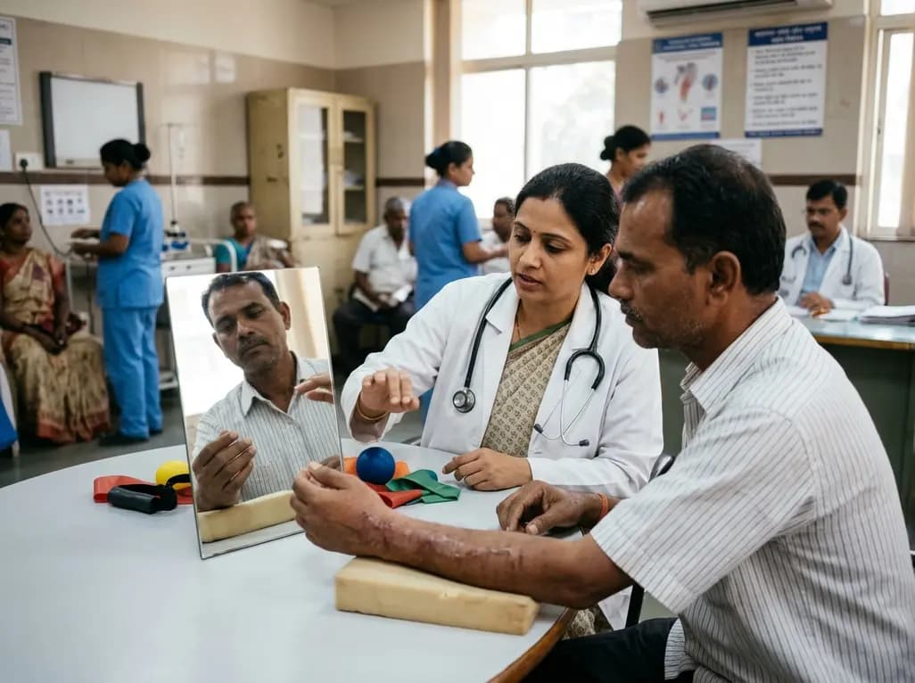
A meta-analysis shows that movement representation techniques significantly enhance upper limb and wrist function in distal radius fracture rehabilitation. While effects on pain and grip strength remain inconclusive, early low-cost telerehabilitation and mirror therapy provide promising clinical benefits.
Today
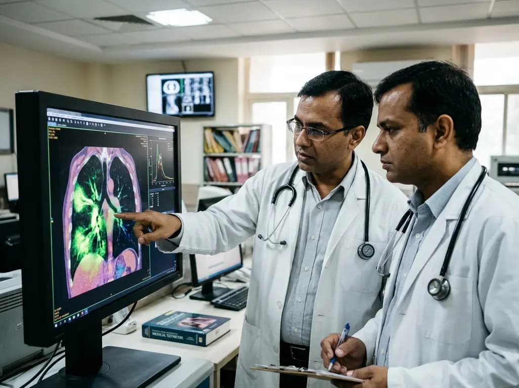
A novel fluorine-functionalized near-infrared copper fluorescent probe selectively illuminates lung tissue, revealing critical links between hyperglycemia, ER stress, and pulmonary malignancy for improved early diagnosis.
Today
