

Loading, please wait...
Loading, please wait...
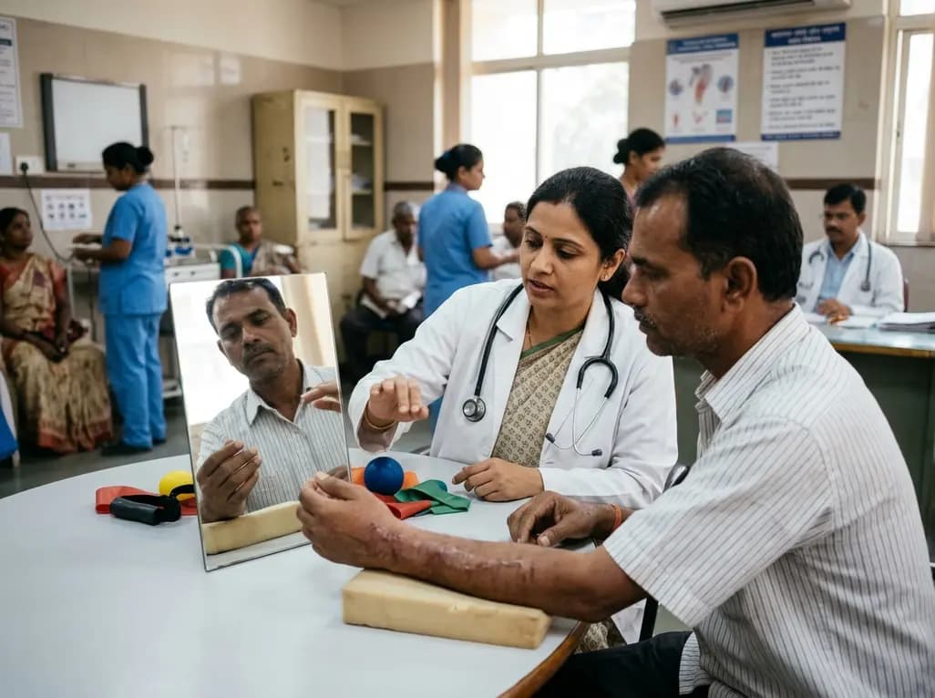
Distal radius fractures represent one of the most frequent skeletal injuries treated in orthopedic and emergency clinics worldwide. Following fracture stabilization, patients often endure prolonged wrist stiffness, muscle weakness, and debilitating functional limitations. Consequently, traditional physical therapy focuses primarily on peripheral joint mechanics and muscular strengthening during recovery. However, prolonged limb disuse substantially impairs cortical motor networks and sensorimotor representation. To counteract these central neuroplastic deficits, clinicians increasingly explore movement representation techniques as promising cognitive adjuncts in orthopedic care. These innovative strategies help preserve central motor pathways before formal mechanical loading can safely begin.
Prolonged wrist immobilization disrupts somatosensory input and degrades primary motor cortex representation. As a result, neural circuitry controlling fine motor skills becomes functionally altered through cortical smudging. Movement representation techniques actively counteract this neuroplastic decline by stimulating cortical motor areas without requiring physical movement of the fractured bone. These cognitive approaches encompass graded motor imagery, visual mirror therapy, and implicit motor practice. During mirror therapy, patients observe the reflection of their unaffected wrist performing painless, coordinated motions. Consequently, the brain receives vivid visual feedback signaling successful, pain-free movement in the immobilized extremity. Neuroimaging confirms that mental motor imagery activates premotor and supplementary motor areas similarly to actual joint kinematics. Therefore, engaging these pathways preserves corticospinal excitability during the initial weeks of healing. Furthermore, these techniques maintain sensorimotor congruence, protecting patients against maladaptive pain sensitization. Clinicians find these non-invasive modalities appealing because they protect delicate orthopedic fixation while preparing the brain for active recovery.
Recent meta-analytic evidence demonstrates significant clinical gains when clinicians incorporate cognitive motor interventions into fracture rehabilitation. Specifically, pooled results across randomized controlled trials reveal that movement representation modalities produce meaningful gains in upper extremity performance. Patients engaging in these mental exercises achieved a statistically significant reduction in disability scores compared with standard care alone. In addition, objective wrist function scores showed marked improvement across evaluated patient cohorts. These functional benefits emerge because structured mental practice preserves motor planning and coordination during immobilization phases. Consequently, patients exhibit less motor hesitation and fear-avoidance behavior when formal joint mobilization commences. The absence of statistical heterogeneity across trials underscores the consistency of these functional improvements. Furthermore, early preservation of kinematic templates facilitates active joint range of motion, particularly in wrist flexion and extension. By maintaining central sensorimotor maps, patients regain dexterity and purposeful hand coordination much faster than with passive therapy alone.
Although functional mobility scores show marked enhancement, meta-analytic findings present a more nuanced picture regarding pain relief and handgrip strength. The pooled data demonstrated no statistically significant overall effect on resting pain intensity or maximal isometric grip force. Nevertheless, individual trials investigating graded motor imagery reported notable pain reductions among patients with heightened baseline sensitivity. Handgrip strength recovery relies heavily on muscular hypertrophy and mechanical tendon glide, which mental practice cannot fully replicate. However, movement representation therapies effectively modulate central pain processing, thereby reducing movement-related apprehension. When patients rehearse pain-free movement patterns mentally, they dampen hyperactive central nociceptive circuits. Therefore, while grip dynamometry may not change dramatically in short-term analyses, patient willingness to perform daily grasping tasks improves noticeably. In addition, the current evidence exhibits low to very low GRADE certainty due to modest sample sizes. Consequently, orthopedic surgeons should view these cognitive techniques as complementary functional tools rather than replacements for progressive resistance exercise.
A major practical advantage of cognitive representation protocols lies in their remarkable simplicity and affordability. Clinical trials demonstrate that these interventions require only basic, low-cost equipment, such as mirrors, printed lateralization cards, or digital tablet screens. Moreover, therapists have successfully deployed these programs through supervised face-to-face sessions as well as structured telerehabilitation platforms. Typical effective regimens span three to eight weeks, delivering thirty-five to sixty minutes per session three times weekly. Clinicians can safely initiate motor imagery within two weeks following surgical fixation. In conservatively managed cases, therapists introduce these exercises immediately after six weeks of cast immobilization or during early immobilization. Furthermore, telerehabilitation offers substantial logistical advantages for elderly patients facing travel barriers in community settings. Through remote video consultations, physiotherapists guide patients through mirror exercises and left-right hand discrimination drills at home. Consequently, digital delivery ensures high patient compliance while optimizing clinical resources across diverse socioeconomic settings.
Integrating movement representation into standard orthopedic workflows requires minimal adjustments to traditional physical therapy routines. Initially, clinicians assess the patient for movement fear, acute swelling, and baseline wrist range of motion. Subsequently, therapists introduce implicit motor imagery, asking patients to identify hand laterality using simple visual recognition flashcards. As lateralization accuracy improves, patients advance to explicit motor imagery, mentally visualizing smooth wrist flexion and extension without muscle contraction. Finally, therapists incorporate mirror visual feedback, enabling patients to execute symmetrical bilateral hand drills behind a vertical mirror. Because these exercises require no mechanical joint loading, patients practice them safely during early bone healing. In addition, educating patients on the neurobiology of recovery diminishes fear-avoidance behaviors and improves exercise adherence. However, clinicians must monitor cognitive fatigue, as intense mental rehearsal demands substantial concentration. Ultimately, integrating central cognitive retraining with peripheral biomechanical rehabilitation provides a modern, holistic approach to distal radius fracture care.
Clinicians can safely introduce movement representation techniques within the first two weeks following stable surgical fixation. For patients managed conservatively with cast immobilization, gentle cognitive training can start during immobilization or immediately after cast removal at six weeks. Because these mental exercises involve no physical loading across the fracture site, they protect healing bone while preserving cortical motor maps and mitigating joint stiffness early.
Current meta-analytic evidence shows no statistically significant overall improvements in resting pain intensity or isometric grip strength across pooled cohorts. Handgrip strength relies heavily on muscular hypertrophy and mechanical tissue loading, which require progressive physical resistance. Nevertheless, individual studies show that graded motor imagery reduces movement-related pain sensitivity. Clinicians should therefore combine cognitive representation techniques with structured physical loading to optimize both strength and functional mobility.
Yes, telerehabilitation represents an effective and accessible delivery model for movement representation programs. Since these interventions rely on inexpensive mirrors, digital image recognition, and mental practice, therapists can easily instruct patients via video calls. Supervised remote sessions conducted three times weekly over three to eight weeks ensure high treatment adherence, reduce travel burdens for recovering individuals, and provide clinical outcomes comparable to face-to-face clinic visits.
Disclaimer: This content is for informational and educational purposes only and does not substitute professional clinical judgment. Refer to the latest local and national guidelines for clinical practice.
References
Atencio-Aguilar JL et al. Effectiveness of movement representation techniques on function, pain intensity, and handgrip strength in patients with distal radius fracture: A systematic review and meta-analysis. J Hand Ther. 2026 Nov 12. doi: undefined. PMID: 43020758.
Bayon-Calatayud M, Benavente-Valdepeñas AM, Del Prado Vazquez-Muñoz M. Mirror therapy for distal radial fractures: A pilot randomized controlled study. J Rehabil Med. 2016;48(9):829-832.
McGee C, Skye J, Van Heest A. Graded motor imagery for women at risk for developing type I CRPS following closed treatment of distal radius fractures: a randomized comparative effectiveness trial protocol. BMC Musculoskelet Disord. 2018;19(1):164.

Read summarized clinical updates, watch expert medical content, and earn CME certifications right from your smartphone.
Open your smartphone camera to scan and install.





A meta-analysis shows that movement representation techniques significantly enhance upper limb and wrist function in distal radius fracture rehabilitation. While effects on pain and grip strength remain inconclusive, early low-cost telerehabilitation and mirror therapy provide promising clinical benefits.
Today
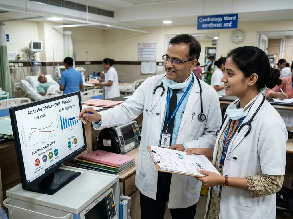
The VOCAL-AHF study reveals that most acute heart failure patients across reduced and non-reduced ejection fraction categories are eligible for in-hospital GDMT initiation. Notably, nearly two-thirds of HFrEF patients qualify for quadruple therapy, while non-reduced EF patients show high eligibility for dual therapy.
Today
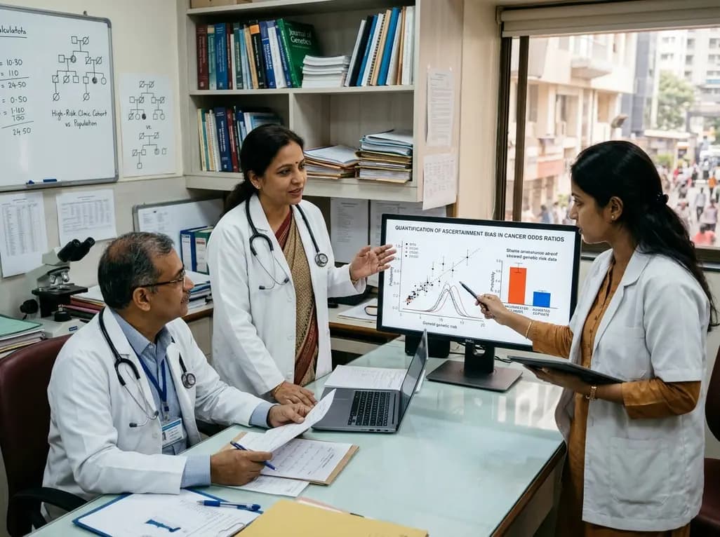
Ascertainment bias in cancer case-control studies inflates odds ratios and penetrance estimates when cases are drawn from high-risk clinics. A landmark modeling study demonstrates substantial bias ranging up to 23.72, highlighting the critical need for ascertainment adjustment in genetic risk counseling.
Today
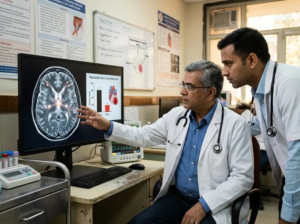
Researchers demonstrate that central induction of synthetic torpor in non-hibernating rats provides striking cardioprotection, reducing myocardial infarct size by ~40% during ischaemia-reperfusion injury through endogenous survival signaling, independent of hypothermia.
Today
A novel fluorine-functionalized near-infrared copper fluorescent probe selectively illuminates lung tissue, revealing critical links between hyperglycemia, ER stress, and pulmonary malignancy for improved early diagnosis.
Today
